Mission Microbe Created By - Science Olympiad Student Center
Mission Microbe Created By - Science Olympiad Student Center
Mission Microbe Created By - Science Olympiad Student Center

You also want an ePaper? Increase the reach of your titles
YUMPU automatically turns print PDFs into web optimized ePapers that Google loves.
<strong>Mission</strong> <strong>Microbe</strong><br />
Sample Test 2010-2011<br />
NC <strong>Science</strong> <strong>Olympiad</strong><br />
<strong>Created</strong> <strong>By</strong>: Rick Mencer<br />
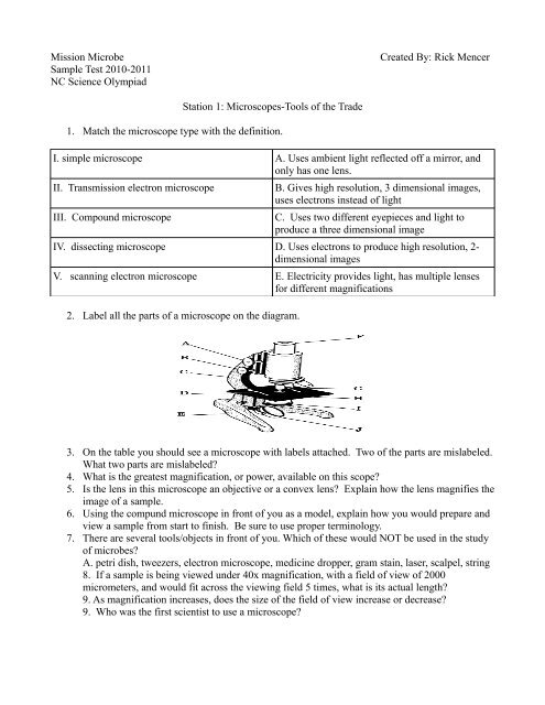
Station 1: Microscopes-Tools of the Trade<br />
1. Match the microscope type with the definition.<br />
I. simple microscope A. Uses ambient light reflected off a mirror, and<br />
only has one lens.<br />
II. Transmission electron microscope<br />
III. Compound microscope<br />
B. Gives high resolution, 3 dimensional images,<br />
uses electrons instead of light<br />
C. Uses two different eyepieces and light to<br />
produce a three dimensional image<br />
IV. dissecting microscope D. Uses electrons to produce high resolution, 2-<br />
dimensional images<br />
V. scanning electron microscope E. Electricity provides light, has multiple lenses<br />
for different magnifications<br />
2. Label all the parts of a microscope on the diagram.<br />
3. On the table you should see a microscope with labels attached. Two of the parts are mislabeled.<br />
What two parts are mislabeled<br />
4. What is the greatest magnification, or power, available on this scope<br />
5. Is the lens in this microscope an objective or a convex lens Explain how the lens magnifies the<br />
image of a sample.<br />
6. Using the compund microscope in front of you as a model, explain how you would prepare and<br />
view a sample from start to finish. Be sure to use proper terminology.<br />
7. There are several tools/objects in front of you. Which of these would NOT be used in the study<br />
of microbes<br />
A. petri dish, tweezers, electron microscope, medicine dropper, gram stain, laser, scalpel, string<br />
8. If a sample is being viewed under 40x magnification, with a field of view of 2000<br />
micrometers, and would fit across the viewing field 5 times, what is its actual length<br />
9. As magnification increases, does the size of the field of view increase or decrease<br />
9. Who was the first scientist to use a microscope
<strong>Mission</strong> <strong>Microbe</strong><br />
Sample Test 2010-2011<br />
NC <strong>Science</strong> <strong>Olympiad</strong><br />
<strong>Created</strong> <strong>By</strong>: Rick Mencer<br />
Station 2: Using Microscopes to Identify Samples as Bacterial, Fungal, or Viral<br />
(This sample test uses images from sources on the internet (see the Works Cited page). The actual event<br />
may use sample on already prepared slides, which would require students to know how to set the slide<br />
on the microscope, adjust the lighting, magnification, and focus as necessary).<br />
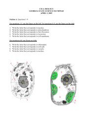
1. Identify each the two drawings below as eukarotic or prokaryotic cells. Label each part. (images<br />
from www.phschool.com. Copyright Prentice Hall 2010.<br />
Illustration 1: Cell A<br />
Illustration 2: Cell B<br />
2. Give one example of a eukaryotic cell, and one example of a prokaryotic cell. Explain how the<br />
two are different.<br />
3. List five main types of microbes.<br />
4. The following examples are all different kinds of microbes. Identify each microbe as one of the<br />
five main types of microbes.<br />
Images from www.soinc.com, from “<strong>Microbe</strong> <strong>Mission</strong> 2011, created by Karen Lancour, National<br />
Supervisor- National Rules committee, Chairman-Life <strong>Science</strong>s unless otherwise noted.<br />
5. Use the microscope to view the slide. Sketch what you see, and identify it as an amoeba, bacterium,<br />
or virus.<br />
(For information on calculating viewing fields and sample sizes, go to<br />
http://www.saskschools.ca/curr_content/biology20/unit1/. Click Lessons 2 and 4
<strong>Mission</strong> <strong>Microbe</strong><br />
Sample Test 2010-2011<br />
NC <strong>Science</strong> <strong>Olympiad</strong><br />
<strong>Created</strong> <strong>By</strong>: Rick Mencer<br />
Population Growth<br />
Answer the following questions using the graphs/charts below.<br />
1. Which axis shows the number of viable cells<br />
2. At about what hour did exponential growth begin and end<br />
3. Explain why the population of cells increased exponetially.<br />
4. Identify at least two reasons why the population of viable cells became stationary, then began to<br />
decline<br />
5. If more food source is added to the sample culture, predict how the population will change.<br />
6. Would the population growth curve for the deer population in an area be exponential (as in the<br />
chart above) or linear<br />
7. Use the table below to sketch a graph showing the change in population over 5 hours for a<br />
colony of Protozoa. Assume no death occurs.<br />
Time (Hours)<br />
Population (thousands)<br />
1 10<br />
2 30<br />
3 90<br />
4 360<br />
5 1080<br />
8. Predict what the population will be in 7 hours, 10 hours, and 12 hours, assuming none of the<br />
organisms die.<br />
9. Explain why unlimited population growth in microbes is very unlikely or impossible.
<strong>Mission</strong> <strong>Microbe</strong><br />
Sample Test 2010-2011<br />
NC <strong>Science</strong> <strong>Olympiad</strong><br />
<strong>Created</strong> <strong>By</strong>: Rick Mencer<br />
Station 4: <strong>Microbe</strong>s and Diseases<br />
(At a competition event, I would have these as matching flashcards: Five of them would be numbered<br />
1-5, and show a specific kind of microbe- image and/or word. The next five would be labeled A-E,<br />
accompanied by a single specific disease/symptoms. Competitors would match these, and transfer the<br />
answers to the answer sheet. I give examples here in chart form due to time constraints.)<br />
1.Protozoa<br />
<strong>Microbe</strong><br />
Disease<br />
A. Influenza<br />
2. Fungus B Tubercolosis<br />
3. Bacteria C. Dysentery<br />
4. Virus D. Mad Cow Disease<br />
5. Prions E. Athletes Foot<br />
2. Identify at least two symptoms for each kind of disease listed above.<br />
3. Identify at least one mode of entry (one way the disease can be caught) for each example listed<br />
above.<br />
4. Of the microbes and diseases listed above, which two are the most difficult to treat and why
<strong>Mission</strong> <strong>Microbe</strong><br />
Sample Test 2010-2011<br />
NC <strong>Science</strong> <strong>Olympiad</strong><br />
<strong>Created</strong> <strong>By</strong>: Rick Mencer<br />
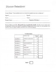
Part A<br />
1. Label each organelle in the cell below.<br />
Station 5: Organelles and Cells<br />
2. Is this microbe a bacteria or a virus<br />
3. Label each organelle in the cell below.<br />
(Insert protozoa diagram for labeling)<br />
4. Is this microbe an fungus or a bacteria<br />
Part B<br />
1. Set up the microscope to view the slide. Make a quick sketch of what you see.<br />
2. Identify the sample as a protozoa or a fungus.<br />
3. What organelles are visible<br />
Part C<br />
1. Create a double Venn Diagram showing how protozoa and cyanobacteria.
<strong>Mission</strong> <strong>Microbe</strong><br />
Sample Test 2010-2011<br />
NC <strong>Science</strong> <strong>Olympiad</strong><br />
<strong>Created</strong> <strong>By</strong>: Rick Mencer<br />
Station 6: Positive Uses of <strong>Microbe</strong>s<br />
Part A<br />
Match the product or outcome with the associated microbe.<br />
Product/Benefit<br />
Associated <strong>Microbe</strong><br />
1. Petroleum A. Saccharomyces cerevisiae<br />
2. Penicillin Lactobacillus bulgaricus<br />
3.Yogurt<br />
4 Bread D.<br />
C. Clostridun<br />
5 Gene splicing E. penecillium notatum<br />
6. Intestinal Tract (Food digestion) F. Botryococcus braunii<br />
Part B<br />
1. Why are people taking antibiotics often feel nauseus<br />
2. What organelle found in all human cells is thought to have evolved from a symbiotic<br />
relationship between host cells and a bacteria<br />
3. When BP applied oil-eating microbes to the recent Gulf Oil Spill, what were scientists<br />
concerned about Were these microbes aerobic or anaerobic
<strong>Mission</strong> <strong>Microbe</strong><br />
Sample Test 2010-2011<br />
NC <strong>Science</strong> <strong>Olympiad</strong><br />
<strong>Created</strong> <strong>By</strong>: Rick Mencer