Biological activity of substrate-bound basic fibroblast growth factor ...
Biological activity of substrate-bound basic fibroblast growth factor ...
Biological activity of substrate-bound basic fibroblast growth factor ...

Create successful ePaper yourself
Turn your PDF publications into a flip-book with our unique Google optimized e-Paper software.
3890<br />
<strong>Biological</strong> <strong>activity</strong> <strong>of</strong> immobilized FGF2<br />
E Tanghetti et al<br />
(Brooks et al., 1994; Friedlander et al., 1995; Eliceiri et<br />
al., 1998). Interestingly, FGFRs, integrins, and intracellular<br />
transducers may co-localize in focal adhesion<br />
contacts (Plopper et al., 1995, Miyamoto et al.,<br />
1996). Despite these observations, the molecular<br />
mechanism(s) underlying the relationship between the<br />
FGF2/FGFR system and the cell adhesion machinery<br />
are not fully elucidated.<br />
Large amounts <strong>of</strong> FGF2 are present in ECM both in<br />
vivo and in vitro (Vlodavsky et al., 1987; Folkman et<br />
al., 1988). Collagen-<strong>bound</strong> FGF2 is mitogenically<br />
active in situ for BALB/c-3T3 ®broblasts (Smith et<br />
al., 1982) and FGF2 immobilized onto heparin-coated<br />
surfaces promotes endothelial cell adhesion (Baird et<br />
al., 1988) and PC12 cell adhesion and di€erentiation<br />
(Schubert et al., 1987). Thus, ECM-<strong>bound</strong> FGF2 may<br />
induce endothelial cell adhesion and act at the same<br />
time as a localized, persistent stimulus for angiogenesis<br />
by interacting with di€erent cell-surface molecules.<br />
Indeed, previous data obtained in our laboratory had<br />
shown that FGF2 interacts also with a v b 3 integrin and<br />
that this interaction mediates the capacity <strong>of</strong> the<br />
angiogenic <strong>growth</strong> <strong>factor</strong> to induce cell adhesion,<br />
mitogenesis, and urokinase-type plasminogen activator<br />
upregulation in endothelial cells (Rusnati et al., 1997).<br />
Interestingly, the endothelial cell-adhesive capacity <strong>of</strong><br />
FGF2 does not require the interaction <strong>of</strong> the <strong>growth</strong><br />
<strong>factor</strong> with FGFRs and/or HSPGs that are instead<br />
able to mediate a FGF2-dependent cell-cell interaction<br />
via the formation <strong>of</strong> a ternary FGFR/FGF2/HSPG<br />
complex (Richard et al., 1995).<br />
In the present study we investigated the molecular<br />
mechanisms mediating the mitogenic <strong>activity</strong> <strong>of</strong><br />
<strong>substrate</strong>-<strong>bound</strong> FGF2 in endothelial cells. The results<br />
demonstrate that immobilized FGF2 induces focal<br />
adhesion plaque formation and the activation <strong>of</strong><br />
extracellular signal-regulated kinases (ERK 1/2 ) in<br />
bovine aortic endothelial GM7373 cells. This is<br />
paralleled by a signi®cant increase in the proliferation<br />
rate <strong>of</strong> adherent cells. GM7373 cells transfected with a<br />
dominant negative, truncated TK 7 FGFR1 mutant<br />
adhere and spread but have lost the capacity to<br />
proliferate on immobilized FGF2. Intact FGFR1, but<br />
not the truncated receptor, localizes in the focal<br />
adhesion contacts induced by FGF2. These data shed<br />
a new light on the cross-talk between endothelial cell<br />
adhesion and proliferative events during angiogenesis.<br />
and the localization <strong>of</strong> vinculin (Figure 1B) and<br />
paxillin (not shown) demonstrate that immobilized<br />
FGF2 induces the formation <strong>of</strong> focal adhesion contacts<br />
in adherent GM7373 cells. Similar results were<br />
obtained for cells adherent to FN or VN (data not<br />
shown). Under the same experimental conditions, GM<br />
7373 cells attach (Figure 1A) but do not spread (not<br />
shown) on polylysine (PLL)-coated plastic.<br />
To con®rm these observations, GM7373 cells were<br />
seeded on FGF2, FN, VN, or PLL, allowed to adhere<br />
for 6 h, and stripped by PBS/EDTA washes (Culp,<br />
1976; Del Rosso et al., 1992). Cell-substratum contact<br />
sites, that represent 3 ± 4% <strong>of</strong> the surface membrane <strong>of</strong><br />
monolayered cells (Del Rosso et al., 1992), were then<br />
extracted and analyzed by Western blotting. As shown<br />
in Figure 2, FGF2-adherent plasma membrane remnants<br />
contain paxillin, a v b 3 integrin, focal adhesion<br />
kinase (FAK), and pp60 src . Similar results were<br />
obtained with cells adherent to FN and VN but not<br />
with cells adherent to PLL.<br />
Taken together the results demonstrate that immobilized<br />
FGF2 promotes focal adhesion plaque formation<br />
in endothelial cells with the recruitment <strong>of</strong> signal<br />
transducing molecules including FAK and pp60 src .<br />
Immobilized FGF2 induces endothelial cell proliferation<br />
Next, we evaluated the capacity <strong>of</strong> plastic-<strong>bound</strong> FGF2<br />
to promote cell proliferation in endothelial GM 7373<br />
cells. To this purpose, cells were allowed to adhere for<br />
2 h on di€erent substrata, including native or heatinactivated<br />
FGF2, FN, or VN. Then, non-adherent<br />
cells were removed and adherent cells were incubated<br />
in low serum. After a 24 h incubation, cells were<br />
trypsinized and counted. As shown in Figure 3, only<br />
immobilized native FGF2 is able to exert a rapid and<br />
signi®cant mitogenic response in adherent cells that<br />
Results<br />
Substrate-<strong>bound</strong> FGF-2 promotes focal adhesion plaque<br />
formation in endothelial cells<br />
Previous observations had shown that <strong>substrate</strong>-<strong>bound</strong><br />
FGF2 promotes endothelial cell adhesion via a v b 3<br />
integrin engagement (Rusnati et al., 1997). Accordingly,<br />
fetal bovine aortic endothelial GM 7373 cells<br />
adhere onto non-tissue culture plates coated with<br />
native or heat-inactivated FGF2, FN, or VN (Figure<br />
1A). Also, the distribution and organization <strong>of</strong> F-actin<br />
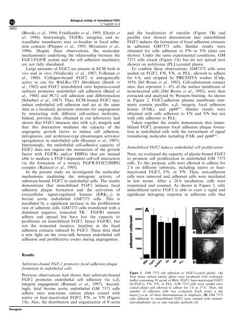
Figure 1 GM 7373 cell adhesion to FGF2-coated plastic. (A)<br />
Non tissue culture plastic plates were incubated with carbonate<br />
bu€er containing 20 mg/ml <strong>of</strong> BSA, FGF2, heat-inactivated FGF2<br />
(hi-FGF2), FN, VN, or PLL. GM 7373 cells were seeded onto<br />
coated plates and allowed to adhere for 2 h at 378C. Then, the<br />
number <strong>of</strong> adherent cells was evaluated. Each point is the<br />
mean+s.e.m. <strong>of</strong> three determinations in duplicate. (B) GM 7373<br />
cells adherent to immobilized FGF2 were stained with rodaminate-phalloidin<br />
(a) or anti-vinculin antibody (b)<br />
Oncogene