Biological activity of substrate-bound basic fibroblast growth factor ...
Biological activity of substrate-bound basic fibroblast growth factor ...
Biological activity of substrate-bound basic fibroblast growth factor ...

Create successful ePaper yourself
Turn your PDF publications into a flip-book with our unique Google optimized e-Paper software.
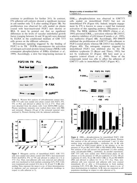
continue to proliferate for further 24 h. In contrast,<br />
FN adherent cell cultures showed a signi®cant increase<br />
in cell number only 72 h after seeding (Figure 3B). No<br />
proliferation was observed for cells seeded on plastic<br />
coated with heat-inactivated FGF2 (not shown) or<br />
BSA. It must be pointed out that no signi®cant<br />
di€erences in the levels <strong>of</strong> vascular endothelial <strong>growth</strong><br />
<strong>factor</strong> (ranging between 16 and 30 pg/ml) were detected<br />
by ELISA in the conditioned medium <strong>of</strong> GM 7373<br />
cells grown on the di€erent substrata.<br />
Downstream signaling triggered by the binding <strong>of</strong><br />
FGF2 to its TK + FGFRs encompasses the activation<br />
<strong>of</strong> mitogen-activated protein kinase kinase (MEK) with<br />
consequent phosphorylation <strong>of</strong> ERKs (Giuliani et al.,<br />
1999). Accordingly, a slow but long-lasting increase in<br />
<strong>Biological</strong> <strong>activity</strong> <strong>of</strong> immobilized FGF2<br />
E Tanghetti et al<br />
ERK 1/2<br />
phosphorylation was observed in GM7373<br />
cells seeded on immobilized FGF2 but not on<br />
immobilized FN (Figure 4A). Indeed, integrin engagement<br />
by FN is known to cause a rapid but transient<br />
activation <strong>of</strong> this signaling pathway (Miyamoto et al.,<br />
1996). The MEK inhibitor PD 098059 (Alessi et al.,<br />
1995) prevented ERK 1/2 activation whereas SB 210313,<br />
a selective inhibitor <strong>of</strong> p38 kinase (Cuenda et al., 1995),<br />
was ine€ective (Figure 4B). Accordingly, PD 098059<br />
inhibited the proliferation <strong>of</strong> GM7373 cells adherent to<br />
FGF2-coated plastic whereas SB 210313 was ine€ective<br />
(Figure 4D). The mitogenic response triggered by<br />
immobilized FGF2 was inhibited also by the TK<br />
inhibitor tyrphostin 23 (Boyer and Thiery, 1993), but<br />
not by tyrphostin 63 (Figure 4D) here used as a<br />
negative control (Gazit et al., 1989). None <strong>of</strong> the<br />
compounds tested was able to a€ect the adhesion <strong>of</strong><br />
GM7373 cells to immobilized FGF2 (Figure 4C).<br />
3891<br />
Figure 2 Western blot analysis <strong>of</strong> cell-substratum contact sites.<br />
GM 7373 cells were seeded onto plates coated with FGF2, FN,<br />
VN, or PLL and allowed to adhere for 6 h at 378C. Then cells<br />
were detached from the plastic with 3 mM EDTA/PBS washes.<br />
Plasma membrane remnants were washed three times with PBS<br />
and extracted. Aliquots (30 mg) <strong>of</strong> the extracted material were<br />
analysed by Western blotting with the indicated antibodies<br />
respect to cells adherent at T 0 population doublings in respect to cells adherent at T 0<br />
Figure 4 ERK 1/2 phosphorylation by immobilized FGF2. GM<br />
7373 cells were seeded onto FN- or FGF2-coated plastic. Western<br />
blot analysis <strong>of</strong> the cell extracts was performed 1 and 2 h after<br />
seeding using anti-phospho-ERK 1/2 antibodies (A). In B, cells<br />
were seeded on FGF2-coated plastic in the absence or in the<br />
presence <strong>of</strong> 50 mM SB 210313 (SB) or PD 098059 (PD). Western<br />
blot analysis <strong>of</strong> the cell extracts was performed 2 h after seeding<br />
using anti-phospho-ERK 1/2 antibodies. In parallel experiments,<br />
Figure 3 Mitogenic <strong>activity</strong> <strong>of</strong> immobilized FGF2. GM 7373 GM 7373 cells were seeded onto FGF2-coated plates in the<br />
cells were seeded onto plates coated with FGF2, heat-inactivated<br />
FGF2 (hi-FGF2), FN, or VN and allowed to adhere for 2 h at<br />
378C (T 0 ). Then, non-adherent cells were removed and adherent<br />
cells were incubated in fresh medium containing 0.4% FCS. Cells<br />
were trypsinized and counted 24 h (A) or 24, 48 and 72 h (B) after<br />
seeding. Data represent the mean+s.e.m. <strong>of</strong> three determinations<br />
in duplicate and are expressed as cell population doublings in<br />
absence or in the presence <strong>of</strong> 100 mM tyrphostin 23 (t23), 100 mM<br />
tyrphostin 63 (t63), 50 mM SB 210313 (SB) or 50 mM PD 098059<br />
(PD) and allowed to adhere for 2 h at 378C (T 0 ). Then, nonadherent<br />
cells were removed and adherent cells were counted<br />
immediately (C) or after a 24 h-incubation in fresh medium<br />
containing 0.4% FCS (D). Data represent the mean+s.e.m. <strong>of</strong><br />
three determinations in duplicate. In D, data are expressed as cell<br />
Oncogene