Abdomen and Pelvis objectives - Surgery - Severn Deanery
Abdomen and Pelvis objectives - Surgery - Severn Deanery
Abdomen and Pelvis objectives - Surgery - Severn Deanery
You also want an ePaper? Increase the reach of your titles
YUMPU automatically turns print PDFs into web optimized ePapers that Google loves.
1<br />
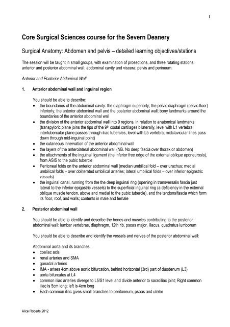
Core Surgical Sciences course for the <strong>Severn</strong> <strong>Deanery</strong><br />
Surgical Anatomy: <strong>Abdomen</strong> <strong>and</strong> pelvis – detailed learning <strong>objectives</strong>/stations<br />
The session will be taught in small groups, with examination of prosections, <strong>and</strong> three rotating stations:<br />
anterior <strong>and</strong> posterior abdominal wall; abdominal cavity <strong>and</strong> viscera; pelvis <strong>and</strong> perineum.<br />
Anterior <strong>and</strong> Posterior Abdominal Wall<br />
1. Anterior abdominal wall <strong>and</strong> inguinal region<br />
You should be able to describe:<br />
the boundaries of the abdominal cavity: the diaphragm superiorly; the pelvic diaphragm (pelvic floor)<br />
inferiorly; the anterior abdominal wall <strong>and</strong> the posterior abdominal wall; bony l<strong>and</strong>marks around the<br />
boundaries of the anterior abdominal wall<br />
the division of the anterior abdominal wall into 9 regions, in relation to anatomical l<strong>and</strong>marks<br />
(transpyloric plane joins the tips of the 9 th costal cartilages bilaterally, level with L1 vertebra;<br />
intertubercular plane passes through iliac tubercles, level with L5 vertebra; midclavicular lines pass<br />
down through mid-inguinal point)<br />
the cutaneous innervation of the anterior abdominal wall<br />
the layers of the anterolateral abdominal wall (NB. No deep fascia over thorax or abdomen)<br />
the attachments of the inguinal ligament (the inferior free edge of the external oblique aponeurosis),<br />
from ASIS to the pubic tubercle<br />
Peritoneal folds on the anterior abdominal wall (median umbilical fold – over urachus; medial<br />
umbilical folds – over obliterated umbilical arteries; lateral umbilical folds – over inferior epigastric<br />
vessels)<br />
the inguinal canal, running from the the deep inguinal ring (opening in transversalis fascia just<br />
lateral to the inferior epigastric vessels) to the superficial inguinal ring (a deficiency in the external<br />
oblique muscle tendon, above <strong>and</strong> medial to the pubic tubercle), <strong>and</strong> the tendons/fascia which form<br />
its floor, roof, <strong>and</strong> walls; contents in male <strong>and</strong> female<br />
2. Posterior abdominal wall<br />
You should be able to identify <strong>and</strong> describe the bones <strong>and</strong> muscles contributing to the posterior<br />
abdominal wall: lumbar vertebrae, diaphragm, 12th rib, psoas major, iliacus, quadratus lumborum<br />
You should be able to describe <strong>and</strong> identify the vessels <strong>and</strong> nerves of the posterior abdominal wall:<br />
Abdominal aorta <strong>and</strong> its branches:<br />
coeliac axis<br />
renal arteries <strong>and</strong> SMA<br />
gonadal arteries<br />
IMA - arises 4cm above aortic bifurcation, behind horizontal (3rd) part of duodenum (L3)<br />
aorta bifurcates at L4<br />
common iliac arteries diverge to L5/S1 level <strong>and</strong> divide anterior to sacroiliac joint; Right common<br />
iliac is 5cm long; left is 4cm long<br />
Each common iliac gives small branches to peritoneum, psoas <strong>and</strong> uteter<br />
Alice Roberts 2012
2<br />
<br />
<br />
<br />
External iliac artery forms principal supply to lower limb<br />
Internal iliac artery supplies pelvic viscera, walls <strong>and</strong> gluteal region<br />
common iliac <strong>and</strong> gonadal arteries<br />
IVC <strong>and</strong> its tributaries: common iliac, gonadal, renal <strong>and</strong> hepatic veins.<br />
Branches of the lumbar plexus emerging around <strong>and</strong> through psoas: subcostal, ilioinguinal <strong>and</strong><br />
iliohypogastric, genitofemoral, lateral femoral cutaneous, femoral, obturator nerves <strong>and</strong> the lumbosacral<br />
trunk.<br />
You should be able to describe <strong>and</strong> identify the kidneys:<br />
Position <strong>and</strong> relations of the kidneys: extend from T12 to L3 with the hila at the level of L1; lie 3<br />
finger breadths from the midline; right kidney slightly lower than the left<br />
The three ‘capsules’ of the kidney (fibrous capsule; perinephric fat; renal/extraperitoneal fascia)<br />
Posterior relations of each kidney (diaphragm, 12 th rib, quadratus lumborum, psoas, transversus<br />
abdominis; subcostal, iliohypogastric, ilioinguinal nerves)<br />
Anterior relations of the right kidney (suprarenal gl<strong>and</strong>, liver, 2 nd part of duodenum, ascending<br />
colon) <strong>and</strong> left kidney (suprarenal gl<strong>and</strong>, stomach, pancreas, spleen, descending colon)<br />
You should be able to describe <strong>and</strong> identify the suprarenal gl<strong>and</strong>s:<br />
Right adrenal gl<strong>and</strong> lies posterior to liver; left lies posterior to stomach <strong>and</strong> pancreas; both lie within<br />
the coverings of the perinephric fat <strong>and</strong> renal fascia<br />
Structure: outer cortex <strong>and</strong> inner medulla<br />
Right adrenal gl<strong>and</strong> is pyramidal; left is crescent-shaped <strong>and</strong> larger)<br />
Supplied by inferior phrenic arteries, aorta <strong>and</strong> renal arteries;<br />
You should be able to describe the external appearance <strong>and</strong> internal architecture of the kidneys:<br />
poles, hilum (order of structures entering/exiting)<br />
cortex; medullary pyramids; major <strong>and</strong> minor calyces; renal pelvis <strong>and</strong> hilum of the kidney<br />
lymph from the kidney drains to para-aortic nodes<br />
sympathetic innervation via renal plexus; afferents enter the spinal cord in the tenth, eleventh <strong>and</strong><br />
twelth thoracic nerves<br />
You should be able to discuss how <strong>and</strong> why an intravenous urogram (IVU) may be obtained, <strong>and</strong> the<br />
appearance of common problems on the IVU (eg: cystic or neoplastic disease may be seen, reduced<br />
renal blood flow, obstruction, bladder filling defects <strong>and</strong> emptying). There are three constrictions along<br />
the course of the ureter: pelvi-ureteric junction (PUJ), pelvic brim, vesico-ureteric junction (VUJ)<br />
You should underst<strong>and</strong> the anatomical basis of renal clinical problems, including:<br />
Developmental anomalies (polycystic kidneys, horseshoe kidney, duplication of the ureter, ectopic<br />
ureters, pelvic kidney)<br />
Urinary tract calculi – complications <strong>and</strong> treatment<br />
Referred pain from the kidney produces typical ‘loin to groin’ pain: visceral afferents in the least<br />
splanchnic nerve enter the spinal cord at T12, so pain is referred to T12 dermatome, corresponding<br />
to the flanks <strong>and</strong> pubic region.<br />
Alice Roberts 2012
3<br />
You should be able to describe <strong>and</strong> identify the abdominal ureters:<br />
25cm long; lined with uroepithelium/transitional epithelium<br />
<strong>Pelvis</strong> of ureter/renal pelvis variably embedded in kidneys – from intrarenal to extrarenal<br />
Abdominal ureter lies retroperitoneally (adherent to peritoneum), on the medial border of psoas<br />
major (anterior to the tips of transverse processes of lumbar vertebrae); crosses pelvic brim at<br />
bifurcation of common iliac artery<br />
Ureter crosses pelvic brim, over bifurcation of common iliac artery, turns forward <strong>and</strong> medially at<br />
level of ischial spine; lies close to seminal vesicle in the male; passes lateral to the cervix <strong>and</strong> under<br />
the uterine vessels in the female<br />
Blood supply to the ureter is derived from local arteries along its course (venous drainage via<br />
corresponding veins): aorta, renal artery, gonadal artery, internal iliac artery, inferior vesical artery<br />
Lymph drains to para-aortic nodes<br />
Nerve supply via autonomic plexuses<br />
You should be able to describe the normal anatomy of the bladder, <strong>and</strong> mechanisms of continence <strong>and</strong><br />
micturition:<br />
The empty bladder lies in the true pelvis, posterior to the pubic symphysis; exp<strong>and</strong>s out of the pelvis<br />
as it fills, to become intra-abdominal; ureteric peristalsis pumps urine into bladder; the network of<br />
detrusor fibres gives the bladder wall a trabeculated appearance – becomes more prominent in<br />
obstruction<br />
Supplied by superior <strong>and</strong> inferior vesical/vaginal artery<br />
Lymph drains to external <strong>and</strong> internal iliac nodes<br />
Parasympathetic motor innervation <strong>and</strong> sensation of distension<br />
Sympathetic efferents are vasomotor, motor to internal urethral sphincter <strong>and</strong> muscle of trigone<br />
Pain is carried by both parasympathetic <strong>and</strong> sympathetic afferents<br />
The external urethral sphincter (striated muscle, supplied by pudendal nerve) is also important in<br />
continence; midflow stop is achieved by contraction of external urethral sphincter with delayed<br />
reflex relaxation of detrusor muscle; external sphincter assisted by pelvic floor (levator ani muscle)<br />
in active continence<br />
You should be able to discuss how <strong>and</strong> why urethral vs suprapubic catheterisation may be carried out.<br />
You should underst<strong>and</strong> the anatomical basis of common lower urinary tract pathologies, including:<br />
Urinary tract calculi - may lodge inside external urethral meatus<br />
Straddle injuries – urine collects in the superficial pouch<br />
Benign prostatic hyperplasia (BPH) - commonest urinary tract pathology in men<br />
Urinary tract infection (UTI) - commonest pathology in women<br />
Incontinence: stress incontinence; urge incontinence; overflow incontinence; incontinence after<br />
childbirth<br />
Alice Roberts 2012
4<br />
Abdominal cavity <strong>and</strong> viscera<br />
1. Peritoneum, peritoneal cavity <strong>and</strong> compartments<br />
You should be able to describe the peritoneum:<br />
A thin serous membrane (similar to pleura <strong>and</strong> serous pericardium);<br />
Parietal <strong>and</strong> visceral layers; forms folds through which nerves <strong>and</strong> vessels reach the viscera<br />
Entire embryonic gut tube possesses a mesentery; some gut remains intraperitoneal <strong>and</strong> retains a<br />
mesentery, other parts become secondarily retroperitoneal<br />
Peritoneal fluid lubricates gut movement <strong>and</strong> expansion<br />
Visceral peritoneum innervated by autonomic afferents from viscera; parietal by somatic nerves<br />
Describe the omenta, greater <strong>and</strong> lesser sacs:<br />
Lesser omentum - double fold of peritoneum joining the lesser curvature of the stomach to the<br />
visceral surface of the liver (part of the embryonic ventral mesogastrium)<br />
Lesser sac (omental bursa) - peritoneal pouch behind the lesser omentum <strong>and</strong> stomach;<br />
communicating with the greater sac (peritoneal cavity) at the epiploic foramen; formed by rotation of<br />
stomach during embryological development<br />
Dorsal mesentery of stomach forms greater omentum, gastrosplenic <strong>and</strong> splenorenal ligaments<br />
The greater omentum – a quadruple fold of peritoneum hanging down over the intestines, <strong>and</strong><br />
attaching from the greater curvature of the stomach to the transverse colon; its two posterior layers<br />
adhere to the transverse mesocolon to form the posterior wall of lesser sac (surgically separable);<br />
contains fat <strong>and</strong> macrophages<br />
Splenic vessels <strong>and</strong> tail of pancreas in splenorenal lig<br />
Epiploic foramen (of Winslow) - vertical slit about 2.5cm long; anterior border formed by free edge of<br />
lesser omentum (containing portal vein posteriorly, bile duct <strong>and</strong> hep artery anteriorly, & autonomic<br />
nerves, lymphatics)<br />
Describe the peritoneal compartments <strong>and</strong> pouches:<br />
Supracolic compartment above attachment of transverse mesocolon (over body of pancreas);<br />
infracolic below (NB. traction on root of mesentery stimulates pressure receptors <strong>and</strong> causes reflex<br />
drop in BP)<br />
Supracolic compartment divided into right <strong>and</strong> left subphrenic/diaphragmatic spaces by falciform<br />
ligament – extend up as far as sup layer of coronary ligament on right <strong>and</strong> anterior layer of left<br />
triangular ligament on the left<br />
Right subhepatic space (or hepatorenal pouch) between lower layer of coronary ligament <strong>and</strong><br />
transverse mesocolon attachment (lowest part of cavity when supine – fluid accumulates here)<br />
Right paracolic gutter <strong>and</strong> right infracolic space lateral <strong>and</strong> medial to ascending colon, respectively<br />
Left paracolic gutter <strong>and</strong> left infracolic space lateral <strong>and</strong> medial to descending colon, respectively<br />
Left infracolic space cont with pelvic cavity<br />
Sigmoid mesocolon attaches down to S3 – then rectum<br />
Alice Roberts 2012
5<br />
2. Digestive tract<br />
You should be able to identify <strong>and</strong> describe the following parts of the digestive tract, within the<br />
abdominal cavity:<br />
Oesophagus<br />
25cm long<br />
Lies anterior to lower cervical vertebrae <strong>and</strong> prevertebral fascia, posterior to trachea in the neck;<br />
anterior to thoracic vertebrae, posterior to trachea, arch of the aorta, left bronchus, pericardium <strong>and</strong><br />
left atrium in chest; passes through right crus of diaphragm; 1-2cm of abdominal oesophagus, with<br />
vagal trunks lying anterior <strong>and</strong> posterior; enters cardiac orifice of stomach<br />
Factors guarding against gastric reflux: contraction of right crus, angle of entry of oesophagus,<br />
longitudinal folds of oesophageal mucosa, high pressure zone in the last few cm, effect of increased<br />
intra-abdominal pressure on abdominal oesophagus<br />
Constrictions (may hold up endoscope) at cricopharyngeal sphincter; crossing of the left bronchus;<br />
at the diaphragm<br />
Outer longitudinal <strong>and</strong> inner circular muscle layers - upper third is striated muscle, becoming<br />
smooth muscle<br />
Lined with SSNKE, with mucous gl<strong>and</strong>s<br />
Innervation: autonomic afferents <strong>and</strong> efferents; parasympathetic efferents stimulate contraction<br />
Blood supply: upper third: inferior thyroid artery <strong>and</strong> vein; middle third: branches of thoracic aorta,<br />
drains to azygos vein; lower third: left gastric artery <strong>and</strong> vein (tributaries of the left gastric vein <strong>and</strong><br />
azygos vein form portosystemic anastomosis)<br />
Lymph drainage: posterior mediastinal nodes<br />
Stomach<br />
Relations: anteriorly: abdominal wall, left costal margin, left lobe of liver; posteriorly: lesser sac;<br />
superiorly: left dome of diaphragm<br />
Body, fundus, greater <strong>and</strong> lesser curvatures, pyloric antrum, pyloric canal, cardiac <strong>and</strong> pyloric<br />
sphincters<br />
Muscular wall: longitudinal, circular <strong>and</strong> oblique fibres<br />
Mucosa: thick <strong>and</strong> vascular, thrown into rugae; SSNKE replaced by simple columnar epithelium at<br />
cardia; forms gastric pits<br />
Intrinsic innervation from enteric nervous system; extrinsic innervation: parasympathetic from vagal<br />
trunks (large gastric branches run in lesser omentum near curvature); sympathetic from sympathetic<br />
trunks via coeliac plexus<br />
Blood supply coeliac axis (left gastric artery; right gastric artery from hepatic artery; right<br />
gastroepiploic artery from gastroduodenal branch of hepatic artery; left gastroepiploic <strong>and</strong> short<br />
gastric arteries from splenic artery); veins drain to portal system (portal vein; splenic <strong>and</strong> SM<br />
tributaries)<br />
Lymph drains from lesser curvature <strong>and</strong> upper body drains to right <strong>and</strong> left gastric nodes; lymph<br />
from greater curvature <strong>and</strong> lower body drains to pancreaticosplenic nodes (at splenic hilum) <strong>and</strong><br />
gastroepiploic nodes; lower pylorus drains to hepatic nodes<br />
Prepyloric vein of Mayo (indicates position of pyloric sphincter) drains into right gastric vein<br />
Alice Roberts 2012
6<br />
Duodenum<br />
25cm long; First 2.5cm is intraperitoneal – between layers of omenta<br />
Gallbladder lies anterior to duodenal cap (no plicae circulares)<br />
4 parts:<br />
o 1 st part lies at L1, on transpyloric plane<br />
o 2nd part loops around head of pancreas; lies over hilum of right kidney, with coils of jejunum<br />
in front; receives hepatopancreatic ampulla of vater on major duodenal papilla about 10cm<br />
from pylorus (this also marks the division between foregut <strong>and</strong> midgut)<br />
o 3rd part crossed anteriorly by SM vessels (lies in both infracolic compartments)<br />
o 4th part breaks free from peritoneum at DJ flexure; fixed to psoas by fibrous tissue <strong>and</strong> by<br />
suspensory ligament ligament (of Treitz) to left crus<br />
Paraduodenal recess between 4th part <strong>and</strong> IMV (hernia may become incarcerated here)<br />
Secretin <strong>and</strong> parasympathetic fibres (from vagus) stimulate secretions <strong>and</strong> motility<br />
Blood supply from coeliac axis (superior pancreaticoduodenal artery) <strong>and</strong> SMA (inferior<br />
pancreaticoduodenal artery); first 2cm – usual site of ulceration – also receives blood from hepatic,<br />
gastroduodenal, <strong>and</strong> epiploic arteries; venous drainage to portal <strong>and</strong> SMV<br />
Lymph drains along arteries to coeliac <strong>and</strong> superior mesenteric nodes<br />
Jejunum <strong>and</strong> ileum<br />
Both have plicae circulares <strong>and</strong> villous mucosa<br />
Jejunum (around 2m long) is thicker (wall feels ‘double’) <strong>and</strong> more vascular than ileum (around 3m<br />
long); jejunum has ‘windows’ in its arterial arcade; ileum has more layers of arcades <strong>and</strong> shorter<br />
straight arteries; jejunum lies in upper infracolic compartment (umbilical region); ileum lies in lower<br />
infracolic compartment <strong>and</strong> pelvic cavity (suprapubic region)<br />
Mucosa: columnar epithelium forms villi <strong>and</strong> intestinal gl<strong>and</strong>s (crypts of Lieberkuhn)<br />
In terminal ileum, lymphoid follicles congregate to form Peyer’s patches; gut-associated lymphoid<br />
tissue (GALT) comprises Peyer's patches, peripheral lymphoid tissues <strong>and</strong> appendix<br />
Meckel’s diverticulum: classically found in 2%, 2 feet from ileocaecal junction, <strong>and</strong> 2 inches long<br />
(but may be more proximal <strong>and</strong> length varies); may contain gastric, hepatic or pancreatic tissue;<br />
may ulcerate<br />
Jejunal <strong>and</strong> ileal branches of the SMA; vasa recta/straight arteries are end-arteries – occlusion may<br />
cause infarction<br />
Corresponding veins drain into portal system<br />
Lymph drains along the arteries to the superior mesenteric lymph nodes<br />
Autonomic nerves travel with blood vessels; parasympathetic stimulates contraction <strong>and</strong> secretion;<br />
sympathetic vasocontricts <strong>and</strong> inhibits peristalsis; sympathetic pain fibres enter spinal cord at T9<br />
<strong>and</strong> 10 (referred umbilical pain)<br />
Large intestine<br />
Large diameter compared with small bowel;<br />
Mucosa: no villi, only intestinal gl<strong>and</strong>s only – with many goblet cells; numerous lymphoid follicles in<br />
submucosa <strong>and</strong> mucosa; columnar epithelium changes to SSNKE in anal canal<br />
Taeniae (3 b<strong>and</strong>s of longitudinal muscle) gather the bowel into sacculations, separated by narrower<br />
parts or haustra; taeniae coli converge on appendix; exp<strong>and</strong> to coat the terminal sigmoid colon<br />
Ascending colon is retroperitoneal; transverse colon is intraperitoneal (the transverse mesocolon<br />
varies in length so that the transverse may even hang down to the pelvis); descending colon is<br />
retroperitoneal; the sigmoid colon is intraperitoneal (with its own sigmoid mesocolon)<br />
Alice Roberts 2012
7<br />
<br />
<br />
<br />
<br />
<br />
<br />
<br />
Splenic flexure attached to diaphragm by phrenicocolic ligament; risk of ischaemia in splenic flexure<br />
if arc of Riolan (inner arcade) not well developed; gut supplied by vagi to splenic flexure, then by<br />
pelvic splanchnic nerves; sympathetic supply to colon from T10-L2 - referred umbilical pain<br />
Appendices epiploicae (fatty tags on antimesenteric border; mucosa herniates into them in<br />
diverticulosis) common in descending <strong>and</strong> sigmoid colon<br />
Caceum usually intraperitoneal; appendix may lie in retrocaecal recess; The ileocaecal valve<br />
prevents reflux from the caecum to the ileum<br />
Vermiform appendix usually 6-9cm long; opens into posteromedial wall of caecum; the fold of<br />
peritoneum from terminal ileum to mesoappendix is termed the ‘bloodless fold of Treves’ – but may<br />
contain vessels; Anterior <strong>and</strong> posterior caecal arteries arise from ileocolic; mesenteric artery from<br />
posterior caecal - end-artery, may become thrombosed in appendicitis; position varies: may be<br />
retrocaecal, subcaecal, pelvic (hanging down over pelvic brim), anterior or posterior to the terminal<br />
ileum (preileal or postileal)<br />
Branches of the SMA <strong>and</strong> IMA anastomose to form a continuous arcade along the length of the<br />
bowel; SMA provides: ileocolic artery (supplies terminal ileum, caecum & appendix), right colic<br />
artery (to ascending colon) <strong>and</strong> middle colic artery (to proximal 2/3 of transverse colon); IMA<br />
provides: left colic artery (supplies distal 1/3 of transverse colon <strong>and</strong> descending colon), sigmoid<br />
artery (to sigmoid colon)<br />
Corresponding veins drain to the SMV <strong>and</strong> IMV, <strong>and</strong> then to the portal vein (portosystemic<br />
anastomoses may exist between the mesenteric veins <strong>and</strong> retroperitoneal systemic veins such as<br />
the renal, phrenic <strong>and</strong> lumbar veins)<br />
Lymphatics follow arteries – to superior or inferior mesenteric nodes; lymph from caecum <strong>and</strong><br />
appendix drains to nodes along ileocolic artery<br />
3. Abdominal organs<br />
You should be able to identify <strong>and</strong> describe the following organs:<br />
Liver<br />
Large, wedge-shaped organ occupying most of right hypochondrium & epigastrium; upper margin<br />
level with xiphisternal joint, lower margin a h<strong>and</strong>’s breadth below; reaches to 5th intercostals space<br />
in midclavicular line on left; from ribs 7-11 on right in the midaxillary line<br />
Diaphragmatic & visceral surfaces separated by sharp inferior border<br />
Covered in visceral peritoneum apart from bare area; falciform ligament – to anterior abdominal<br />
wall; ligamentum teres (obliterated umbilical vein) <strong>and</strong> ligamentum venosum (obliterated ductus<br />
venosus)<br />
Portal vein, hepatic artery, bile duct from posterior to anterior at porta hepatis<br />
Bile canaliculi collect bile from hepatocytes…bile ductules in portal triad….intrahepatic ducts… right<br />
<strong>and</strong> left hepatic ducts….common hepatic duct, joined by cystic duct: bile duct<br />
Common hepatic artery may arise directly from aorta or from SMA; right hepatic may arise from<br />
SMA <strong>and</strong> left from left gastric; occlusion of hepatic arteries leads to infarction<br />
Right, left, quadrate <strong>and</strong> caudate lobes (physiologically – left <strong>and</strong> right lobes)<br />
4 sectors, 8 segments by blood supply<br />
In liver transplant – liver plus IVC removed & replaced<br />
Portal vein divisions follow arteries; anastomoses with azygos vv in bare area; venous return mixes<br />
right to left; 3 hepatic veins drain to IVC<br />
3-4 hepatic nodes at porta – drain to coeliac nodes<br />
Sympathetic innervation from coeliac ganglia; parasympathetic from anterior vagal trunk<br />
Alice Roberts 2012
8<br />
Gallbladder<br />
Fibromuscular sac; stores & concentrates bile; 50ml capacity; bound to liver by connective tissue<br />
(may be embedded or hang free); fundus, body <strong>and</strong> neck; fundus contacts parietal peritoneum at tip<br />
of 9th costal cartilage<br />
Lined with columnar epithelium<br />
Cystic duct 2-3cm long, 2-3mm wide; spiral valve (of heister) in neck & cystic duct<br />
Cystic artery from right hepatic artery; multiple cystic veins drain to liver <strong>and</strong> ultimately into hepatic<br />
veins<br />
CCK from small intestine more imp than parasympathetic stimulation for emptying<br />
Afferents from T7-9 <strong>and</strong> some in phrenic – shoulder tip pain<br />
Bile duct 8cm long; may be embedded in pancreas below<br />
Pancreas<br />
Structure <strong>and</strong> function:Head (lying in the curve of the duodenum), neck, body <strong>and</strong> tail (abutting the<br />
splenic hilum); retroperitoneal, behind lesser sac<br />
Uncinate process hooks behind SM vessels; transverse mesocolon attaches across head & body;<br />
splenic artery runs along upper border; tail runs in splenorenal ligament<br />
Main pancreatic duct (of Wirsung) begins in tail <strong>and</strong> runs the length of the gl<strong>and</strong>; accessory (of<br />
Santorini) drains lower head <strong>and</strong> uncinate – 2 ducts communicate<br />
Composite exocrine <strong>and</strong> endocrine gl<strong>and</strong>; acinar cells secrete trypsin, lipase, bicarbonate; islet cells<br />
secrete glucagon (alpha cells), insulin (beta cells) <strong>and</strong> somatostatin (gamma cells)<br />
Mainly supplied by splenic artery; head by superior <strong>and</strong> inferior pancreaticoduodenal arteries;<br />
corresponding veins drain to portal system<br />
Lymph drains to coeliac <strong>and</strong> SM nodes<br />
Secretin <strong>and</strong> CCK more important than parasympathetic stimulation for secretion<br />
Sympathetic vasoconstrictor & pain fibres<br />
Spleen<br />
Intraperitoneal – peritoneal folds form gastrosplenic <strong>and</strong> lienorenal ligaments; anteriorly – the<br />
stomach <strong>and</strong> splenic flexure; posteriorly – the left kidney; laterally – the diaphragm (separating it<br />
from the 9 th , 10 th <strong>and</strong> 11 th ribs)<br />
Contains red <strong>and</strong> white pulp<br />
Extremely friable (it is the commonest abdominal organ to be injured by blunt trauma)<br />
Supplied by splenic artery <strong>and</strong> vein<br />
Alice Roberts 2012
9<br />
<strong>Pelvis</strong> <strong>and</strong> perineum<br />
1. Bones, ligaments <strong>and</strong> muscles of the pelvis<br />
You should be able to:<br />
Identify <strong>and</strong> describe the innominate bones (pubis, ilium <strong>and</strong> ischium) with particular reference to bony<br />
l<strong>and</strong>marks, <strong>and</strong> define the following:<br />
The pelvic inlet (pelvic brim), comprising: pubic crest, iliopectineal line, sacral promontory<br />
The false pelvis (part of the abdominal cavity) lying above the pelvic inlet, <strong>and</strong> the true pelvis below<br />
the inlet<br />
The pelvic outlet, comprising: coccyx, ischial tuberosities, ischiopubic rami, pubic symphysis (these<br />
are also the boundaries of the perineum)<br />
Describe the functions of the bony pelvis: protection of pelvic viscera; supporting body weight; muscle<br />
attachment; in women, Supporting a gravid uterus <strong>and</strong> forming the birth canal<br />
Identify <strong>and</strong> describe the actions <strong>and</strong> innervation of the muscles of the pelvic sidewall - obturator<br />
internus <strong>and</strong> piriformis<br />
Identify <strong>and</strong> describe levator ani (forming the pelvic floor/diaphragm):<br />
Pubococcygeus (arising from posterior surface of pubis), including fibres which form slings around<br />
the prostate (levator prostatae) or vagina (pubovaginalis) <strong>and</strong> the rectum (puborectalis)<br />
Iliococcygeus (arising from the fascia over obturator internus)<br />
Supplied by S4 fibres from the sacral plexus<br />
Supports the pelvic viscera, <strong>and</strong> acts as a sphincter on the rectum <strong>and</strong> vagina - reflexively contracts<br />
to counter raised abdominal pressure, <strong>and</strong> relaxes to allow defaecation, micturition <strong>and</strong> parturition<br />
NB. (Ischio) coccygeus is a functionally separate muscle from levator ani, <strong>and</strong> is represented merely by<br />
a few muscle fibres on the inner surface of the sacrospinous ligament<br />
Describe <strong>and</strong> identify the layers of pelvic fascia:<br />
Parietal pelvic fascia - over obturator internus <strong>and</strong> piriformis<br />
Endopelvic fascia - between levator ani <strong>and</strong> peritoneum, condenses around neurovascular bundles<br />
to form ligaments, eg: sacrouterine <strong>and</strong> pubocervical ligaments, which provide additional support for<br />
pelvic viscera<br />
Alice Roberts 2012
10<br />
2. Neurovascular supply to the pelvis<br />
You should be able to:<br />
Identify <strong>and</strong> describe the sacral <strong>and</strong> pelvic plexuses:<br />
The roots of the sacral plexus lie on the deep surface of piriformis; most of its branches pass out of<br />
the pelvis via the greater sciatic foramen. The pudendal nerve exits the greater sciatic foramen,<br />
wraps around the sacrospinous ligament, enters to the lesser sciatic foramen to lie in the pudendal<br />
(Alcock’s) canal<br />
The inferior hypogastric (pelvic) plexuses lie within the endopelvic fascia, flanking (<strong>and</strong> supplying)<br />
the pelvic viscera; parasympathetic input derives from the pelvic splanchnic nerves (S2,3,4) <strong>and</strong><br />
sympathetic from the superior hypogastric plexus <strong>and</strong> sympathetic chain<br />
Describe the arterial supply to the pelvis: from the internal iliac, median sacral, ovarian <strong>and</strong> inferior<br />
mesenteric (provides superior rectal) arteries.<br />
Identify the internal iliac artery <strong>and</strong> its main branches:<br />
From the posterior division:<br />
Iliolumbar artery – supplies psoas, quadratus lumborum, vertebrae, ilium, iliacus<br />
Lateral sacral artery – supplies sacrum, sacral nerve roots, piriformis<br />
Superior gluteal artery – supplies gluteal muscles <strong>and</strong> skin of buttock<br />
From the anterior division:<br />
Superior vesical artery (continues as obliterated umbilical artery)<br />
Obturator artery – supplies medial compartment of thigh<br />
Uterine artery – supplies uterus, oviduct, upper vagina, <strong>and</strong> vaginal artery in the female<br />
Middle rectal <strong>and</strong> inferior vesical arteries in the male<br />
Internal pudendal artery – supplies perineum<br />
Inferior gluteal artery – supplies gluteus maximus <strong>and</strong> obturator internus<br />
Corresponding veins form plexuses around the viscera, <strong>and</strong> drain to the internal iliac vein.<br />
Alice Roberts 2012
11<br />
3. Pelvic viscera<br />
You should be able to<br />
Identify <strong>and</strong> describe female organs:<br />
Ovary:<br />
3cm long (smaller post menopause)<br />
Lies vertically, in angle between internal <strong>and</strong> external iliac arteries (against parietal perironeum –<br />
supplied by obturator nerve; ovarian pain may be referred to medial thigh)<br />
Mesovarium attaches anterior border of ovary to broad ligament; ovarian ligament attaches to cornu<br />
of uterus; suspensory ligament of ovary contains ovarian artery <strong>and</strong> pampiniform plexus of veins<br />
(resolve into uterine veins)<br />
Lymph drains to para-aortic nodes<br />
Sympathetic vasoconstrictor <strong>and</strong> pain fibres<br />
Thin capsule (tunica albuginea); inner, vascular medulla; outer cortex containing follicles<br />
1 million primordial follicles at birth; 40k by puberty, 400 ovulated<br />
Oviducts:<br />
10cm long, lies in free, upper edge of broad ligament<br />
Infundibulum, ampulla, isthmus, intramural (medial 1cm)<br />
Outer long, inner circular muscle wall; Lined with ciliated, columnar epithelium, highly folded<br />
Supplied by uterine <strong>and</strong> ovarian arteries; drained by corresponding veins<br />
Lymph drains to para-aortic <strong>and</strong> internal iliac nodes<br />
Uterus:<br />
Flattened pear-shape; 10cm long; lies in free, upper edge of broad ligament<br />
Fundus, cornua (round ligament, oviduct <strong>and</strong> ovarian ligaments attach here), body, cervix; intestinal<br />
<strong>and</strong> vesical surfaces<br />
3 layers of wall: endometrium (mucosa), myometrium (smooth muscle <strong>and</strong> connective tissue),<br />
perimetrium (visceral peritoneum - forms broad ligament laterally)<br />
Normally anteverted, anteflexed<br />
Uterine artery runs in base of broad ligament<br />
Lymph drains to internal <strong>and</strong> external iliac nodes, to para-aortic nodes (upper body) <strong>and</strong> superficial<br />
inguinal nodes (cornua – along round ligament)<br />
The uterus <strong>and</strong> vagina are supported by levator ani, but also by endopelvic fascia (uterosacral <strong>and</strong><br />
cardinal ligaments); both levator ani <strong>and</strong> the endopelvic fascia may be damaged in childbirth, leading to<br />
prolapse of the uterus<br />
Alice Roberts 2012
12<br />
Vagina:<br />
10cm long, fibromuscular (outer longitudinal, inner circular muscle) tube; lined with SSNKE; rugose<br />
mucosa<br />
Rectovaginal septum below rectouterine pouch<br />
Anterior, posterior <strong>and</strong> lateral fornices<br />
Urethra is embedded in anterior vaginal wall (intact vaginal support important in continence)<br />
Supplied by vaginal <strong>and</strong> uterine arteries; vaginal plexus of veins drains to internal iliac vein<br />
Lymph drains to external <strong>and</strong> internal iliac nodes (upper part) <strong>and</strong> to superficial inguinal nodes<br />
(lower part)<br />
Upper vagina supplied by autonomic nerves; sensation from lower vagina by pudendal <strong>and</strong><br />
ilioinguinal nerves<br />
Identify <strong>and</strong> describe internal male organs:<br />
Testis, epididymis <strong>and</strong> scrotum<br />
<br />
<br />
<br />
<br />
<br />
<br />
<br />
<br />
Tough fibrous capsule: tunica albuginea<br />
Contains 200-300 lobules, which in turn contain the seminiferous tubules<br />
Seminiferous tubules drain into the rete testis… ~12 efferent ducts…head of the epididymis<br />
Epididymis (head, body <strong>and</strong> tail) lies on posterolateral surface of the testis (6m long - uncoiled)<br />
Coverings of testis: tunica vaginalis (remnant of the processus vaginalis, covering anterior, medial<br />
<strong>and</strong> lateral surfaces of each testis); then: internal spermatic fascia; cremasteric fascia & cremaster<br />
muscle; external spermatic fascia - same coverings as the cord; scrotum (subcutaneous tissue is<br />
continuous with Camper’s fascia <strong>and</strong> contains Dartos muscle<br />
Testis supplies by testicular arteries; drain to pampiniform plexus which resolves into testicular<br />
veins (right drains into IVC, left into renal vein)<br />
Lymphatic drainage to para-aortic nodes (while scrotum drains to superficial inguinal nodes)<br />
T10 sympathetic fibres via renal <strong>and</strong> aortic plexus<br />
Ductus deferens <strong>and</strong> the spermatic cord<br />
Fibromuscular tube, beginning at the tail of the epididymis, running superiorly in the spermatic cord,<br />
through the inguinal canal (deep inguinal ring lies just lateral to the inferior epigastric vessels; the<br />
superficial inguinal ring lies above <strong>and</strong> medial to the pubic tubercle), across the external iliac<br />
vessels <strong>and</strong> pelvic side wall (under to parietal peritoneum), turning medially near ischial spine<br />
towards the bladder, joining the duct of the seminal vesicle to form the ejaculatory duct - which<br />
opens into the prostatic urethra.<br />
Spermatic cord contains ductus deferens, testicular vessels, <strong>and</strong> sympathetic nerves; it is covered<br />
by layers of spermatic fascia, <strong>and</strong> accompanied by the ilioinguinal nerve<br />
Prostate gl<strong>and</strong><br />
<br />
<br />
<br />
<br />
Fibromuscular <strong>and</strong> gl<strong>and</strong>ular; produces a third of seminal fluid<br />
True capsule (fibrous sheath) <strong>and</strong> false capsule (extraperitoneal fascia)<br />
Supplied by inferior vesical artery; venous drainage to prostatic plexus; lymph to internal iliac nodes<br />
Sympathetic innervation to smooth muscle<br />
Seminal vesicles - coiled tubes (~5cm long uncoiled); produce ~60% of seminal fluid<br />
Bulbourethral gl<strong>and</strong>s<br />
Alice Roberts 2012
13<br />
Identify <strong>and</strong> describe the rectum (curved in humans):<br />
The rectum is a dilatable ‘holding area’ above levator ani – with the lowest part dilated as the rectal<br />
ampulla<br />
The rectum has three lateral flexures (middle flexure bulges to left); no mesentery, no sacculations,<br />
no appendices epiploicae; peritoneum covers front <strong>and</strong> sides of upper rectum<br />
Presacral (Waldeyer’s) fascia between sacrum <strong>and</strong> rectum; rectovaginal (Denonvillier’s) fascia<br />
Branches of superior rectal artery (continuation of IMA at pelvic brim) enter rectal wall, anastomose<br />
with branches of inferior rectal artery in anal canal; middle rectal vessels often small or absent<br />
Internal rectal venous plexus (in submucosa) <strong>and</strong> external plexus (external to muscle wall)<br />
Sympathetic innervation from hypogastric plexuses; parasympathetic from pelvic splanchnic nerves<br />
Identify <strong>and</strong> describe the anal canal:<br />
4cm long, from anorectal ring (level with coccyx) to the anus; mucosa, submucosa <strong>and</strong> muscle layer<br />
(all circular fibres – inner layer thickened as internal sphincter); 6 to 10 anal columns lie in upper<br />
anal canal, joined as anal valves inferiorly (pectinate line); columnar epithelium over anal columns,<br />
NKSSE below; KSSE at anus; submucosal fibroelastic tissue forms anal cushions<br />
Anal canal passes through funnel formed by levator ani <strong>and</strong> external anal sphincter; fibromuscular<br />
str<strong>and</strong>s from sphincter pass to coccyx, forming anococcygeal ligament; sphincter attached to<br />
perineal body anteriorly<br />
Inferior rectal nerve (from pudendal) supplies anal canal below pectinate line <strong>and</strong> external sphincter<br />
Sympathetic nerves are motor to internal sphincter<br />
The anal canal is supplied by superior <strong>and</strong> inferior rectal arteries (drains to corresponding veins -<br />
portosystemic anastomosis)<br />
Lymph drains to internal iliac <strong>and</strong> inguinal nodes<br />
4. Perineum <strong>and</strong> external genitalia<br />
You should be able to:<br />
Identify the boundaries of the perineum, its divisions <strong>and</strong> structures within them:<br />
Urogenital triangle<br />
Bounded by symphysis pubis, pubic rami <strong>and</strong> a line between the ischial tuberosities<br />
Perineal membrane - free posterior edge anchored in the midline at the perineal body; pierced by<br />
urethra (<strong>and</strong> vagina); provides attachment for external genitalia, superficial <strong>and</strong> deep perineal<br />
muscles<br />
The penile root consists of the bulb (becomes the corpus spongiosum, containing the urethra) <strong>and</strong><br />
paired crura (become corpora cavernosa); bulbourethral gl<strong>and</strong>s empty into the penile urethra<br />
The root of the clitoris includes a pair of bulbs <strong>and</strong> crura; the body has two corpora cavernosa (but<br />
no corpus spongiosum); the glans is partially covered by the prepuce<br />
The perineum is supplied by branches of the internal pudendal artery; corresponding veins drain<br />
into the internal pudendal vein<br />
Penile skin drains to superficial inguinal nodes; glans <strong>and</strong> corpora drain to deep inguinal nodes<br />
The perineum, penis <strong>and</strong> clitoris are supplied by branches of the pudendal nerve:<br />
o Erection (of penis or clitoris) is mediated by parasympathetic stimulation: smooth muscle in<br />
the corpora cavernosa relaxes, allowing increased arterial flow, which impedes venous return,<br />
so that the corpora cavernosa become turgid<br />
Alice Roberts 2012
14<br />
o Emission is produced by sympathetic stimulation: Dartos muscle in the scrotum contracts, as<br />
does smooth muscle in vas deferens, prostate & seminal vesicle - to expel semen<br />
(spermatozoa & seminal plasma) into the urethra<br />
o Ejaculation is produced by both sympathetic & somatic (pudendal nerve) stimulation: the<br />
smooth muscle of urethra & striated bulbospongiosus contracts to expel semen; the internal<br />
urethral sphincter at bladder neck contracts to prevent retrograde ejaculation, <strong>and</strong> the perineal<br />
muscles (supplied by pudendal nerve) around crura tighten to increase turgor as ejaculation<br />
approaches<br />
NB. The superficial pouch is the area enclosed between the perineal membrane <strong>and</strong> the superficial<br />
fascia of the scrotum.<br />
Anal triangle<br />
Bounded by coccyx, sacrotuberous ligament <strong>and</strong> line joining the ischial tuberosities<br />
Contains anal canal <strong>and</strong> external anal sphincter; ischianal fossae laterally <strong>and</strong> levator ani superiorly<br />
Alice Roberts 2012


