5-Hydroxymethylcytosine Antibodies Enhance DNA ... - Active Motif
5-Hydroxymethylcytosine Antibodies Enhance DNA ... - Active Motif
5-Hydroxymethylcytosine Antibodies Enhance DNA ... - Active Motif
Create successful ePaper yourself
Turn your PDF publications into a flip-book with our unique Google optimized e-Paper software.
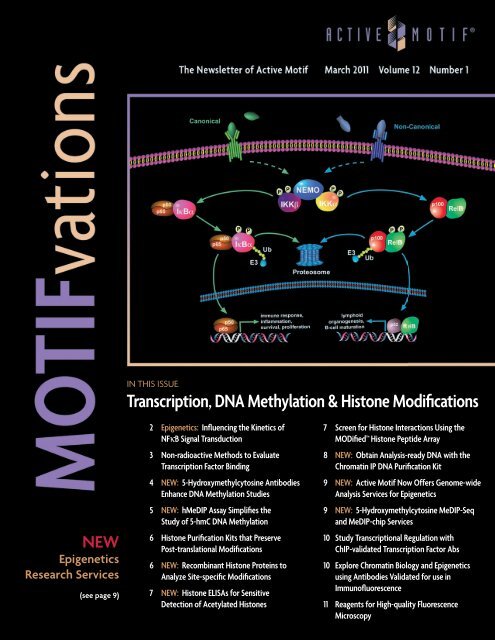
IN THIS ISSUE<br />
Transcription, <strong>DNA</strong> Methylation & Histone Modifications<br />
NEW<br />
Epigenetics<br />
Research Services<br />
(see page 9)<br />
2 Epigenetics: Influencing the Kinetics of<br />
NFκB Signal Transduction<br />
3 Non-radioactive Methods to Evaluate<br />
Transcription Factor Binding<br />
4 NEW: 5-<strong>Hydroxymethylcytosine</strong> <strong>Antibodies</strong><br />
<strong>Enhance</strong> <strong>DNA</strong> Methylation Studies<br />
5 NEW: hMeDIP Assay Simplifies the<br />
Study of 5-hmC <strong>DNA</strong> Methylation<br />
6 Histone Purification Kits that Preserve<br />
Post-translational Modifications<br />
6 NEW: Recombinant Histone Proteins to<br />
Analyze Site-specific Modifications<br />
7 NEW: Histone ELISAs for Sensitive<br />
Detection of Acetylated Histones<br />
7 Screen for Histone Interactions Using the<br />
MODified Histone Peptide Array<br />
8 NEW: Obtain Analysis-ready <strong>DNA</strong> with the<br />
Chromatin IP <strong>DNA</strong> Purification Kit<br />
9 NEW: <strong>Active</strong> <strong>Motif</strong> Now Offers Genome-wide<br />
Analysis Services for Epigenetics<br />
9 NEW: 5-<strong>Hydroxymethylcytosine</strong> MeDIP-Seq<br />
and MeDIP-chip Services<br />
10 Study Transcriptional Regulation with<br />
ChIP-validated Transcription Factor Abs<br />
10 Explore Chromatin Biology and Epigenetics<br />
using <strong>Antibodies</strong> Validated for use in<br />
Immunofluorescence<br />
11 Reagents for High-quality Fluorescence<br />
Microscopy
NFκ B Activation & Signaling<br />
Epigenetics: Influencing the Kinetics of NFκB Signal Transduction<br />
The nuclear factor-κB (NFκB)<br />
signaling pathway is widely studied<br />
due to its involvement in the<br />
regulation of genes that control<br />
inflammation, immune response,<br />
cell survival, cell proliferation and<br />
human disease.<br />
The NFκB Rel family of transcription<br />
factors consists of a set of<br />
evolutionarily conserved <strong>DNA</strong>binding<br />
proteins that includes p65,<br />
p50, p52, RelB and c-Rel. Inactive<br />
NFκB is sequestered in the cytoplasm<br />
by the IκB family of inhibitory<br />
proteins. Activation of the<br />
NFκB signaling pathway induces<br />
phosphorylation of IκB proteins<br />
by IκB kinases (IKK). Depending on<br />
the type of stimulation, the classical<br />
(also referred to as ‘canonical’)<br />
or the ‘non-canonical’ NFκB signaling<br />
pathways can be activated<br />
(Figure 1). Phosphorylation by the<br />
IKK complex leads to ubiquitination<br />
and degradation of the<br />
inhibitor IκB molecules by the<br />
proteasome. The liberated NFκB<br />
complexes translocate to the<br />
nucleus where they bind <strong>DNA</strong> and<br />
regulate gene expression by binding<br />
to κB <strong>DNA</strong>-binding sites.<br />
Figure 1: The NFκB signaling pathway.<br />
Depiction of the NFκB canonical and non-canonical signaling pathways.<br />
Evidence is building that epigenetic<br />
modifications play a critical role<br />
in orchestrating NFκB-mediated<br />
transcriptional activation. There<br />
is a significant understanding of<br />
the contribution of the synergistic<br />
effects of various cofactors and<br />
signaling networks on the spatiotemporal<br />
coordination of NFκBmediated<br />
transcriptional events<br />
at the cellular level. However, the<br />
fine-tuning and kinetics of the response<br />
to those signals at the genome<br />
level can best be explained<br />
by analyzing how histone modification<br />
patterns influence and are, in<br />
turn, influenced by the activity of<br />
NFκB. In particular, recent studies<br />
have focused on how epigenetic<br />
events influence the accessibility<br />
of the NFκB transcription factor/<br />
cofactor enhanceosome complex<br />
to target κB <strong>DNA</strong>-binding sites<br />
within the chromatin structure.<br />
<strong>Active</strong> <strong>Motif</strong> offers a variety of<br />
products to analyze the complex<br />
dynamics of NFκB transcription<br />
factor binding and associated<br />
epigenetic events. To learn more<br />
about <strong>Active</strong> <strong>Motif</strong>’s NFκB-related<br />
products, please give us a call<br />
or visit us on our website at<br />
www.activemotif.com/nfkb.<br />
Save 10% on products for NFκB research.<br />
For a limited time, get 10% off select products for NFκB.<br />
Just cite NFKB11 when you order. For complete details and a list of<br />
eligible products, please visit www.activemotif.com/promo.<br />
2 North America 877 222 9543 Europe +32 (0)2 653 0001 Japan +81 (0)3 5225 3638 www.activemotif.com
March 2011 • volume 12 • number 1<br />
NFκ B Activation & Signaling<br />
Non-radioactive Methods to Evaluate Transcription Factor Binding<br />
To facilitate the study of <strong>DNA</strong>-binding activity, <strong>Active</strong> <strong>Motif</strong> offers its TransAM Kits, which are highly-sensitive,<br />
non-radioactive transcription factor ELISAs that enable detection of even the smallest changes in the levels of<br />
activated transcription factors. In addition, the 96-stripwell format allows for both high and low throughput screening<br />
of samples. Its sensitivity, flexibility and specificity make the TransAM method the most published alternative<br />
to electrophoretic mobility shift assays (EMSA). To view a complete list of available TransAM Kits, please visit us at<br />
www.activemotif.com/transam. Recombinant proteins are also available for<br />
use as standard curves in the TransAM NFκB p50 and TransAM NFκB p65 Kits.<br />
<strong>Active</strong> <strong>Motif</strong> offers the Gelshift Chemiluminescent EMSA Kit as a simple,<br />
convenient, non-radioactive solution for assessing variant transcription factor<br />
binding to <strong>DNA</strong> targets. The non-radioactive format displays enhanced sensitivity<br />
when compared to traditional 32 P or digoxigenin EMSA methods.<br />
A<br />
NFκB activation (OD450 nm)<br />
2.5<br />
2<br />
1.5<br />
1<br />
0.5<br />
0<br />
IL-1α stimulated<br />
Unstimulated<br />
IL-1α stimulated<br />
0.5 1 2 5 10 25 50<br />
Cell extract/well (µg)<br />
Unstimulated<br />
0.5 1 5 10 25 50 0.5 1 5 10 25 50<br />
Product Format Catalog No.<br />
TransAM NFκB Family (p50, p52, p65, c-Rel & RelB) 2 x 96 rxns 43296<br />
TransAM NFκB p50 1 x 96 rxns 41096<br />
TransAM NFκB p52 1 x 96 rxns 48196<br />
TransAM NFκB p65 1 x 96 rxns 40096<br />
Gelshift Chemiluminescent EMSA 100 rxns 37341<br />
B<br />
µg of cell extract<br />
Figure 1: Comparison of non-radioactive TransAM Kits<br />
and radioactive gelshift.<br />
Activated NFκB p50/<strong>DNA</strong>-binding results are shown for<br />
human fibroblast WI-38 cells that were stimulated with<br />
IL-1α for 30 minutes. Increasing amounts of whole-cell<br />
extracts were assayed using either TransAM NFκB p50<br />
(A) or gelshift (B). The TransAM method is 10-fold more<br />
sensitive and provides quantitative data.<br />
Additional Tools to Aid your NFκB Research (for Custom Services, see page 9)<br />
Phospho-protein Detection<br />
• FACE NFκB p65 Profiler (Ser468 & Ser536)<br />
• FunctionELISA IκBα<br />
Luciferase Reporter Assays<br />
• RapidReporter® pRR-High-NFκB vector<br />
• RapidReporter® pRR-High-NFκB Assay<br />
• RapidReporter® Gaussia Luciferase Assay<br />
In Vivo Fluorescent Labeling<br />
• LigandLink pLL-1-NFκB p65<br />
SUMOylation<br />
• SUMOlink SUMO-1<br />
• SUMOlink SUMO-2/3<br />
<strong>Antibodies</strong><br />
• Highly characterized antibodies including: IκBα, IKKα,<br />
IKKβ, IKKγ, NFκB p50, NFκB p65, NFκB p100 and more<br />
www.activemotif.com/abs<br />
Extracts<br />
• Variety of untreated and stimulated nuclear, cytoplasmic<br />
and whole-cell extracts to study NFκB signaling<br />
www.activemotif.com/extracts<br />
Recombinant Proteins<br />
• NFκB-related recombinant proteins, including NFκB p50,<br />
NFκB p65, IKKβ and others<br />
www.activemotif.com/proteins<br />
To learn more about all of <strong>Active</strong> <strong>Motif</strong>’s NFκB-related products, please visit www.activemotif.com/nfkb.<br />
www.activemotif.com Japan +81 (0)3 5225 3638 Europe +32 (0)2 653 0001 North America 877 222 9543<br />
3
5-Hydroxymethylcytidine Abs & Standards<br />
NEW: 5-<strong>Hydroxymethylcytosine</strong> <strong>Antibodies</strong> <strong>Enhance</strong> <strong>DNA</strong> Methylation Studies<br />
Conventional methods for studying <strong>DNA</strong> methylation (enrichment by antibody or methylated-<strong>DNA</strong> binding protein,<br />
methylation sensitive enzyme digestion and bisulfite sequencing) cannot distinguish 5-hydroxymethylcytosine (5-hmC)<br />
from 5-methylcytosine (5-mC). To help study this novel form of <strong>DNA</strong> methylation, <strong>Active</strong> <strong>Motif</strong> offers two<br />
polyclonal antibodies and a new monoclonal antibody that are specific for the 5-hmC modification. Like all of<br />
our antibodies for epigenetics research, they are highly specific and have been validated for use in various practical<br />
applications, including methylated <strong>DNA</strong> immunoprecipitation (MeDIP), immunofluorescence and dot blot.<br />
The importance of <strong>DNA</strong> methylation<br />
<strong>DNA</strong> methylation is an epigenetic<br />
event in which <strong>DNA</strong> methyltransferases<br />
(DNMTs) catalyze the reaction of a<br />
methyl group to the fifth carbon of<br />
cytosine in a CpG dinucleotide. This<br />
modification helps to regulate gene expression<br />
and is also involved in genomic<br />
imprinting, while aberrant <strong>DNA</strong> methylation<br />
is often associated with disease.<br />
5-methylcytosine (5-mC) is a type of<br />
<strong>DNA</strong> methylation found in plants and<br />
vertebrates. A second type of <strong>DNA</strong><br />
methylation exists, 5-hydroxymethylcytosine<br />
(5-hmC), which results from the<br />
enzymatic conversion of 5-methylcytosine<br />
into 5-hydroxymethylcytosine by<br />
the TET family of cytosine oxygenases<br />
(Figure 1). Elevated levels of 5-hydroxymethylcytosine<br />
have been observed in<br />
neurons and embryonic stem cells. It is<br />
possible that 5-hmC represents a pathway<br />
by which <strong>DNA</strong> is demethylated, as<br />
5-hydroxymethylcytosine is recognized<br />
by the <strong>DNA</strong> mismatch repair system and<br />
replaced with unmethylated cytosine.<br />
Figure 2: 5-Hydroxymethylcytidine monoclonal<br />
antibody tested by dot blot analysis.<br />
<strong>DNA</strong> samples were spotted (indicated in ng on the left)<br />
and blotted with 5-Hydroxymethylcytidine monoclonal<br />
antibody (Clone 59.1) at a dilution of 0.2 µg/ml.<br />
Lanes 1-3: double-stranded <strong>DNA</strong>.<br />
Lanes 4-6: single-stranded <strong>DNA</strong>.<br />
Lanes 1 & 4: <strong>DNA</strong> containing 5-hydroxymethylcytosine.<br />
Lanes 2 & 5: <strong>DNA</strong> containing 5-methylcytosine.<br />
Lanes 3 & 6: unmethylated <strong>DNA</strong>.<br />
Methylated <strong>DNA</strong> Standard Kit<br />
In addition to the antibodies to study<br />
<strong>DNA</strong> methylation, <strong>Active</strong> <strong>Motif</strong> offers<br />
our Methylated <strong>DNA</strong> Standard Kit. This<br />
kit includes three recombinant <strong>DNA</strong><br />
standards derived from the APC gene<br />
regulatory region: unmethylated <strong>DNA</strong>,<br />
5-methylcytosine methylated <strong>DNA</strong> and<br />
5-hydroxymethylcytosine methylated<br />
<strong>DNA</strong>. This kit (which also includes PCR<br />
primers specific to the APC gene) can be<br />
used to provide controls for experiments<br />
studying the different types of <strong>DNA</strong><br />
methylation (see Figure 1 on page 5).<br />
<strong>Antibodies</strong> to study <strong>DNA</strong> methylation<br />
<strong>Active</strong> <strong>Motif</strong> has three antibodies specific<br />
for the 5-hydroxymethylcytosine<br />
form of <strong>DNA</strong> methylation. Our newest<br />
release, the 5-Hydroxymethylcytidine<br />
mouse monoclonal antibody (Clone 59.1)<br />
has been validated for use in MeDIP and<br />
dot blot (Figure 2). Like our polyclonal<br />
antibodies, the new monoclonal does<br />
not require <strong>DNA</strong> to be denatured to<br />
its single-stranded form in order to<br />
work in a MeDIP experiment. This can<br />
be advantageous when analyzing the<br />
resulting <strong>DNA</strong> using high-throughput<br />
Next-Gen sequencing. We also offer<br />
the more traditional 5-Methylcytidine<br />
mouse monoclonal antibody to study<br />
5-mC <strong>DNA</strong> methylation in a variety of<br />
applications.<br />
Full line of <strong>DNA</strong> Methylation products<br />
<strong>Active</strong> <strong>Motif</strong> offers a variety of antibodies,<br />
assay kits and services for the<br />
epigenetics and chromatin research<br />
community. To learn more about the different<br />
<strong>DNA</strong> methylation tools available,<br />
visit www.activemotif.com/dnamt. For<br />
a complete listing of <strong>DNA</strong> methylation<br />
antibodies, including application data,<br />
visit www.activemotif.com/dnamethabs.<br />
Figure 1: Schematic of conversion of 5-mC to 5-hmC.<br />
5-methylcytosine is converted to 5-hydroxymethylcytosine<br />
by the TET family of enzymes.<br />
Product Format Catalog No.<br />
5-Hydroxymethylcytidine mAb (Clone 59.1) 100 µg 39999<br />
5-Hydroxymethylcytidine antibody (rabbit IgG) 100 µg 39791<br />
5-Hydroxymethylcytidine antibody (rabbit serum) 100 µl 39769<br />
5-Methylcytidine antibody mAb (Clone 33D3) 50 µg 39649<br />
Methylated <strong>DNA</strong> Standard Kit 3 x 2.5 µg 55008<br />
4 North America 877 222 9543 Europe +32 (0)2 653 0001 Japan +81 (0)3 5225 3638 www.activemotif.com
March 2011 • volume 12 • number 1<br />
MeDIP & hMeDIP Assays<br />
NEW: hMeDIP Assay Simplifies the Study of 5-hmC <strong>DNA</strong> Methylation<br />
<strong>Active</strong> <strong>Motif</strong>’s new hMeDIP Kit helps simplify the analysis of 5-hydroxymethylcytosine methylation. Methylated <strong>DNA</strong><br />
Immunoprecipitation (MeDIP) is an immunocapture technique in which an antibody specific for methylated cytosines<br />
is used to immunoprecipitate methylated genomic <strong>DNA</strong> fragments. The hMeDIP Assay uses a highly specific, purified<br />
5-hydroxymethylcytidine (5-hmC) antibody to selectively enrich for <strong>DNA</strong> fragments with 5-hydroxymethylcytosine<br />
methylation from the rest of the genomic <strong>DNA</strong> population. Some advantages of the hMeDIP Kit are that it not only<br />
differentiates between 5-hydroxymethylcytosine <strong>DNA</strong> and 5-methylcytosine <strong>DNA</strong>, but also that the fragmented<br />
genomic <strong>DNA</strong> used in the assay can be double-stranded <strong>DNA</strong>, which prevents the problems associated with linker<br />
bias when preparing the enriched <strong>DNA</strong> for downstream Next-Gen sequencing.<br />
Why use hMeDIP over other <strong>DNA</strong><br />
methylation enrichment techniques<br />
Unlike traditional methods used to study<br />
<strong>DNA</strong> methylation, such as enzymatic<br />
approaches & bisulfite conversion that<br />
cannot directly differentiate between<br />
5-mC and 5-hmC <strong>DNA</strong> methylation, or<br />
methyl CpG binding protein enrichment<br />
which can only recognize and bind 5-mC<br />
methylation, hMeDIP is able to use the<br />
selective affinity of the immunocapture<br />
antibody to distinguish between<br />
5-methylcytosine and 5-hydroxymethylcytosine<br />
<strong>DNA</strong> methylation.<br />
Additionally, the hMeDIP technique is<br />
able to enrich for <strong>DNA</strong> fragments containing<br />
cytosine methylation regardless<br />
of the sequence context. While most<br />
<strong>DNA</strong> methylation in mammalian tissues<br />
occurs in a CpG context, studies have<br />
found that 15-20% of total cytosine<br />
methylation in embryonic stem (ES) cells<br />
occurs at sequences other than CpG.<br />
Kit Antibody <strong>DNA</strong> Input Input Range<br />
hMeDIP<br />
5-hmC purified rabbit pAb<br />
Table 1: hMeDIP Kit sample requirements.<br />
Included methylated <strong>DNA</strong> controls<br />
The hMeDIP Kit also includes methylated<br />
<strong>DNA</strong> controls for use in determining the<br />
efficiency of the enrichment. The controls<br />
are 338 base pair <strong>DNA</strong> fragments<br />
that are completely unmethylated,<br />
5-methylcytosine methylated or<br />
5-hydroxymethylcytosine methylated.<br />
When these control <strong>DNA</strong>s are spiked<br />
into sample <strong>DNA</strong> the specificity of the<br />
capture antibody can be confirmed via<br />
real time PCR with the included PCR<br />
primers (Figure 1). The Methylated <strong>DNA</strong><br />
Standards used as controls in the<br />
hMeDIP Kit can also be purchased<br />
separately (see page 4).<br />
double-stranded or<br />
single-stranded <strong>DNA</strong><br />
% of Input<br />
20<br />
18<br />
16<br />
14<br />
12<br />
10<br />
8<br />
6<br />
4<br />
2<br />
0<br />
Sample 1<br />
Sample 2<br />
100 ng – 1 µg<br />
un<strong>DNA</strong> me<strong>DNA</strong> hme<strong>DNA</strong> rabbit IgG<br />
Figure 1: hMeDIP results using “spiked” control <strong>DNA</strong>.<br />
Mse I digested human genomic <strong>DNA</strong> (500 ng) was spiked<br />
with 25 pg of either methylated (me<strong>DNA</strong>), hydroxymethylated<br />
(hme<strong>DNA</strong>) or unmethylated (un<strong>DNA</strong>) <strong>DNA</strong><br />
controls. These samples were processed using the<br />
hMeDIP Kit with the 5-hmC antibody and compared to a<br />
negative control rabbit IgG antibody. Real time PCR with<br />
the included APC PCR primers was run and the recovered<br />
<strong>DNA</strong> was plotted as a percentage of the total input<br />
<strong>DNA</strong> (500 ng). The results show the hMeDIP Kit is specific<br />
for enrichment of 5-hydroxymethylcytosine <strong>DNA</strong>.<br />
These features make the hMeDIP Kit<br />
ideal for researchers interested in identification<br />
of total cytosine methylation,<br />
or in differentiating between 5-mC and<br />
5-hmC methylation.<br />
Visit www.activemotif.com/dnamt to<br />
learn more about the wide variety of<br />
<strong>DNA</strong> methylation analysis tools and<br />
assay kits offered by <strong>Active</strong> <strong>Motif</strong>.<br />
Product Format Catalog No.<br />
hMeDIP 10 rxns 55010<br />
MeDIP COMING SOON 10 rxns 55009<br />
As part of its recent acquisition<br />
of Genpathway, <strong>Active</strong> <strong>Motif</strong> now<br />
offers a 5-<strong>Hydroxymethylcytosine</strong><br />
MeDIP-Seq service. For details,<br />
please see page 9 or visit<br />
www.genpathway.com.<br />
www.activemotif.com Japan +81 (0)3 5225 3638 Europe +32 (0)2 653 0001 North America 877 222 9543<br />
5
Analysis of Histone Post-translational Modifications<br />
Complete Analysis of Histone Post-translational Modifications<br />
Whether you are studying transcription factors such as NFκB or chromatin-modifying proteins, understanding the<br />
implications of histone post-translational modifications as they relate to gene regulation and chromatin context will<br />
be critical to determining how these modifications affect or contribute to disease. <strong>Active</strong> <strong>Motif</strong>’s unique portfolio<br />
of histone technologies provides researchers with a complete solution for analysis. To either download or request a<br />
copy of our Histone Analysis Products brochure by mail, please visit www.activemotif.com/info.<br />
Histone Purification Kits that Preserve Post-translational Modifications<br />
Histone Purification Kit advantages<br />
<strong>Active</strong> <strong>Motif</strong>’s unique Histone Purification<br />
and Histone Purification Mini Kits<br />
enable the isolation of core histones<br />
from any cell culture or tissue sample<br />
while preserving their post-translational<br />
modifications (Figure 1). Unlike standard<br />
acid extraction techniques, it is possible<br />
to isolate core histones as either a single<br />
fraction or to further separate them into<br />
H2A/H2B and H3/H4 fractions.<br />
Acetylation<br />
Methylation<br />
Which kit is right for you<br />
• Histone Purification – can process<br />
samples of up to 2.5 mg which can<br />
be eluted as either a single fraction<br />
with all four histones or as separate<br />
H2A/H2B and H3/H4 fractions<br />
• Histone Purification Mini – has a<br />
capacity of up to 0.5 mg per sample<br />
which is eluted as a single fraction<br />
containing H2A, H2B, H3 and H4<br />
Phosphorylation<br />
Figure 1: <strong>Active</strong> <strong>Motif</strong>’s Histone Purification Kits preserve post-translational modifications.<br />
Acetylation, methylation and phosphorylation states are preserved as well or better with the Histone Purification Kit than<br />
standard acid extraction methods.<br />
Product Format Catalog No. Price ($US)<br />
Histone Purification Kit 10 rxns 40025 275<br />
Histone Purification Mini Kit 20 rxns 40026 275<br />
NEW: Recombinant Histone Proteins to Analyze Site-specific Modifications<br />
Recombinant histone proteins with<br />
site-specific modifications<br />
<strong>Active</strong> <strong>Motif</strong> continues to expand its<br />
line of recombinant methylated and<br />
acetylated histones. We have added<br />
16 new recombinant proteins to our<br />
portfolio, including methylation of lysine<br />
14, 18 and 23 on histone H3 and methylation<br />
of lysine 5 and 16 on histone H4.<br />
Additionally, we now offer recombinant<br />
histone H3 acetylated at lysine 23.<br />
Why use recombinant histones<br />
These recombinant proteins more<br />
closely mimic “natural” histones than<br />
peptides, making them ideal substrates<br />
for functional assays and activity<br />
screens. The recombinant proteins are<br />
suitable for use with <strong>Active</strong> <strong>Motif</strong>’s<br />
histone modifying enzymes and are even<br />
included in our Histone Modification<br />
ELISAs to generate standard curves for<br />
histone quantification.<br />
New biotinylated recombinant H3<br />
To expand the versatility of our recombinant<br />
histones, <strong>Active</strong> <strong>Motif</strong> now offers<br />
Recombinant Histone H3 biotinylated at<br />
the N-terminus.<br />
To see an up-to-date list of the more<br />
than 40 recombinant histone proteins<br />
currently available, please visit us at<br />
www.activemotif.com/recombhis.<br />
6 North America 877 222 9543 Europe +32 (0)2 653 0001 Japan +81 (0)3 5225 3638 www.activemotif.com
March 2011 • volume 12 • number 1<br />
Analysis of Histone Post-translational Modifications<br />
NEW: Histone ELISAs for Sensitive Detection of Acetylated Histones<br />
Quantify your histone modifications<br />
The Histone Modification ELISA Kits<br />
offer a quick and easy way to screen and<br />
quantify histone modifications within<br />
your sample. The kits are designed as<br />
sandwich ELISAs that utilize a histone<br />
H3 capture antibody and a detecting<br />
antibody that is specific for the modified<br />
residue of interest.<br />
<strong>Active</strong> <strong>Motif</strong> currently offers more<br />
than ten different Histone ELISAs for<br />
methylation and phosphorylation marks<br />
on histone H3. Now, <strong>Active</strong> <strong>Motif</strong> has<br />
expanded its line of ELISAs to include<br />
assays to detect acetylation of histone<br />
H3 lysine 9 and lysine 14. Each kit also<br />
includes a positive control recombinant<br />
protein that can be used to generate a<br />
standard curve for histone quantification<br />
(Figure 1). The Total Histone H3 ELISA is<br />
available to measure total histone levels<br />
or to normalize quantities of modified<br />
histones from the sample.<br />
Figure 1: Detection of histone H3 lysine 9 acetylation in purified core histones and acid extracted histones.<br />
The Histone H3 acetyl Lys9 ELISA was used to quantitate the amount of histone H3 lysine 9 acetylation present in HeLa<br />
core histones made using <strong>Active</strong> <strong>Motif</strong>’s Histone Purification Mini Kit (Cat. No. 40026, page 6) and HeLa acid extracts. The<br />
Recombinant Histone H3 acetyl Lys 9 protein that is provided in the kit was assayed from 0.039 - 2.5 ng/well as a standard<br />
curve. Data was quantified against the linear range of the standard curve. Data shown are from wells assayed in duplicate.<br />
Product Format Catalog No.<br />
Histone H3 acetyl Lys9 ELISA 1 x 96 rxns 53114<br />
Histone H3 acetyl Lys14 ELISA 1 x 96 rxns 53115<br />
Total Histone H3 ELISA 1 x 96 rxns 53110<br />
To see a complete list of the over 10 Histone Modification ELISAs available, including<br />
methylation and phosphorylation ELISAs, please visit www.activemotif.com/hiselisa.<br />
Screen for Histone Interactions Using the MODified Histone Peptide Array<br />
Unique array to screen interactions<br />
The MODified Histone Peptide Array<br />
enables analysis of antibody, protein and<br />
enzyme interactions with histones and<br />
their post-translational modifications.<br />
The array contains 384 different combinations<br />
of acetylation, phosphorylation,<br />
methylation and citrullination modifications<br />
on the N-terminal tails of histones<br />
H2A, H2B, H3 and H4.<br />
To learn more about the MODified<br />
Histone Peptide array, or to download<br />
the free Array Analyse software, please<br />
visit www.activemotif.com/modified.<br />
MODified Array advantages<br />
• Histone specific – unique array<br />
panel tests for specific histone<br />
modifications<br />
• Study neighboring effects – each<br />
peptide contains up to four modification<br />
combinations, enabling the<br />
analysis of the effects of neighboring<br />
modifications<br />
• Detects like a Western blot – fast<br />
and easy to use; works with either<br />
ECL-based or colorimetric detection<br />
• Free software for analysis – measures<br />
spot intensity and generates<br />
Excel-compatible files for analysis<br />
Product Format Catalog No.<br />
MODified Histone Peptide Array<br />
1 array<br />
5 arrays<br />
13001<br />
13005<br />
MODified Array Labeling Kit 5 rxns 13006<br />
Histone H3 Acetyl Lys9 (OD 450nm)<br />
2<br />
1.8<br />
1.6<br />
1.4<br />
1.2<br />
0.8<br />
0.6<br />
0.4<br />
0.2<br />
1<br />
0<br />
Histone H3 Acetyl Lys9 ELISA<br />
2.5 1.25 0.625 0.313 0.156 0.078 0.039 0 50 50<br />
Purified HeLa<br />
Recombinant Histone H3 Acetyl Lys9 Protein<br />
Core Acid<br />
(provided positive control)<br />
Histones Extract<br />
Sample (ng/well)<br />
A<br />
B<br />
C<br />
Figure 1: Detection of G9a histone methyltransferase<br />
activity using the MODified Histone Peptide Array.<br />
MODified Histone Peptide Arrays were treated with<br />
A) 25 µM G9a methyltransferase, B) 25 µM G9a mutant<br />
methyltransferase or C) a no enzyme control overnight<br />
in the presence of 1 mM AdoMet. The arrays were then<br />
labeled with a Histone H3 dimethyl Lys9 antibody. Novel<br />
methylation sites were observed on the array treated<br />
with wild-type G9a histone methyltransferase, showing<br />
the activity of G9a enzyme on the peptide substrate.<br />
www.activemotif.com Japan +81 (0)3 5225 3638 Europe +32 (0)2 653 0001 North America 877 222 9543<br />
7
Chromatin IP <strong>DNA</strong> Purification<br />
NEW: Obtain Analysis-ready <strong>DNA</strong> with the Chromatin IP <strong>DNA</strong> Purification Kit<br />
Chromatin Immunoprecipitation (ChIP) is a powerful, well-established technique for studying interactions between<br />
chromatin-associated proteins and specific regions of the genome. While the use of ChIP in combination with<br />
genome-wide analysis techniques can yield a tremendous amount of information regarding the distribution of<br />
transcription factors and histone modifications, most downstream analysis techniques require <strong>DNA</strong> that has been<br />
purified away from the components and contaminants present in your eluted ChIP sample. <strong>Active</strong> <strong>Motif</strong>’s Chromatin<br />
IP <strong>DNA</strong> Purification Kit enables you to quickly and easily clean up your ChIP <strong>DNA</strong> samples to make them ready for<br />
analysis, eliminating the need for labor intensive and time consuming phenol/chloroform extraction.<br />
What is the Chromatin IP <strong>DNA</strong><br />
Purification Kit process<br />
Once your ChIP experiments are complete,<br />
the <strong>DNA</strong> purification procedure<br />
can be started immediately. The entire<br />
procedure takes only five to ten minutes,<br />
depending upon the number of samples<br />
to purify. The Chromatin IP <strong>DNA</strong> Purification<br />
Kit is compatible with samples<br />
from all of <strong>Active</strong> <strong>Motif</strong> ChIP-IT Kits,<br />
or from any standard chromatin IP kit or<br />
procedure, whether they use mechanical<br />
or enzymatic shearing of chromatin,<br />
agarose or paramagnetic bead enrichment.<br />
It can also be used to purify <strong>DNA</strong><br />
from other <strong>Active</strong> <strong>Motif</strong> kits, including<br />
the new hMeDIP Kit, MethylCollector<br />
Ultra and UnMethylCollector. The new<br />
Chromatin IP <strong>DNA</strong> Purification Kit is<br />
designed for use with a microcentrifuge<br />
for sample processing, but can also be<br />
used with a vacuum manifold.<br />
Because binding of <strong>DNA</strong> to the silica<br />
matrix of the <strong>DNA</strong> purification column<br />
is pH-dependant, the kit’s Binding Buffer<br />
contains a convenient pH indicator dye.<br />
This makes it possible for you to see<br />
the pH of your samples before applying<br />
them to the column, helping ensure<br />
successful purification of your ChIP <strong>DNA</strong><br />
(Figure 1). After binding, the <strong>DNA</strong> on the<br />
column is washed, then eluted using the<br />
included Elution Buffer.<br />
<strong>DNA</strong> recovery and yield<br />
After elution of the <strong>DNA</strong>, you now<br />
have samples of high quality and purity,<br />
suitable for a number of downstream<br />
analysis techniques, including PCR (endpoint<br />
or quantitative), Southern blotting,<br />
microarray analysis or Next-Gen sequencing<br />
(Figure 2). Depending upon the<br />
number of cell equivalents of chromatin<br />
used in each ChIP reaction, the amount<br />
of recovered <strong>DNA</strong> will be from 100 ng to<br />
1 µg. <strong>DNA</strong> can be successfully recovered<br />
from ChIP experiments starting with<br />
as few as 10,000 cells. <strong>DNA</strong> fragments<br />
below 50 base pairs in length are not recovered<br />
efficiently, but this shouldn’t be<br />
a problem as the recommended average<br />
size for chromatin fragments in a typical<br />
ChIP reaction is 500 base pairs.<br />
Figure 1: <strong>DNA</strong> Binding Buffer pH indicator dye.<br />
The <strong>DNA</strong> Purification Binding Buffer has a pH indicator<br />
dye so the pH of the solution can be easily determined.<br />
The <strong>DNA</strong> should only be applied to the column if the<br />
solution is bright yellow (left), indicating a pH under 7.5.<br />
<strong>DNA</strong> will not bind to the column if the pH is higher than<br />
7.5. (NaOAc is included to reduce sample pH, if needed.)<br />
Figure 2: Quantitative PCR performed on ChIP <strong>DNA</strong>.<br />
Quantitative PCR was performed using <strong>DNA</strong> purified<br />
with the Chromatin IP <strong>DNA</strong> Purification Kit after ChIP<br />
using the indicated antibodies (ChIP Abs). Chromatin<br />
IP experiments were performed using the ChIP-IT<br />
Express Kit (Catalog No. 53008) and Ready-to-ChIP HeLa<br />
Chromatin (Catalog No. 53015, 7.5 x 10 5 cell equivalents<br />
per ChIP). Quantitative PCR was carried out using primers<br />
specific for the indicated gene (Locus). The normalized<br />
data represent fold enrichment over ChIP experiments<br />
carried out with control IgG.<br />
Chromatin IP <strong>DNA</strong> Purification Kit<br />
advantages<br />
• Optimized for use with epigenetics<br />
applications<br />
• Compatible with all of <strong>Active</strong> <strong>Motif</strong>’s<br />
ChIP-IT Kits, as well as with kits<br />
from other manufacturers<br />
• Purify <strong>DNA</strong> ChIP’d using agarose or<br />
paramagnetic bead methods<br />
• Convenient pH indicator to ensure<br />
proper binding of your samples<br />
• Can also be used to purify <strong>DNA</strong><br />
from our hMeDIP, MethylCollector<br />
and UnMethylCollector Kits<br />
To find out more about our new Chromatin<br />
IP <strong>DNA</strong> Purification Kit, please visit<br />
us at www.activemotif.com/dnapure.<br />
Product Format Catalog No.<br />
Chromatin IP <strong>DNA</strong> Purification Kit 50 rxns 58002<br />
8 North America 877 222 9543 Europe +32 (0)2 653 0001 Japan +81 (0)3 5225 3638 www.activemotif.com
March 2011 • volume 12 • number 1<br />
Epigenetic Services<br />
NEW: <strong>Active</strong> <strong>Motif</strong> Now Offers Genome-wide Analysis Services for Epigenetics<br />
As part of its recent acquisition of Genpathway, genome-wide analysis services are now offered by <strong>Active</strong> <strong>Motif</strong>.<br />
This is the perfect combination of products and know-how. The leader in kits and reagents for the study of<br />
chromatin biology and epigenetics now offers services and expertise to facilitate your research into transcription<br />
factor distribution and function, histone modifications and <strong>DNA</strong> methylation.<br />
• FactorPath – discovery, identification<br />
and quantitation of transcription<br />
factor and cofactor binding<br />
sites across the genome<br />
• TranscriptionPath – discovery and<br />
identification of actively transcribed<br />
genes at the <strong>DNA</strong> level to detect<br />
genome-wide changes in gene<br />
expression within minutes of<br />
treatment<br />
• MethylPath – discovery, identification<br />
and quantitation of methylated<br />
<strong>DNA</strong> regions; also includes our new<br />
5-hmC MeDIP-Seq and bisulfite<br />
sequencing services<br />
• HistonePath – map histone<br />
modifications and/or enzymes<br />
that regulate histone modifications<br />
across the genome<br />
• Bisulfite Sequencing – determine<br />
the locations and methylation<br />
status of cytosines<br />
H3K27me3<br />
EZH2<br />
genes strand<br />
genes - strand<br />
Figure 1: ChIP-Seq data for histone H3 lysine 27 methylation and EZH2.<br />
Chromatin IP was performed using antibodies specific for H3K27me3 and EZH2, the H3K27 methyltransferase, followed by<br />
high-throughput sequencing of the ChIP <strong>DNA</strong>. The data are presented in an 8 million base pair window and shows nearly<br />
identical genomic localization for histone H3 lysine 27 methylation and the enzyme responsible for depositing the mark.<br />
Shown is a region of chromosome 10q surrounding the HPSE2 gene.<br />
In addition to the services listed to the left, we also offer customized assay development<br />
and specific services to optimize ChIP-based procedures, including antibody qualification<br />
and data analysis. To learn more about our genome-wide analysis services for<br />
chromatin biology, transcription, histones and <strong>DNA</strong> methylation, please visit our website<br />
at www.activemotif.com/services or email us at services@activemotif.com.<br />
NEW: 5-<strong>Hydroxymethylcytosine</strong> MeDIP-Seq and MeDIP-chip Services<br />
Due to the importance of <strong>DNA</strong> methylation<br />
in biology and disease, <strong>Active</strong> <strong>Motif</strong><br />
now offers 5-hydroxymethylcytosine<br />
MeDIP-Seq and MeDIP-chip as custom<br />
services. Just provide us with frozen<br />
biological samples or purified <strong>DNA</strong> and<br />
we will perform the hMeDIP-Seq or<br />
hMeDIP-chip experiments, then provide<br />
you with, and help you interpret, the data.<br />
is that our proprietary monoclonal<br />
antibody recognizing 5-hydroxymethylcytosine<br />
methylation does not require<br />
that the <strong>DNA</strong> be made single-stranded<br />
before immunoprecipitation. The ability<br />
to use double-stranded <strong>DNA</strong> eliminates<br />
linker bias in the downstream processing<br />
steps prior to Next-Gen sequencing.<br />
Elevated signal in gene bodies<br />
Our hMeDIP genome-wide analysis services<br />
allow you to study 5-hmC methylation<br />
on a genome-wide scale utilizing<br />
<strong>Active</strong> <strong>Motif</strong>’s expertise and research<br />
tools without having to be an expert in<br />
the techniques yourself. The advantage<br />
<strong>Active</strong> <strong>Motif</strong> has over other companies<br />
genes strand<br />
Figure 1: 5-hMeDIP-chip performed on human brain <strong>DNA</strong>.<br />
Human brain <strong>DNA</strong> (2 µg) was immunoprecipitated with 10 µg of 5-Hydroxymethylcytidine antibody (Clone 59.1, Catalog No.<br />
39999). Following hMeDIP, the <strong>DNA</strong> was amplified, labeled and hybridized to an Affymetrix Human Tiling 2.0R Array. Shown<br />
is a region from chromosome 6q containing the ARID1B, ZDHHC14 and SNX9 genes. The results show that 5-hydroxymethylcytosine<br />
is enriched primarily in the coding region of genes, rather than the promoter or regulatory regions.<br />
www.activemotif.com Japan +81 (0)3 5225 3638 Europe +32 (0)2 653 0001 North America 877 222 9543<br />
9
ChIP- & Immunofluorescence-validated <strong>Antibodies</strong><br />
Study Transcriptional Regulation with ChIP-validated Transcription Factor Abs<br />
Gene expression depends on the recognition<br />
of specific promoter sequences<br />
by transcriptional regulatory proteins,<br />
which in turn recruit other proteins, such<br />
as RNA polymerase, in order to effect<br />
transcription. In the human genome,<br />
there are at least 2,600 genes that encode<br />
proteins involved in transcriptional<br />
regulation. These fall into three main<br />
categories: sequence specific <strong>DNA</strong> binding<br />
transcription factors (proteins that<br />
directly bind specific <strong>DNA</strong> regulatory<br />
sequences), general transcription factors<br />
(those associated with RNA polymerase<br />
holoenzyme complex) and transcriptional<br />
co-regulators (proteins recruited<br />
by the <strong>DNA</strong> binding factors).<br />
Because one of the most important<br />
techniques used to study the function<br />
and genomic localization of these pro-<br />
teins is chromatin immunoprecipitation<br />
(ChIP), <strong>Active</strong> <strong>Motif</strong> offers an extensive<br />
line of ChIP-validated antibodies to<br />
important regulatory factors (Figure 1).<br />
As part of its acquisition of<br />
Genpathway, <strong>Active</strong> <strong>Motif</strong> now<br />
offers FactorPath genome-wide<br />
transcription factor binding<br />
identification and analysis as a<br />
custom service. See page 9 for<br />
more details, or visit us at<br />
www.activemotif.com/factorpath.<br />
To find out more about our ChIPvalidated<br />
antibodies to transcriptional<br />
regulators, please call or visit us at<br />
www.activemotif.com/tfchipabs.<br />
Figure 1: Quantitative PCR performed on RNA pol II<br />
CTD phospho Ser5 antibody ChIP’d <strong>DNA</strong><br />
Chromatin IP was performed using the ChIP-IT Express<br />
Kit (Catalog No. 53008) and HeLa chromatin (1.5 x 10 6 cell<br />
equivalents per ChIP) with 10 µg RNA pol II CTD phospho<br />
Ser5 antibody (Catalog No. 39749) or the equivalent<br />
amount of rabbit IgG as a negative control. Real time,<br />
quantitative PCR (RT-qPCR) was performed on <strong>DNA</strong> purified<br />
from each of the ChIP reactions using a primer pair<br />
specific for the PABPC1 gene. Data are presented as fold<br />
enrichment of the ChIP antibody signal versus the negative<br />
control IgG (arbitrarily assigned a value of 1) using<br />
the ddCT method.<br />
Explore Chromatin Biology and Epigenetics using <strong>Antibodies</strong> Validated for use<br />
in Immunofluorescence<br />
Visualizing a protein in its native cellular<br />
environment using immunofluorescence<br />
microscopy requires the use of very<br />
high-quality antibodies. They must be<br />
specific, of high titer and able to recognize<br />
native (non-denatured) protein. To<br />
assist you in your study of chromatin<br />
biology, epigenetics and transcription,<br />
<strong>Active</strong> <strong>Motif</strong> offers antibodies validated<br />
for use in immunofluorescence to a wide<br />
range of protein targets including:<br />
• Chromatin-modifying proteins<br />
• Histones and histone modifications<br />
• Transcription factors<br />
• Nuclear receptors<br />
• Regulators of cell structure and<br />
function<br />
Figure 1: HDAC2 visualized by immunofluorescence.<br />
HeLa cells were stained with HDAC2 monoclonal antibody<br />
(Clone 3F3, Catalog No. 39533) at a 1:1000 dilution.<br />
Red: HDAC2 mAb (Clone 3F3) Green: alpha-Tubulin<br />
mouse monoclonal (Clone 5-B-1-2, Catalog No. 39527)<br />
conjugated to Chromeo 488.<br />
<strong>Active</strong> <strong>Motif</strong> offers a wide range of<br />
primary antibodies validated for use in<br />
immunofluorescence. For more information,<br />
visit www.activemotif.com/ifabs.<br />
Figure 2: Aurora B visualized by immunofluorescence.<br />
HeLa cells were stained with Aurora B polyclonal<br />
antibody (Catalog No. 39261) at a dilution of 1:200 and<br />
alpha-Tubulin monoclonal antibody (Clone 5-B-1-2,<br />
Catalog No. 39527) used at a dilution of 1:500.<br />
Red: Aurora B. Green: alpha-Tubulin. Blue: DAPI.<br />
To find out more about our high-quality<br />
fluorescent secondary antibodies, visit<br />
www.activemotif.com/secondary.<br />
10 North America 877 222 9543 Europe +32 (0)2 653 0001 Japan +81 (0)3 5225 3638 www.activemotif.com
March 2011 • volume 12 • number 1<br />
Reagents for Fluorescence<br />
Reagents for High-quality Fluorescence Microscopy<br />
To produce superior results with our primary antibodies in immunofluorescence (IF) experiments, <strong>Active</strong> <strong>Motif</strong> has<br />
developed a variety of high-quality reagents including Chromeo secondary antibody conjugates and MAX Stain<br />
Immunofluorescence Tools. These optimized reagents can be used in a variety of fluorescence microscopy applications,<br />
even in novel high-resolution fluorescence microscopy (e.g. STimulated Emission Depletion, or STED). We use these<br />
reagents in our daily work to develop novel assays and to evaluate our antibodies in a consistent and reliable manner.<br />
Fluorescent secondary antibodies<br />
<strong>Active</strong> <strong>Motif</strong> offers goat anti-mouse<br />
and goat anti-rabbit secondary antibodies<br />
that are conjugated to a number of<br />
high-quality dyes, including Chromeo<br />
fluorescent dyes (Table 1). Our optimized<br />
conjugation method, coupled with<br />
subsequent purification of the conjugate,<br />
makes our fluorescent secondaries<br />
brighter than other commercially<br />
available conjugates and lowers the<br />
background in many applications. <strong>Active</strong><br />
<strong>Motif</strong>’s antibody conjugates have<br />
been tested in applications such as flow<br />
cytometry and widefield, confocal and<br />
high-resolution fluorescence microscopy.<br />
Fluorescent Dye Absorption Emission Replaces<br />
Chromeo 488 498 nm 524 nm FITC, Alexa 488*<br />
Chromeo 494 489 nm 624 nm unique<br />
Chromeo 505 514 nm 530 nm Oregon Green derivatives<br />
Chromeo 546 550 nm 567 nm Cy3, Alexa 546*<br />
Chromeo 642 647 nm 666 nm Cy5, Alexa 647*<br />
Table 1: Fluorescent properties of dyes when conjugated to secondary antibodies.<br />
* Alexa Fluor dyes are a registered trademark of Life Technologies.<br />
MAX Stain Immunofluorescence Tools<br />
MAX Stain Immunofluorescence Tools<br />
eliminate the challenge of getting<br />
consistent high-quality fluorescent<br />
images by providing a complete set of<br />
optimized reagents. The MAXblock and<br />
MAXwash reagents use non-mammalian<br />
agents to effectively block non-specific<br />
antibody binding. The MAXpack<br />
Immunostaining Media Kit includes<br />
one each of the MAXblock, MAXbind<br />
and MAXwash reagents.<br />
• MAXblock Blocking Medium –<br />
effectively blocks non-specific<br />
antibody binding without reducing<br />
signal intensity or specificity<br />
• MAXbind Staining Medium –<br />
increases the antibody binding<br />
specificity and intensity of your<br />
IF experiments<br />
• MAXwash Washing Medium –<br />
eliminates non-specific binding of<br />
primary and secondary antibodies<br />
Product Format Catalog No.<br />
MAXblock Blocking Medium 150 ml 15252<br />
MAXbind Staining Medium 250 ml 15253<br />
MAXwash Washing Medium 1000 ml 15254<br />
MAXpack Immunostaining Media Kit 1 kit 15251<br />
Figure 1: Chromeo fluorescent secondaries and<br />
MAX Stain Tools provide bright, high-quality images.<br />
HeLa cells were stained with alpha Tubulin mouse mAb<br />
(Catalog No. 39527) and Chromeo 488 Goat anti-mouse<br />
IgG (Catalog No. 15031). The nuclei have been counterstained<br />
with DAPI. In all steps of slide preparation, MAX<br />
Stain Immunofluorescence Tools have been used.<br />
Why use Chromeo conjugated<br />
secondary antibodies<br />
• Brightness – high fluorescent<br />
intensity improves sensitivity<br />
• Limited photobleaching – enables<br />
multiple exposures and increased<br />
exposure times<br />
• Flexibility – conjugates work under<br />
multiple fixation conditions<br />
• Specificity – low fluorescent<br />
background<br />
Fluorescent Chromeo Dyes<br />
In addition to the secondary antibody<br />
conjugates, Chromeo Dyes are also<br />
available as NHS-Esters, Carboxylic Acids,<br />
Azides and Alkynes for Click-Chemistry.<br />
Biotin and Streptavidin conjugates are<br />
also available. To see a complete list of<br />
available fluorescent dyes, please visit us<br />
at www.activemotif.com/dyes.<br />
www.activemotif.com Japan +81 (0)3 5225 3638 Europe +32 (0)2 653 0001 North America 877 222 9543<br />
11
Visit us at www.activemotif.com<br />
<strong>Active</strong> <strong>Motif</strong> develops validated antibodies<br />
and assays that enable the discovery and<br />
characterization of key epigenetic processes.<br />
Our products are developed in-house and<br />
supported by scientists with expertise in<br />
chromatin biology. For a complete product<br />
listing please visit www.activemotif.com.<br />
Superior <strong>Antibodies</strong> & Kits for<br />
EPIGENETICS RESEARCH<br />
CHROMATIN ANALYSIS<br />
ChIP kits and ChIP-validated antibodies<br />
HISTONE MODIFICATION<br />
<strong>Antibodies</strong> & ELISAs, Arrays, HAT/HDAC assays,<br />
Histone purification and Recombinant Histones<br />
<strong>DNA</strong> METHYLATION<br />
Methylated <strong>DNA</strong> enrichment, bisulfite conversion,<br />
DNMT assays, whole genome amplification,<br />
hMeDIP, 5-hmC & 5-mC antibodies


