Can denaturing gradient gel electrophoresis (DGGE) analysis of ...
Can denaturing gradient gel electrophoresis (DGGE) analysis of ...
Can denaturing gradient gel electrophoresis (DGGE) analysis of ...

Create successful ePaper yourself
Turn your PDF publications into a flip-book with our unique Google optimized e-Paper software.
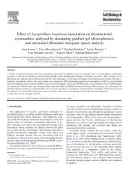
1190<br />
A. Lerner et al. / Soil Biology & Biochemistry 38 (2006) 1188–1192<br />
(TAE, pH 8.5) TAE buffer. The polyacrylamide <strong>gel</strong>s were<br />
prepared with a <strong>denaturing</strong> <strong>gradient</strong> ranging from 30 to 60%<br />
(where 80% denaturant contained 7 M urea and 40%<br />
formamide). The <strong>electrophoresis</strong> was run for 20 h at 85 V<br />
at 60 8C. After the runs, <strong>gel</strong>s were removed from the set up and<br />
stained for 30 min with 2 l <strong>of</strong> 1!TAE and 100 ml <strong>of</strong><br />
10 mg ml K1 EtBr solution followed by washing with 1!<br />
TAE for 15 min. The stained <strong>gel</strong>s were immediately<br />
photographed using an AlphaImagere System (Labtrade<br />
Inc., FL, USA).<br />
2.8. Cluster <strong>analysis</strong><br />
The <strong>analysis</strong> was performed as blind test in which the<br />
operator was unaware <strong>of</strong> the origin <strong>of</strong> the samples. Cluster<br />
<strong>analysis</strong> <strong>of</strong> pr<strong>of</strong>ile similarity was performed using the<br />
Discovery Series Quantity One 1-D Analysis S<strong>of</strong>tware Version<br />
4.4.1, PC (Bio-Rad, Rishon Le Zion, Israel) and UPGMA.<br />
3. Results<br />
3.1. DNA extraction<br />
All the protocols used in this study were based on direct<br />
DNA extraction. Direct DNA protocols include three main<br />
elements: chemical, physical and enzymatic lysis (Miller et al.,<br />
1999). Each protocol, which had been tested herein, included<br />
one or more <strong>of</strong> these elements. Of the methods tested, only<br />
protocols T, F and Z were successful in extracting DNA from a<br />
Rehovot and a Kfar Menachem maize rhizosphere soil.<br />
DNA was also successfully extracted from a coconut<br />
residues medium and from Hula soil, two highly organic<br />
soils, using the T and F methods (Table 1). The use <strong>of</strong> the DNA<br />
Isolation kit (Biological Ind., Israel), failed to remove humic<br />
acids and resulted in brownish samples that could not be<br />
amplified by PCR using primers for the 16S rRNA gene. After<br />
a second purification step, including electrophoretic separation<br />
<strong>of</strong> humic substances from DNA in agarose, DNA excision from<br />
the <strong>gel</strong> and purification using QIAquick Gel Extraction kit<br />
(QIAGEN GmbH, Hilden, Germany), PCR products were<br />
obtained.<br />
As a lesser amount <strong>of</strong> impurities co-extracted with DNA,<br />
protocol T was selected as the DNA extraction method to be<br />
used for the evaluation <strong>of</strong> soil microbial community <strong>analysis</strong><br />
for forensic purposes.<br />
3.2. Microbial community <strong>analysis</strong><br />
Soil samples were taken at various places at and around<br />
the murder scene, at the alibi area and near the family<br />
parking lot at the main suspect’s home. In some forensic<br />
cases, the amount <strong>of</strong> material collected is so low that no<br />
replicate samples can be obtained (for example, soil<br />
adhering to a sole, or to a piece <strong>of</strong> cloth). In order to<br />
reflect this situation, the presented <strong>analysis</strong> was performed:<br />
(i) without prior knowledge <strong>of</strong> the origin <strong>of</strong> the samples,<br />
and; (ii) with only one replicate per location.<br />
Therefore, to mimic the amount found on the suspect’s shoe<br />
(around 0.2 g), which was not available, and to evaluate the<br />
efficiency <strong>of</strong> the protocol T, 0.2–0.6 g <strong>of</strong> soil was extracted. DNA<br />
was retrieved from all samples. In some instances, amplicons<br />
were only obtained after dilution <strong>of</strong> the DNA samples.<br />
<strong>DGGE</strong> was performed on the samples originating from the<br />
crime scene, from the alibi scene, from the suspect’s home and<br />
from different geographical places in Israel in a blind test.<br />
Banding patterns were compared using cluster <strong>analysis</strong>. All<br />
samples collected from the crime scene and surroundings<br />
clustered together. All the samples from the alibi scene and<br />
surroundings were clearly separated from the crime scene<br />
samples. They clustered closer to the samples from Rehovot<br />
and Kfar Menahem, two soils with a texture similar to that <strong>of</strong><br />
the alibi scene samples. Also, a Beit Dagan and a Hula soil,<br />
both richer in organic matter appeared to be closer to the crime<br />
scene samples. However, a sample from the suspect’s home<br />
also clustered with crime scene samples (Fig. 1).<br />
4. Discussion<br />
4.1. Use <strong>of</strong> soils in forensic science<br />
Molecular approaches have become a common tool for the<br />
<strong>analysis</strong> <strong>of</strong> the effect <strong>of</strong> plant cover, agricultural amendments,<br />
pollutants and environmental disorders on soil microbial<br />
communities (Torsvik et al., 1998; Marilley and Aragno,<br />
1999; Smit et al., 2001; Johnsen et al., 2001). Such tools may<br />
also prove useful in criminal investigations to link a suspect to<br />
a crime scene. The first requirement for implementing such<br />
methods is a reliable, convenient and reproducible method <strong>of</strong><br />
DNA extraction from soils.<br />
A number <strong>of</strong> methods for extracting DNA from diverse<br />
environments such as soils are available. Thus, the first step <strong>of</strong><br />
this study was to compare the efficacy <strong>of</strong> DNA retrieval with<br />
various methods on a particular soil type (Haploxeralfs brown–<br />
red <strong>of</strong> Rehovot). The amount <strong>of</strong> DNA extracted was evaluated<br />
by running the DNA obtained on <strong>gel</strong> agarose and comparing<br />
the band intensity after EtBr staining. Zhou et al. (1996) found<br />
a negative correlation between cell lysis efficiency and clay<br />
content, in contrast to Ranjard et al. (2000) who found no such<br />
significant correlation. Our results are in better agreement with<br />
those <strong>of</strong> Ranjard et al. (1998) as the ‘Z’ protocol (Zhou et al.,<br />
1996) applied to an Haploxeralfs brown–red soil with a low<br />
clay content only yielded low amounts <strong>of</strong> DNA. Bead beating,<br />
although recommended for sandy soils (Yeates et al., 1998),<br />
was found to be rather inefficient for this Haploxeralfs soil.<br />
Furthermore, only three out <strong>of</strong> the five methods evaluated<br />
successfully extracted DNA from a loamy sand test soil<br />
(Chromoxererts brown alluvial <strong>of</strong> Kfar Menachem). Further<br />
comparison with other soil types clearly showed that the timeconsuming<br />
Tsai and Olson (1991) protocol (protocol T) was<br />
the most suitable. The combination <strong>of</strong> physical, chemical and<br />
enzymatic attack <strong>of</strong> the cell wall in addition to initial grinding<br />
<strong>of</strong> the sample enabled efficient recovery <strong>of</strong> DNA from<br />
small samples (0.2 g) and from all soil types. Furthermore,<br />
DNA could also be extracted and amplified from pieces <strong>of</strong>