Disinfection/Sterilization of Extracted Teeth for ... - ResearchGate
Disinfection/Sterilization of Extracted Teeth for ... - ResearchGate
Disinfection/Sterilization of Extracted Teeth for ... - ResearchGate
Create successful ePaper yourself
Turn your PDF publications into a flip-book with our unique Google optimized e-Paper software.
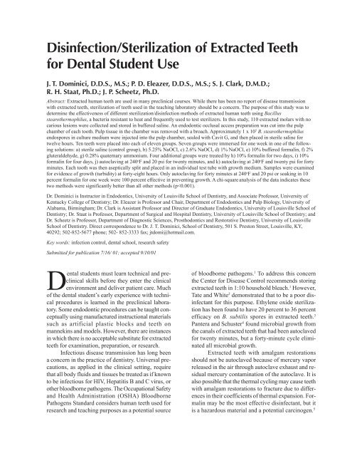
<strong>Disinfection</strong>/<strong>Sterilization</strong> <strong>of</strong> <strong>Extracted</strong> <strong>Teeth</strong><br />
<strong>for</strong> Dental Student Use<br />
J. T. Dominici, D.D.S., M.S.; P. D. Eleazer, D.D.S., M.S.; S. J. Clark, D.M.D.;<br />
R. H. Staat, Ph.D.; J. P. Scheetz, Ph.D.<br />
Abstract: <strong>Extracted</strong> human teeth are used in many preclinical courses. While there has been no report <strong>of</strong> disease transmission<br />
with extracted teeth, sterilization <strong>of</strong> teeth used in the teaching laboratory should be a concern. The purpose <strong>of</strong> this study was to<br />
determine the effectiveness <strong>of</strong> different sterilization/disinfection methods <strong>of</strong> extracted human teeth using Bacillus<br />
stearothermophilus, a bacteria resistant to heat and frequently used to test sterilizers. In this study, 110 extracted molars with no<br />
carious lesions were collected and stored in buffered saline. An endodontic occlusal access preparation was cut into the pulp<br />
chamber <strong>of</strong> each tooth. Pulp tissue in the chamber was removed with a broach. Approximately 1 x 10 5 B. stearothermophilus<br />
endospores in culture medium were injected into the pulp chamber, sealed with Cavit G, and then placed in sterile saline <strong>for</strong><br />
twelve hours. Ten teeth were placed into each <strong>of</strong> eleven groups. Seven groups were immersed <strong>for</strong> one week in one <strong>of</strong> the following<br />
solutions: a) sterile saline (control group), b) 5.25% NaOCl, c) 2.6% NaOCl, d) 1% NaOCl, e) 10% buffered <strong>for</strong>malin, f) 2%<br />
gluteraldehyde, g) 0.28% quaternary ammonium. Four additional groups were treated by h) 10% <strong>for</strong>malin <strong>for</strong> two days, i) 10%<br />
<strong>for</strong>malin <strong>for</strong> four days, j) autoclaving at 240 o F and 20 psi <strong>for</strong> twenty minutes, and k) autoclaving at 240 o F and twenty psi <strong>for</strong> <strong>for</strong>ty<br />
minutes. Each tooth was then aseptically split and placed in an individual test tube with growth medium. Samples were examined<br />
<strong>for</strong> evidence <strong>of</strong> growth (turbidity) at <strong>for</strong>ty-eight hours. Only autoclaving <strong>for</strong> <strong>for</strong>ty minutes at 240 o F and 20 psi or soaking in 10<br />
percent <strong>for</strong>malin <strong>for</strong> one week were 100 percent effective in preventing growth. A chi-square analysis <strong>of</strong> the data indicates these<br />
two methods were significantly better than all other methods (p
The purpose <strong>of</strong> this study was to evaluate the<br />
effectiveness <strong>of</strong> various disinfection/sterilization<br />
methods <strong>for</strong> extracted human teeth.<br />
Methods and Materials<br />
Noncarious, unrestored molar teeth were collected<br />
and stored in saline until the start <strong>of</strong> the study<br />
(n = 110). Approval <strong>for</strong> the use <strong>of</strong> human teeth in<br />
this study was obtained from the University <strong>of</strong> Louisville<br />
Human Studies Committee. An endodontic<br />
access was prepared through the occlusal enamel and<br />
dentin into the pulp chamber using a high-speed #4<br />
round bur. The pulp tissue from all three canals was<br />
extirpated, and gross pulpal tissue was removed with<br />
a fine broach (Union Broach, NY). The canals were<br />
then instrumented with a # 25 Hedstrom file (Kerr,<br />
Romulus, MI) to further remove additional tissue.<br />
The canals and pulp chamber were irrigated with<br />
normal saline to remove dentinal and pulpal debris.<br />
The pulp chamber and canals were dried with absorbent<br />
paper points and then inoculated with 0.05ml<br />
Bacillus stearothermophilus endospores (approximately<br />
1 x 10 5 ). The occlusal access was then sealed<br />
with Cavit G (ESPE, Germany). The teeth were<br />
placed in sterile saline <strong>for</strong> twelve hours and then assigned<br />
to one <strong>of</strong> the following treatment groups (10<br />
teeth /group): 5.25 percent, 2.6 percent, or 1.3 percent<br />
NaOCl (Clorox Bleach, Clorox Co., Oakland,<br />
CA) <strong>for</strong> one week, 10 percent <strong>for</strong>malin (Baxter Scientific<br />
Products, IL) <strong>for</strong> one week, four days, or two<br />
days, 2 percent Glutaraldehyde (JT Baker,<br />
Phillipsburg, NJ) <strong>for</strong> one week, 0.28 percent Quaternary<br />
ammonium (Envirocide, Metrex Research Co.,<br />
Parker, CO) <strong>for</strong> one week, autoclaving at 240 o F at 20<br />
psi <strong>for</strong> twenty minutes or <strong>for</strong>ty minutes, and buffered<br />
saline (control) <strong>for</strong> one week. Groups <strong>of</strong> ten teeth<br />
were immersed in separate containers filled with 30<br />
ml <strong>of</strong> solution. The teeth placed in the autoclave treatment<br />
groups were placed in a glass beaker with 100<br />
ml <strong>of</strong> saline (10 teeth/group) to prevent the teeth from<br />
dehydrating when some <strong>of</strong> the saline solution evaporated<br />
during the autoclave cycle. (See Table 1.)<br />
Fresh solutions were prepared on the day <strong>of</strong><br />
use. Solutions <strong>of</strong> NaOCl were changed after four days<br />
when any evidence <strong>of</strong> oxidizing effervescence had<br />
subsided.<br />
Following the assigned treatment procedure and<br />
time period, teeth from each group were aseptically<br />
split with sterile extraction <strong>for</strong>ceps, and placed into<br />
separate test tubes containing trypticase soy broth<br />
(BBL, Baltimore, MD) growth medium. The test<br />
tubes containing individual teeth were incubated <strong>for</strong><br />
<strong>for</strong>ty-eight hours at 54 o C. Evidence <strong>of</strong> sporulation<br />
and growth was observed using the McFarland turbidity<br />
indicator. Data was collected, and statistical<br />
analysis was per<strong>for</strong>med using a chi-squared (x 2 )<br />
analysis <strong>of</strong> contingency table.<br />
Results<br />
<strong>Teeth</strong> immersed in 10 percent <strong>for</strong>malin <strong>for</strong> one<br />
week or autoclaved at 240 o F at 20 psi <strong>for</strong> <strong>for</strong>ty minutes<br />
were the only methods that prevented the growth<br />
<strong>of</strong> B. stearothermophilus spores (Table 2). Autoclaving<br />
at 240 o F at 20 psi <strong>for</strong> twenty minutes and immersion<br />
in 10 percent <strong>for</strong>malin <strong>for</strong> two days were effective<br />
in preventing microbial growth in nine <strong>of</strong> ten<br />
cases. Immersion in 10 percent <strong>for</strong>malin <strong>for</strong> four days<br />
was effective in 80 percent <strong>of</strong> the samples. Full<br />
strength household bleach (5.25 percent NaOCl) <strong>for</strong><br />
one week prevented growth <strong>of</strong> spores in six <strong>of</strong> ten<br />
samples studied. Only half <strong>of</strong> the teeth immersed in<br />
2 percent glutaraldehyde showed no growth All other<br />
treatments were effective in less than 50 percent <strong>of</strong><br />
the test samples. Positive controls displayed 100 per-<br />
Table 1. <strong>Sterilization</strong>/disinfection methods<br />
Treatment Concentration Time Period<br />
NaOCl 5.25% 1 week<br />
NaOCl 2.6% 1 week<br />
NaOCl 1.3% 1 week<br />
Formalin 10% 1 week<br />
Formalin 10% 4 days<br />
Formalin 10% 2 days<br />
Quaternary Ammonium 0.28% 1 week<br />
Gluteraldehyde 2% 1 week<br />
Autoclave @240@20 psi - 20 minutes<br />
Autoclave @240@20 psi - 40 minutes<br />
Saline (control) - 1 week<br />
Table 2. <strong>Sterilization</strong>/disinfection methods<br />
Number Percent No<br />
Treatment <strong>of</strong> <strong>Teeth</strong> Growth<br />
5.25% NaOCl 1 wk 10 60%<br />
2.6% NaOCl 1 wk 10 10%<br />
1% NaOCl 1 wk 10 0<br />
10% Formalin 2 d 10 90%<br />
10% Formalin 4 d 10 80%<br />
10% Formalin 1 wk 10 100%<br />
Quaternary Am.1 wk 10 30%<br />
2% Gluteraldehyde 10 50%<br />
Autoclave 20 min 10 90%<br />
Autoclave 40 min 10 100%<br />
Saline control 1 wk 10 0%
cent growth. Chi-square analysis <strong>of</strong> the data indicates<br />
a statistically significant difference in the outcomes<br />
when comparing the different methods <strong>of</strong> disinfection<br />
and sterilization. Immersion in 10 percent<br />
<strong>for</strong>malin <strong>for</strong> one week or autoclaving at 240 o F at 20<br />
psi <strong>for</strong> <strong>for</strong>ty minutes were significantly better than<br />
all other methods tested (p< 0.001).<br />
Discussion/Conclusions<br />
<strong>Sterilization</strong> <strong>of</strong> extracted teeth used in the teaching<br />
laboratory should be a concern to educators and<br />
students alike. Potentially pathogenic microorganisms<br />
have been recovered after drilling on extracted teeth<br />
in the dental technique laboratory. 6 The results <strong>of</strong> this<br />
study indicate that extracted teeth inoculated with B.<br />
stearothermophilus then autoclaved at 240 o F <strong>for</strong> <strong>for</strong>ty<br />
minutes or soaked in 10 percent <strong>for</strong>malin <strong>for</strong> seven<br />
days did not have spore growth. All the other methods<br />
tested were less effective in preventing spore<br />
growth. B. stearothermophilus is a biologic testing<br />
standard <strong>for</strong> evaluating sterilization. This microorganism<br />
is very resistant to steam sterilization. Also,<br />
teeth can be particularly difficult to sterilize. The requirement<br />
<strong>for</strong> a longer autoclave cycle (<strong>for</strong>ty minutes<br />
vs. twenty minutes) confirmed the difficulty in<br />
sterilizing extracted teeth. The difference in positive<br />
growth with various time periods in <strong>for</strong>malin may<br />
have occurred because the <strong>for</strong>malin did not adequately<br />
penetrate through the tooth into the pulp<br />
space. Dentinal debris from the root canal instrumentation<br />
may have occluded the tubules. Also, an intact<br />
cemental surface may provide a barrier to diminish<br />
the penetration <strong>of</strong> some <strong>of</strong> the treatment agents. 7<br />
Some <strong>of</strong> the solutions tested have not been recommended<br />
<strong>for</strong> use against spores. These treatment solutions<br />
were tested because they are common disinfectants<br />
found in the dental <strong>of</strong>fice and are solutions<br />
in which extracted teeth have been stored by students.<br />
These solutions (5.25 percent NaOCl, 2.6 percent<br />
NaOCl, 1.3 percent NaOCl, 0.28 percent quaternary<br />
ammonium and 2 percent glutaraldehyde) should not<br />
be relied on to disinfect teeth <strong>for</strong> laboratory/research<br />
use.<br />
Infection control measures to protect students<br />
and faculty are not limited just to the disinfection/<br />
sterilization <strong>of</strong> extracted teeth. Instrument sterilization<br />
as well as the use <strong>of</strong> gloves, eye protection, and<br />
masks should also be used in the preclinical laboratory.<br />
If <strong>for</strong>malin is chosen <strong>for</strong> treatment <strong>of</strong> teeth <strong>for</strong><br />
preclinical laboratory use, the container holding the<br />
teeth should be opened only under a fume hood, and<br />
the teeth should be rinsed prior to their use. Impervious<br />
gloves and goggles should be used to prevent<br />
skin and eye exposure. 5<br />
This study demonstrates the effectiveness <strong>of</strong><br />
one week immersion in 10 percent <strong>for</strong>malin or a <strong>for</strong>ty<br />
minute autoclave cycle at 240 o F and 20 psi. The sterilization<br />
procedures should not affect physical properties<br />
<strong>of</strong> the dentin and enamel to the extent that the<br />
“feel” and cutting characteristics are noticeably different<br />
from the clinical situation, as this is one <strong>of</strong> the<br />
major advantages in using extracted teeth. Further<br />
studies on the physical properties <strong>of</strong> sterilized extracted<br />
teeth are in progress.<br />
REFERENCES<br />
1. Centers <strong>for</strong> Disease Control. Recommended infectioncontrol<br />
practices <strong>for</strong> dentistry. MMWR 1993;42:8-9.<br />
2. Tate WH, White RR. <strong>Disinfection</strong> <strong>of</strong> human teeth <strong>for</strong> education<br />
purposes. J Dent Educ 1991;55:583-5.<br />
3. White RR, Hays GL. Failure <strong>of</strong> ethylene oxide to sterilize<br />
extracted human teeth. Dent Mater 1995;11:321-3.<br />
4. Pantera EA, Schuster GS. <strong>Sterilization</strong> <strong>of</strong> extracted human<br />
teeth. J Dent Educ 1990;54:283-5.<br />
5. Cuny E, Carpenter WM. <strong>Extracted</strong> teeth: decontamination,<br />
disposal and use. Calif Dent Assoc J 1997;25:801-4.<br />
6. Pagniano RP, Scheid RC, Rosen S, Beck TM. Airborne<br />
microorganisms collected in a preclinical dental laboratory.<br />
J Dent Educ 1985;49:653-5.<br />
7. Berutti E, et al. Penetration ability <strong>of</strong> different irrigants<br />
into dentinal tubules. JOE 1997;23:725-7.


