LOWER EXTREMITY RECONSTRUCTION
LOWER EXTREMITY RECONSTRUCTION
LOWER EXTREMITY RECONSTRUCTION

You also want an ePaper? Increase the reach of your titles
YUMPU automatically turns print PDFs into web optimized ePapers that Google loves.
[<strong>LOWER</strong> <strong>EXTREMITY</strong> <strong>RECONSTRUCTION</strong> - BRIAN M. PARRETT ET AL.]<br />
THE RECONSTRUCTIVE LADDER<br />
The reconstructive ladder guides our efforts in lower extremity<br />
reconstruction and describes levels of increasingly complex<br />
management of wounds (Figure 1) (4). The options for reconstruction<br />
from simplest to the most complex are as follows:<br />
Secondary Intention Closure<br />
Closure by secondary intention is the simplest method of<br />
reconstruction and focuses on allowing the wound to naturally<br />
granulate and contract by the use of good local wound care. This<br />
can be performed with wet to dry dressing changes or any number<br />
of advanced wound care dressings. The vacuum-assisted closure<br />
(VAC®; KCI, San Antonio, Texas) device has been useful in promoting<br />
wound granulation and closure by using sub-atmospheric negative<br />
pressure. This has been shown to cause decreased tissue edema,<br />
decreased wound circumference, and increased granulation tissue.<br />
The VAC® device is especially useful in patients who are too ill to<br />
undergo more complex reconstructions. DeFranzo et al. in 2001<br />
demonstrated that the VAC sponge successfully stimulated profuse<br />
granulation tissue over exposed bone, often allowing open fractures<br />
to be closed primarily or with skin grafts thus avoiding more complex<br />
reconstructions (5).<br />
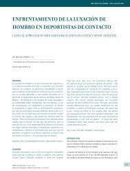
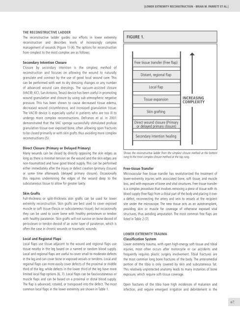
FIGURE 1.<br />
Free tissue transfer (Free flap)<br />
Distant, regional flap<br />
Local flap<br />
Tissue expansion<br />
Skin grafting<br />
Direct wound closure (Primary<br />
or delayed primary closure)<br />
Secondary intention healing<br />
Increasing<br />
Complexity<br />
Direct Closure (Primary or Delayed Primary)<br />
Many wounds can be closed by directly opposing the skin edges as<br />
long as there is minimal tension on the wound and the skin edges are<br />
non-traumatized and have good blood supply. This can be performed<br />
either immediately after the injury or defect creation (primary closure)<br />
or some time afterwards (delayed primary closure). Occasionally<br />
this requires undermining the edges of the wound deep to the<br />
subcutaneous tissue to allow for greater laxity.<br />
Skin Grafts<br />
Full-thickness or split-thickness skin grafts can be used for lower<br />
extremity reconstruction. Skin grafts are best used to cover exposed<br />
muscle or soft tissue (fascia or subcutaneous tissue), but occasionally<br />
they can be used to cover bone with healthy periosteum or tendon<br />
with healthy paratenon. Skin grafts will not survive on bone devoid of<br />
periosteum or tendon devoid of an outer layer of paratenon, which is<br />
often the case in chronic wounds or traumatic wounds.<br />
Local and Regional Flaps<br />
Local flaps use tissue adjacent to the wound and regional flaps use<br />
tissue nearby in the leg based on a named or random blood supply.<br />
Local and regional flaps are useful to cover small to moderate defects<br />
in the leg and can cover bone or exposed vessels or tendons. Local and<br />
regional flaps can more easily cover defects of the proximal or middle<br />
third of the leg; while defects in the lower third of the leg have more<br />
limited local flap options (6, 7). Local flaps can be fasciocutaneous or<br />
muscle flaps and can be based on a proximal or distal blood supply.<br />
The flap is advanced, rotated, or transposed into the defect. The most<br />
common local flaps in the lower extremity are shown in Table 1.<br />
Shows the reconstructive ladder from the simplest closure method at the bottom<br />
rung to the most complex closure method at the top rung.<br />
Free-tissue Transfer<br />
Microvascular free tissue transfer has revolutionized the treatment of<br />
lower-extremity injuries with associated bone, soft tissue, and muscle<br />
loss, and with exposure of bone and vital structures. Free tissue transfer<br />
is a complex procedure that involves removing a piece of tissue with its<br />
blood supply (free flap) from a distal part of the body and placing it over<br />
a defect, reconnecting the artery and vein to vessels at the recipient<br />
site under the microscope. The new tissue acts as an autotransplant,<br />
providing skin or muscle for coverage of otherwise exposed vital<br />
structures, thus avoiding amputation. The most common free flaps are<br />
listed in Table 2 (7).<br />
<strong>LOWER</strong> <strong>EXTREMITY</strong> TRAUMA<br />
Classification System<br />
Lower extremity trauma, with open high-energy soft-tissue and tibial<br />
injuries, most often occurs after motorcycle or car accidents and<br />
frequently requires plastic surgery involvement. Tibial fractures are<br />
the most common long bone fractures of the body. The anteromedial<br />
portion of the tibia is only covered by skin and subcutaneous fat.<br />
This relatively unprotected anatomy leads to many instances of bone<br />
exposure, which require soft-tissue coverage.<br />
Open fractures of the tibia have high incidences of malunion and<br />
infection, and require emergent irrigation and debridement in the<br />
67