Vasectomy reversal using a microsurgical three-layer technique ...
Vasectomy reversal using a microsurgical three-layer technique ...
Vasectomy reversal using a microsurgical three-layer technique ...
Create successful ePaper yourself
Turn your PDF publications into a flip-book with our unique Google optimized e-Paper software.
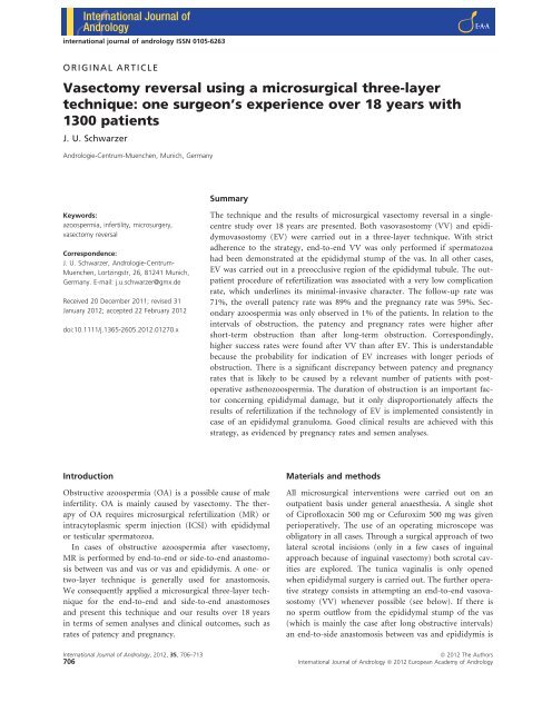
international journal of andrology ISSN 0105-6263<br />
ORIGINAL ARTICLE<br />
<strong>Vasectomy</strong> <strong>reversal</strong> <strong>using</strong> a <strong>microsurgical</strong> <strong>three</strong>-<strong>layer</strong><br />
<strong>technique</strong>: one surgeon’s experience over 18 years with<br />
1300 patients<br />
J. U. Schwarzer<br />
Andrologie-Centrum-Muenchen, Munich, Germany<br />
Summary<br />
Keywords:<br />
azoospermia, infertility, microsurgery,<br />
vasectomy <strong>reversal</strong><br />
Correspondence:<br />
J. U. Schwarzer, Andrologie-Centrum-<br />
Muenchen, Lortzingstr, 26, 81241 Munich,<br />
Germany. E-mail: j.u.schwarzer@gmx.de<br />
Received 20 December 2011; revised 31<br />
January 2012; accepted 22 February 2012<br />
doi:10.1111/j.1365-2605.2012.01270.x<br />
The <strong>technique</strong> and the results of <strong>microsurgical</strong> vasectomy <strong>reversal</strong> in a singlecentre<br />
study over 18 years are presented. Both vasovasostomy (VV) and epididymovasostomy<br />
(EV) were carried out in a <strong>three</strong>-<strong>layer</strong> <strong>technique</strong>. With strict<br />
adherence to the strategy, end-to-end VV was only performed if spermatozoa<br />
had been demonstrated at the epididymal stump of the vas. In all other cases,<br />
EV was carried out in a preocclusive region of the epididymal tubule. The outpatient<br />
procedure of refertilization was associated with a very low complication<br />
rate, which underlines its minimal-invasive character. The follow-up rate was<br />
71%, the overall patency rate was 89% and the pregnancy rate was 59%. Secondary<br />
azoospermia was only observed in 1% of the patients. In relation to the<br />
intervals of obstruction, the patency and pregnancy rates were higher after<br />
short-term obstruction than after long-term obstruction. Correspondingly,<br />
higher success rates were found after VV than after EV. This is understandable<br />
because the probability for indication of EV increases with longer periods of<br />
obstruction. There is a significant discrepancy between patency and pregnancy<br />
rates that is likely to be caused by a relevant number of patients with postoperative<br />
asthenozoospermia. The duration of obstruction is an important factor<br />
concerning epididymal damage, but it only disproportionately affects the<br />
results of refertilization if the technology of EV is implemented consistently in<br />
case of an epididymal granuloma. Good clinical results are achieved with this<br />
strategy, as evidenced by pregnancy rates and semen analyses.<br />
Introduction<br />
Obstructive azoospermia (OA) is a possible cause of male<br />
infertility. OA is mainly caused by vasectomy. The therapy<br />
of OA requires <strong>microsurgical</strong> refertilization (MR) or<br />
intracytoplasmic sperm injection (ICSI) with epididymal<br />
or testicular spermatozoa.<br />
In cases of obstructive azoospermia after vasectomy,<br />
MR is performed by end-to-end or side-to-end anastomosis<br />
between vas and vas or vas and epididymis. A one- or<br />
two-<strong>layer</strong> <strong>technique</strong> is generally used for anastomosis.<br />
We consequently applied a <strong>microsurgical</strong> <strong>three</strong>-<strong>layer</strong> <strong>technique</strong><br />
for the end-to-end and side-to-end anastomoses<br />
and present this <strong>technique</strong> and our results over 18 years<br />
in terms of semen analyses and clinical outcomes, such as<br />
rates of patency and pregnancy.<br />
Materials and methods<br />
All <strong>microsurgical</strong> interventions were carried out on an<br />
outpatient basis under general anaesthesia. A single shot<br />
of Ciprofloxacin 500 mg or Cefuroxim 500 mg was given<br />
perioperatively. The use of an operating microscope was<br />
obligatory in all cases. Through a surgical approach of two<br />
lateral scrotal incisions (only in a few cases of inguinal<br />
approach because of inguinal vasectomy) both scrotal cavities<br />
are explored. The tunica vaginalis is only opened<br />
when epididymal surgery is carried out. The further operative<br />
strategy consists in attempting an end-to-end vasovasostomy<br />
(VV) whenever possible (see below). If there is<br />
no sperm outflow from the epididymal stump of the vas<br />
(which is mainly the case after long obstructive intervals)<br />
an end-to-side anastomosis between vas and epididymis is<br />
International Journal of Andrology, 2012, 35, 706–713<br />
ª 2012 The Authors<br />
706 International Journal of Andrology ª 2012 European Academy of Andrology
J. U. Schwarzer <strong>Vasectomy</strong> <strong>reversal</strong> <strong>using</strong> a <strong>microsurgical</strong> <strong>three</strong>-<strong>layer</strong> <strong>technique</strong><br />
required [epididymovasostomy (EV)]. Both procedures<br />
are carried out <strong>using</strong> a <strong>three</strong>-<strong>layer</strong> <strong>technique</strong>. The wound<br />
is closed with self-dissolving sutures and a pressure dressing<br />
is applied for 1 day.<br />
Intraoperative strategy<br />
At first both ligated stumps of the vas deferens are identified,<br />
prepared and trimmed. If liquid comes out from the<br />
epididymal stump, there is apparently no additional<br />
obstruction in the epididymis, caused by the formation of<br />
an epididymal granuloma. The fluid gushing out of the<br />
vas deferens is examined intraoperatively by microscopic<br />
analysis for the presence of spermatozoa and its viscosity.<br />
If spermatozoa are demonstrated, VV is realizable. Sperm<br />
motility and morphology is of minor importance for the<br />
further surgical strategy according to the authors own<br />
experience and the literature (Belker et al., 1991).<br />
In addition to the presence of spermatozoa, low viscosity<br />
of the fluid is a positive prognostic factor for the outcome<br />
of the procedure. (Belker et al., 1991; Silber & Grotjan,<br />
2004; Schlegel & Margreiter, 2007; Hinz et al., 2009).<br />
If the fluid has a toothpaste like consistence, normally<br />
no or only a few fragments of spermatozoa are found. In<br />
this case, as in the case of missing epididymal fluid, an<br />
anastomosis at the epididymal stump of the vas deferens<br />
does not make sense – a view that is largely non-controversial<br />
(Silber & Grotjan, 2004; Parekattil et al., 2005;<br />
Schlegel & Margreiter, 2007; Hinz et al., 2009; Nagler &<br />
Jund, 2009). Instead, an EV between pre-occlusive<br />
epididymal tubule and abdominal stump of the vas deferens<br />
should be carried out. If spermatozoa cannot be demonstrated<br />
only in case of water clear fluid from the<br />
epididymal stump, it is indicated to carry out a VV.<br />
Patency of the inguinal stump of the vas deferens is<br />
checked by injection of 3 mL saline solution.<br />
Operative <strong>technique</strong> of vasovasostomy<br />
The anastomosis is performed with an end-to-end <strong>technique</strong>.<br />
An absolute precondition for a successful anastomosis<br />
is the possibility of preparing both stumps of the<br />
vas deferens without any tension, so that they can be<br />
fixed in an approximator.<br />
At first the interior (mucosal) <strong>layer</strong> is sutured with<br />
10–12 non-absorbable single-armed 10-0 stitches with a<br />
round needle. So many stitches are necessary to compensate<br />
for the different lumina of both vasal stumps, to<br />
ensure a conical lumen at the point of anastomosis and<br />
to avoid a step-like intraluminal formation and any<br />
shifting of the mucosal <strong>layer</strong>. This adaptation of the<br />
different lumina is crucial for subsequent patency of<br />
the anastomosis. The interior <strong>layer</strong> is a water tight<br />
adaptation of the mucosa, however, without any tensile<br />
strength (Figs 1 & 2).<br />
The second <strong>layer</strong> comprises suturing the muscle walls of<br />
both vasal stumps, which have the same diameter despite<br />
different lumina, if both stumps were cut in the straight<br />
part of the vas deferens. If the vasectomy site is in the<br />
convoluted vas deferens very close to the epididymis, the<br />
muscular <strong>layer</strong> of the epididymal duct stump becomes<br />
significantly thinner with increasing nearness to the<br />
epididymis. About ten 9-0 single stitches are placed with<br />
non-absorbable threads. A sharp spatula needle is necessary<br />
for optimal passage through the compact muscular <strong>layer</strong>.<br />
The closer the cut is to the epididymis, the thinner is<br />
the muscular <strong>layer</strong>, so that stitches should not be placed<br />
too deeply. The muscularis suture provides tension relief<br />
to the fragile internal <strong>layer</strong> (Fig. 3).<br />
The third <strong>layer</strong> consists of adventitial connective tissue<br />
surrounding the duct. About ten 8-0 stitches are placed,<br />
preventing any tensile stress to the internal mucosal <strong>layer</strong>.<br />
Figure 1 Vasovasostomy: internal <strong>layer</strong> between the mucosa of both<br />
stumps of the vas deferens, typically presenting relevant luminar<br />
difference.<br />
Figure 2 Vasovasostomy: finished internal <strong>layer</strong>.<br />
ª 2012 The Authors International Journal of Andrology, 2012, 35, 706–713<br />
International Journal of Andrology ª 2012 European Academy of Andrology 707
<strong>Vasectomy</strong> <strong>reversal</strong> <strong>using</strong> a <strong>microsurgical</strong> <strong>three</strong>-<strong>layer</strong> <strong>technique</strong><br />
J. U. Schwarzer<br />
When preparing the stump of the epididymal duct it is<br />
most important to make sure that the connective tissue<br />
<strong>layer</strong> of the duct is preserved because excessive denudation<br />
involves the risk of secondary hypotrophy (Fig. 4).<br />
Operative <strong>technique</strong> of epididymovasostomy<br />
If there is no outflow or only creamy fluid from the<br />
epididymal stump of the vas deferens, the tunica vaginalis<br />
must be opened for <strong>microsurgical</strong> exploration of the epididymis.<br />
The strategy consists in looking for the duct<br />
obstruction which in most cases is located in the cauda<br />
epididymis.<br />
The pre-occlusive epididymal duct can be identified<br />
under the microscope. Then the dilated pre-occlusive<br />
tubule is tangentially incised in a selective way, which<br />
requires a very subtle operating <strong>technique</strong>. The outflow of<br />
epididymal fluid indicates the preocclusive location. The<br />
outflowing fluid is analysed by the operating surgeon<br />
<strong>using</strong> a lab microscope, with the aim of demonstrating<br />
spermatozoa. If spermatozoa are identified, a side-to-end<br />
anastomosis between epididymal tubule and abdominal<br />
stump of the vas deferens is carried out in a <strong>three</strong>-<strong>layer</strong><br />
<strong>technique</strong>. Crucial to the outcome is an operative procedure<br />
without any tissue tension.<br />
For the internal <strong>layer</strong> between the wall of the laterally<br />
opened epididymal tubule and the mucosa of the vas deferens<br />
8–10 non-absorbable single-armed 10-0 stitches are<br />
placed with a round needle.<br />
This internal <strong>layer</strong>, including the easily tearable structure<br />
of the tubular wall, requires 20–30· magnification<br />
with the operating microscope as well as extensive <strong>microsurgical</strong><br />
experience and utmost concentration of the<br />
surgeon (Fig. 5).<br />
The second <strong>layer</strong> is closed between the muscularis of<br />
the vas and the epididymal serosa with about ten 9-0<br />
stitches with spatula needle. It provides substantial tension<br />
relief to the tearable internal <strong>layer</strong> (Fig. 6).<br />
Complete tension relief is then achieved by suture of<br />
the third <strong>layer</strong>, which is performed between the adventitia<br />
of the vas and the epididymal serosa with about ten 8-0<br />
single stitches (Fig. 7).<br />
For completion of the third <strong>layer</strong> it is most important<br />
that the connective tissue around the vas deferens is wellpreserved;<br />
excessive denudation should therefore be<br />
avoided (see operative <strong>technique</strong> of VV).<br />
Patients<br />
Figure 3 Vasovasostomy: middle <strong>layer</strong> between the muscular <strong>layer</strong> of<br />
both stumps of the vas deferens.<br />
From 10 ⁄ 93 to 06 ⁄ 11, 1429 patients underwent MR by<br />
one surgeon in a single centre for genital microsurgery.<br />
Between 1987 and 1993 the author used a two-<strong>layer</strong><br />
Figure 4 Vasovasostomy: outer <strong>layer</strong> between the adventitia of both<br />
stumps of the vas deferens.<br />
Figure 5 Epididymovasostomy: internal <strong>layer</strong> between mucosa of the<br />
vas deferens and wall of the epididymal tubule.<br />
International Journal of Andrology, 2012, 35, 706–713<br />
ª 2012 The Authors<br />
708 International Journal of Andrology ª 2012 European Academy of Andrology
J. U. Schwarzer <strong>Vasectomy</strong> <strong>reversal</strong> <strong>using</strong> a <strong>microsurgical</strong> <strong>three</strong>-<strong>layer</strong> <strong>technique</strong><br />
Figure 6 Epididymovasostomy: middle <strong>layer</strong> between muscular <strong>layer</strong><br />
of vas deferens and serosal <strong>layer</strong> of epididymis.<br />
thus comprises 1303 patients who underwent vasectomy<br />
<strong>reversal</strong>. Of these, 172 (13.2%) required repeat intervention<br />
after a previous attempt of refertilization.<br />
All patients were physically examined with palpation of<br />
the scrotum, especially for identification of the vasal<br />
stump, and a scrotal sonography.<br />
The age of the patients ranged from 24 to 67 years,<br />
with an average of 41 years. The age of the female partners<br />
ranged from 21 to 45 years, with an average of<br />
34.6 years.<br />
The periods of obstruction ranged between 18 h and<br />
32 years (average 8.2 years). One patient with 18-h<br />
obstructive interval needed immediate <strong>reversal</strong> because of<br />
a non-accepted vasectomy (‘agreement error’). The study<br />
followed ethical guidelines that are established for human<br />
subjects by the Department of Urology of the Technische<br />
Universität München.<br />
Results<br />
Figure 7 Epididymovasostomy: outer <strong>layer</strong> between adventitia of vas<br />
deferens and serosal <strong>layer</strong> of epididymis.<br />
<strong>technique</strong> in several hundred patients, who are not considered<br />
in the database and therefore are not included in<br />
this article. Also excluded are 126 patients with seminal<br />
tract obstruction caused by infection or iatrogenic factors<br />
who were operated during the study period. The study<br />
Perioperative course<br />
Nine-hundred and fifty-eight patients underwent bilateral<br />
VV, 214 patients unilateral VV in combination with contralateral<br />
EV. Another 36 patients underwent unilateral<br />
VV, 84 patients EV bilaterally and 11 patients EV unilaterally<br />
(Table 1). So in 24% of the patients EV had to be<br />
carried out at least at one side according to our strategy<br />
as mentioned above.<br />
The operation time ranged from 90 to 150 min,<br />
110 min on average.<br />
The complication rate was 0.3% (n = 4) for scrotal<br />
haematoma, only one patient had to be reoperated for<br />
evacuation of haematoma. Ten (0.8%) had a superficial<br />
wound infection, no case of epididymitis was seen. Apart<br />
from two cases of allergic reaction to antibiotics, no side<br />
effects or complications were ever seen.<br />
Post-operative course<br />
The follow-up was characterized by special problems, e.g.<br />
that many patients changed their place of residence and<br />
Table 1 <strong>Vasectomy</strong> <strong>reversal</strong> by <strong>microsurgical</strong> <strong>technique</strong>: type of anastomosis in relation to the period of obstruction (total number of patients<br />
n = 1303): epididymovasostomy at least on one side in 24% of patients<br />
Group<br />
no.<br />
Obstruction<br />
period (years)<br />
Patients<br />
(n)<br />
Bilateral<br />
vasovasostomy<br />
Vasovasostomy +<br />
epididymovasostomy<br />
Bilateral<br />
epididymovasostomy<br />
Unilateral<br />
vasovasostomy<br />
Unilateral<br />
epididymovasostomy<br />
1 15 124 n = 74 (59%) n = 22 (18%) n = 23 (18%) n = 2 (2%) n = 3 (3%)<br />
Total 1303 n = 958 (75%) n = 214 (16%) n = 84 (5%) n = 36 (3%) n = 11 (1%)<br />
ª 2012 The Authors International Journal of Andrology, 2012, 35, 706–713<br />
International Journal of Andrology ª 2012 European Academy of Andrology 709
<strong>Vasectomy</strong> <strong>reversal</strong> <strong>using</strong> a <strong>microsurgical</strong> <strong>three</strong>-<strong>layer</strong> <strong>technique</strong><br />
J. U. Schwarzer<br />
Table 2 <strong>Vasectomy</strong> <strong>reversal</strong> by <strong>microsurgical</strong> <strong>technique</strong>: patency and pregnancy rates in relation to the period of obstruction in a follow-up of<br />
n = 924 out of 1303 patients (71% follow-up rate)<br />
Group<br />
no.<br />
Obstruction<br />
period (years)<br />
Patients<br />
(n) Patency rate (%) Pregnancy rate (%)<br />
Average age of the<br />
partner (years)<br />
1 15 108 81 (n = 87) 48 (n = 52) 35.1<br />
Total 924 89 (n = 823) 59 (n = 545) 34.6<br />
Statistical significance<br />
between group (no ⁄ no)<br />
p = 0.0103 (1 ⁄ 2)<br />
p = 0.0155 (2 ⁄ 3)<br />
p = 0.4449 (3 ⁄ 4)<br />
p = 0.7147 (1 ⁄ 2)<br />
p = 0.0015 (2 ⁄ 3)<br />
p = 0.4645 (3 ⁄ 4)<br />
p < 0.05 between<br />
all groups<br />
Table 3 Ejaculate quality after vasectomy <strong>reversal</strong> in relation to the period of obstruction. Follow-up includes 788 patients with semen analyses<br />
according to World Health Organization 2010 (Oligozoospermia:
J. U. Schwarzer <strong>Vasectomy</strong> <strong>reversal</strong> <strong>using</strong> a <strong>microsurgical</strong> <strong>three</strong>-<strong>layer</strong> <strong>technique</strong><br />
procedure of ICSI is a means of artificial reproduction<br />
with a relevant burden to the female partner and higher<br />
costs (Lee et al., 2008).<br />
The follow-up rate in our study is higher after long<br />
periods of obstruction (>10 years) compared with those<br />
<strong>Vasectomy</strong> <strong>reversal</strong> <strong>using</strong> a <strong>microsurgical</strong> <strong>three</strong>-<strong>layer</strong> <strong>technique</strong><br />
J. U. Schwarzer<br />
antisperm antibodies (McDonald, 1996; Marconi et al.,<br />
2008; Légaré et al., 2010).<br />
In Tables 2 and 3 it is shown that with increasing time<br />
of obstruction the sperm quality and pregnancy rates are<br />
decreasing. However, the patency rates did not differ significantly<br />
with the obstruction period.<br />
In our opinion it can be concluded that the main reason<br />
for the decreasing pregnancy rates lies in the decreasing<br />
sperm quality. Directly correlating individual<br />
pregnancies to individual semen analysis is limited,<br />
because for a relevant group of pregnant females (136)<br />
no semen analysis of the male partners was available. In<br />
our study the distribution of the female age was similar<br />
in all the groups independent from the obstructive period.<br />
Besides the female factor no other important factor<br />
was obvious for us. So we conclude that the sperm quality<br />
is the most relevant factor influencing the pregnancy<br />
rates.<br />
Spermatogenesis is not altered by the obstruction for at<br />
least 20–25 years, which was shown by histological studies<br />
in patients with obstructive azoospermia. Only insignificant<br />
alterations of spermatogenesis are described, such as<br />
interstitial fibrosis (Shiraishi et al., 2002) and increased<br />
sperm DNA fragmentation (Smit et al., 2010). However,<br />
the epididymis suffers from obstruction in that it<br />
decreases with time (Srivastava et al., 2000; Lavers et al.,<br />
2006; Yang et al., 2007). Statistically, this deteriorating<br />
effect to the epididymis is related to the interval of<br />
obstruction, which finds expression in the necessity of EV<br />
(Table 1). However, individual differences in the resistance<br />
to time-related epididymal damage must be presumed<br />
because in single cases of normozoospermia<br />
complete recovery of the epididymis after refertilization<br />
may occur.<br />
Female fertility factor<br />
Another relevant factor for the difference between patency<br />
and pregnancy rates may be the relatively high average<br />
age of 34.6 years of the female partners, affecting their<br />
fertility. However the age of the female partner at the<br />
time of operation was not significantly related to the period<br />
of obstruction among the male patients.<br />
Table 2 suggests that the female age being the most<br />
important female fertility factor can be considered as an<br />
independent variable (Fuchs & Burt, 2002; Gerrard et al.,<br />
2007).<br />
Furthermore it should be realized that with increasing<br />
female age abortion rates are most likely increasing. This<br />
should be considered when birth rates are discussed. So<br />
birth rates are probably somewhat lower, as was already<br />
shown in other studies (Belker et al., 1991; Silber & Grotjan,<br />
2004).<br />
Importance of the <strong>three</strong>-<strong>layer</strong> <strong>technique</strong><br />
The advantage of the <strong>three</strong>-<strong>layer</strong> <strong>technique</strong> with a high<br />
number of stitches is a perfect seal of the internal <strong>layer</strong>,<br />
preventing leakage with the possible consequence of granuloma.<br />
In addition, the third <strong>layer</strong> with about ten 8-0<br />
stitches is sufficient to prevent any tension to the internal<br />
<strong>layer</strong> of the anastomosis which secures the liquid-tight<br />
seal. The third <strong>layer</strong> is important for vascularization of the<br />
deferent duct, so that consequent preservation of this connective<br />
tissue coat prevents scarring at the anastomotic<br />
site and secondary occlusion. In the present study, this is<br />
supported by the low rate of 1% of secondary azoospermia<br />
(late failure) and a very low complication rate.<br />
Although excellent results with the two-<strong>layer</strong> <strong>technique</strong><br />
are published in the literature we feel that the <strong>three</strong>-<strong>layer</strong><br />
offers a promising addition the established surgical <strong>technique</strong>s.<br />
In our opinion, the <strong>three</strong>-<strong>layer</strong> anastomosis is no more<br />
time-consuming than a two-<strong>layer</strong> <strong>technique</strong> and the<br />
patient benefit justifies the higher amount of time compared<br />
with the single-<strong>layer</strong> <strong>technique</strong>. According to our<br />
experience, this sophisticated reconstruction of the seminal<br />
tract <strong>using</strong> at least two-<strong>layer</strong>-, better <strong>three</strong>-<strong>layer</strong> <strong>technique</strong>,<br />
in the framework of a minimally invasive<br />
procedure should be the standard of refertilization<br />
surgery against which all other <strong>technique</strong>s, such as the<br />
robotic technology, must be measured.<br />
Acknowledgements<br />
I would like to thank Gudrun Scharfe and Heiko Steinfatt for<br />
their friendly support in drafting the english version of the text.<br />
I would particularly like to thank Prof. Wolfgang Weidner from<br />
Giessen for his generous encouragement of the andrological<br />
microsurgery over many years. I also owe gratitude to my urological<br />
mentor Prof. Rudolf Hartung from Munich who had the<br />
vision to promote my <strong>microsurgical</strong> passion from the beginning.<br />
External sources of support in the form of financial aid, grants<br />
or equipment were not used.<br />
References<br />
Belker AM, Thomas AJ, Fuchs EF, Konnak JW & Sharlip IR. (1991)<br />
Results of 1469 <strong>microsurgical</strong> vasectomy <strong>reversal</strong>s by the vasovasostomy<br />
group. J Urol 145, 505–522.<br />
Bolduc S, Fischer MA, Deceuninck G & Thabet M. (2007) Factors predicting<br />
overall success: a review of 747 <strong>microsurgical</strong> vasovasostomies.<br />
Can Urol Assoc J 1, 388–394.<br />
Boorjian S, Lipkin M & Goldstein M. (2004) The impact of obstructive<br />
interval and sperm granuloma on outcome of vasectomy <strong>reversal</strong>.<br />
J Urol 171, 304–306.<br />
Chawla A, O¢Brien J, Lisi M, Zini A & Jarvi K. (2004) Should all urologists<br />
performing vasectomy <strong>reversal</strong>s be able to perform vasoepididymostomies<br />
if required J Urol 172, 1048–1050.<br />
International Journal of Andrology, 2012, 35, 706–713<br />
ª 2012 The Authors<br />
712 International Journal of Andrology ª 2012 European Academy of Andrology
J. U. Schwarzer <strong>Vasectomy</strong> <strong>reversal</strong> <strong>using</strong> a <strong>microsurgical</strong> <strong>three</strong>-<strong>layer</strong> <strong>technique</strong><br />
Dohle GR & Smit M. (2005) Microsurgical vasovasostomy at the Erasmus<br />
MC, 1998-2002: results and predicitve factors. Ned Tijdschr<br />
Geneeskd 149, 2743–2747.<br />
Eguchi J, Nomata K, Hiose T, Nishimura N, Igawa T, Kanetake H,<br />
Saito Y & Higamy Y. (1999) Clinical experiences of <strong>microsurgical</strong><br />
side-to-end epididymovasostomy for epididymal obstruction. Int J<br />
Urol 6, 271–274.<br />
Fischer MA & Grantmyre JE. (2000) Comparison of modified oneand<br />
two-<strong>layer</strong> <strong>microsurgical</strong> vasovasostomy. BJU International 85,<br />
1085–1088.<br />
Fleming C. (2004) Robot-assisted vasovasostomy. Urol Clin North Am<br />
31, 769–772.<br />
Fuchs EF & Burt RA. (2002) <strong>Vasectomy</strong> <strong>reversal</strong> performed 15 years or<br />
more after vasectomy: correlation of pregnancy outcome with partner<br />
age and with pregnancy results of in vitro fertilization with<br />
intracytoplasmic sperm injection. Fertil Steril 77, 516–519.<br />
Gerrard ER Jr, Sandlow JI, Oster RA, Burns JR, Box LC & Kolettis PN.<br />
(2007) Effect of female partner age on pregnancy rates after vasectomy<br />
<strong>reversal</strong>. Fertil Steril 87, 1340–1344.<br />
Goldstein M & Girardi SK. (1997) <strong>Vasectomy</strong> and vasectomy <strong>reversal</strong>.<br />
Curr Ther Endocrinol Metab 6, 371–385.<br />
Hinz S, Rais-Bahrami S, Kempkensteffen C, Weiske WH, Schrader M<br />
& Magheli A. (2008) Fertility rates following vasectomy <strong>reversal</strong>:<br />
importance of age of the female partner. Urol Int 81, 416–420.<br />
Hinz S, Rais-Bahrami S, Kempkensteffen C, Weiske WH, Schrader M,<br />
Magheli A & Miller K. (2009) Prognostic value of intraoperative<br />
parameters observed during vasectomy <strong>reversal</strong> for predicting postoperative<br />
vas patency and fertility. World J Urol 27, 781–785.<br />
Ho KL, Witte MN, Bird ET & Hakim S. (2005) Fibrin glue assisted<br />
3-suture vasovasostomy. J Urol 174, 1360–1363.<br />
Holman CD, Wisniewski ZS, Semmens JB, Rouse IL & Bass AJ. (2000)<br />
Population-based outcomes after 28.246 in-hospital vasectomies and<br />
1.902 vasovasostomies in Western Australia. BJU Int 86, 1043–1049.<br />
Jee SH & Hong YK. (2010) One-<strong>layer</strong> vasovasostomy: <strong>microsurgical</strong><br />
versus loupe-assisted. Fertil Steril 94, 2308–2311.<br />
Kolettis PN, Burns JR, Nangia AK & Sandlow JI. (2006) Outcomes for<br />
vasovasostomy performed when only sperm parts are present in the<br />
vasal fluid. J Androl 27, 565–567.<br />
Kolettis PN, Fretz P, Burns JR, Dámico AM, Box LC & Sandlow JI. (2005)<br />
Secondary azoospermia after vasovasostomy. Urology 65, 968–971.<br />
Kuang W, Shin PR, Matin S & Thomas AJ Jr. (2004) Initial evaluation<br />
of robotic technology for <strong>microsurgical</strong> vasovasostomy. J Urol 171,<br />
300–303.<br />
Lavers AE, Swanlund DJ, Hunter BA, Tran ML, Pryor JL & Roberts<br />
KP. (2006) Acute effect of vasectomy on the function of the rat epididymal<br />
epithelium and vas deferens. J Androl 27, 826–836.<br />
Lee R, Li PS, Goldstein M, Tanrikut C, Schattmann G & Schlegel PN.<br />
(2008) A decision analysis of treatments for obstructive azoospermia.<br />
Hum Reprod 23, 2043–2049.<br />
Légaré C, Boudreau L, Thimon V, Thabet M & Sullivan R. (2010)<br />
<strong>Vasectomy</strong> affects cysteine-rich secretory protein expression along<br />
the human epididymis and its association with ejaculated spermatozoa<br />
following vasectomy surgical <strong>reversal</strong>. J Androl 31, 573–583.<br />
Lipshultz LI, Rumohr JA & Bennet RC. (2009) Techniques for vasectomy<br />
<strong>reversal</strong>. Urol Clin North Am 36, 375–382.<br />
Magheli A, Rais-Bahrami S, Kempkensteffen C, Weiske WH, Miller K<br />
& Hinz S. (2010) Impact of obstructive interval and sperm granuloma<br />
on patency and pregnancy after vasectomy <strong>reversal</strong>. Int J<br />
Androl 33, 730–735.<br />
Marconi M, Nowotny A, Pantke P, Diemer T & Weidner W. (2008)<br />
Antisperm antibodies detected by mixed agglutination reaction and<br />
immunobead test are not associated with chronic inflammation and<br />
infection of the seminal tract. Andrologia 40, 227–234.<br />
Marmar JL. (2000) Modified vasoepididymostomy with simultaneous<br />
double needle placement, tubulotomy and tubular invagination.<br />
J Urol 163, 483–486.<br />
Matsuda T. (2000) Microsurgical epididymovasostomy. Int J Urol<br />
7(Suppl), 39–41.<br />
Matthews GJ, Schlegel PN & Goldstein M. (1995) Patency following<br />
mircorsurgical vasoepididymstomy and vasovasostomy: temporal<br />
considerations. J Urol 154, 2070–2073.<br />
McDonald SW. (1996) <strong>Vasectomy</strong> review: sequelae in the human epididymis<br />
and ductus deferens. Clin Anat 9, 337–342.<br />
Nagler HM & Jund H. (2009) Factors predicting successful <strong>microsurgical</strong><br />
vasectomy <strong>reversal</strong>. Urol Clin North Am 36, 383–390.<br />
Paick JS, Hong SK, Yun JM & Kim SW. (2000) Microsurgical single<br />
tubular epididymovasostomy: assessment in the era of intracytoplasmic<br />
sperm injection. Fertil Steril 74, 920–924.<br />
Parekattil SJ, Kuang W, Agarwal A & Thomas AJ. (2005) Model to<br />
predict if a vasoepididymostomy will be required for vasectomy<br />
<strong>reversal</strong>. J Urol 173, 1681–1684.<br />
Parekattil SJ, Kuang W, Kolettis PN, Pasqualotto FF, Teloken P, Teloken<br />
C, Nangia AK, Daitch JA, Niederberger C & Thomas AJ Jr.<br />
(2006) Multi-institutional validation of vasectomy <strong>reversal</strong> predictor.<br />
J Urol 175, 247–249.<br />
Parekattil SJ, Ataloh HN & Cohen MS. (2010) Video <strong>technique</strong> for<br />
human robot-assisted <strong>microsurgical</strong> vasovasostomy. J Endourol 24,<br />
511–514.<br />
Patel SR & Sigman M. (2008) Comparison of outcomes of vasovasostomy<br />
performed in the convoluted and straight vas deferens. J Urol<br />
179, 256–259.<br />
Schiff J, Chan P, Li PS, Finkelberg S & Goldstein M. (2005) Outcome<br />
and late failures compared in 4 <strong>technique</strong>s of <strong>microsurgical</strong> vasoepididymostomy<br />
in 153 consecutive men. J Urol 174, 651–654.<br />
Schlegel PN & Margreiter M. (2007) Surgery for male infertility. Eur<br />
Urol Update Series 5, 105–112.<br />
Sheynkin YR, Chen ME & Goldstein M. (2000) Intravasal azoospermia:<br />
a dilemma. BJU International 85, 1089–1092.<br />
Shiraishi K, Takihara H & Naito K. (2002) Influence of interstitial<br />
fibrosis on spermatogenesis after vasectomy and vasovasostomy.<br />
Contraception 65, 245–249.<br />
Sigman M. (2004) The relationship between intravasal sperm quality<br />
and patency rates after vasovasostomy. J Urol 171, 307–309.<br />
Silber SJ & Grotjan HE. (2004) Microscopic vasectomy <strong>reversal</strong> 30<br />
years later: a summary of 4010 cases by the same surgeon. J Androl<br />
25, 845–859.<br />
Smit M, Wissenburg OG, Romijn JC & Dohle GR. (2010) Increased<br />
sperm DNA fragmentation in patients with vasectomy <strong>reversal</strong> has<br />
no prognostic value for pregnancy rate. J Urol 183, 662–665.<br />
Srivastava S, Ansari AS & Lohiya NK. (2000) Ultrastructure of langur<br />
monkey epididymidis prior to and following vasectomy and vasovasostomy.<br />
Eur J Morphol 38, 24–33.<br />
World Health Organization (2010) Laboratory Manual for the Examination<br />
and Processing of Human Semen, 5th edn. WHO Press, Geneva.<br />
Yang G, Walsh TJ, Shefi S & Turek PJ. (2007) The kinetics of the<br />
return of motile sperm to the ejaculate after vasectomy <strong>reversal</strong>.<br />
J Urol 177, 2272–2276.<br />
ª 2012 The Authors International Journal of Andrology, 2012, 35, 706–713<br />
International Journal of Andrology ª 2012 European Academy of Andrology 713


