Squaraine Handout - URI Department of Chemistry
Squaraine Handout - URI Department of Chemistry
Squaraine Handout - URI Department of Chemistry

Create successful ePaper yourself
Turn your PDF publications into a flip-book with our unique Google optimized e-Paper software.
STUDENT HANDOUT<br />
PURPOSE OF THE EXPERIMENT<br />
Synthesize a near-infrared emitting organic fluorophore via multi-step organic synthesis; purify the<br />
product by recrystallization, and characterize the photophysical properties <strong>of</strong> this unique compound.<br />
BACKGROUND REQUIRED<br />
You should be familiar with the set-up <strong>of</strong> standard organic reactions using a reflux condenser,<br />
recrystallization and extraction techniques, and analysis <strong>of</strong> organic compounds by 1 H NMR and FT-IR<br />
spectroscopy. You will learn how to use a Dean-Stark trap to remove water from a reaction, and how to<br />
perform UV-visible and fluorescence analysis.<br />
BACKGROUND INFORMATION<br />
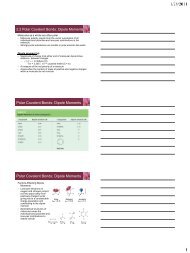
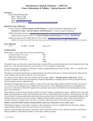
Organic dyes are important for a number <strong>of</strong> applications, including the detection <strong>of</strong> explosive molecules<br />
and the imaging <strong>of</strong> cancer cells and other biological analytes. Some examples <strong>of</strong> commercially available<br />
organic dyes are shown below (Figure S1).<br />
Figure S1: Examples <strong>of</strong> common organic dyes<br />
Researchers are continually working to synthesize new dyes with better properties, such as: (1) a higher<br />
fluorescence quantum yield; (2) better stability in biological environments; and (3) absorption and<br />
emission maxima that are in the red to near-infrared region (because this region will have little<br />
interference from other molecules).<br />
Note: Fluorescence quantum yield is defined as the number <strong>of</strong> photons emitted divided by the number <strong>of</strong><br />
photons absorbed. When a molecule absorbs a photon <strong>of</strong> light, that photon can undergo a variety <strong>of</strong><br />
procedures, one <strong>of</strong> which is emission to cause fluorescence. High quantum yields (near 1) mean that a<br />
high proportion <strong>of</strong> the photons that are absorbed are also emitted.<br />
<strong>Squaraine</strong>s are a class <strong>of</strong> organic dyes that possess a high fluorescence quantum yield, and red to nearinfrared<br />
absorption and emission maxima (2 <strong>of</strong> the 3 properties mentioned above). The general structure<br />
<strong>of</strong> one type <strong>of</strong> squaraines is shown in Figure S2. These compounds possess an electron-deficient<br />
cyclobutenone central ring substituted with two electron-rich aromatic rings. One stable structure <strong>of</strong><br />
squaraine compounds is the zwitterionic form shown, where the cyclobutene has a +2 charge, and both<br />
oxygens are negatively charged.<br />
Figure S2: General structure <strong>of</strong> squaraine compounds<br />
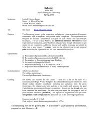
Most red-emitting dyes have large, planar conjugated structures (Figure S3), because the extended<br />
conjugation <strong>of</strong>ten leads to red-shifted absorption and emission maxima. In contrast, squaraines have a<br />
relatively small size, yet still absorb and emit in the red to near-infrared spectral region. This small size<br />
6
enables them to be encapsulated easily in macrocycles, such as cyclodextrins or synthetic rotaxanes, and<br />
allows them to be used in biological detection schemes without interfering with natural cellular pathways.<br />
Figure S3: Examples <strong>of</strong> near-infrared emitting organic dyes<br />
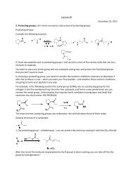
The synthesis <strong>of</strong> squaraines is usually accomplished by reacting two equivalents <strong>of</strong> an electron-rich<br />
aromatic compound with squaric acid (3,4-dihydroxycyclobut-3-ene-1,2-dione). The mechanism <strong>of</strong> this<br />
reaction is shown below:<br />
Scheme S1: Mechanism for the synthesis <strong>of</strong> squaraine compounds<br />
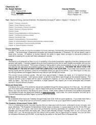
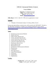
<strong>Squaraine</strong>s are also interesting because <strong>of</strong> their unique absorption and emission spectra. The particular<br />
squaraine you will synthesize in this experiment is shown in Figure S4 (compound 1a) with its<br />
absorbance and emission spectra (measured in chlor<strong>of</strong>orm).<br />
7
Figure S4: Absorption and emission spectra <strong>of</strong> the target squaraine compound<br />
The narrow absorption band around 630 nm and emission band around 650 nm are typical <strong>of</strong> squaraine<br />
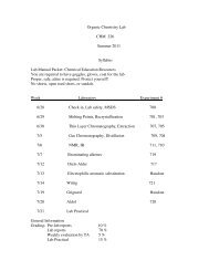
compounds. The squaraine compound is also brightly colored (green-blue solid, bright blue dilute<br />
solution, reddish-blue concentrated solution) (Figure S5).<br />
Figure S5: Colors <strong>of</strong> the squaraine product 1a under various conditions (measured by the authors)<br />
OBJECTIVES<br />
The purpose <strong>of</strong> this experiment is to synthesize squaraine compound 1a in a two-step procedure, and<br />
characterize that compound using UV-Vis and fluorescence spectroscopy, as well as 1 H NMR analysis.<br />
PROCEDURE<br />
Laboratory Period 1:<br />
Using a graduated cylinder, measure 20 mL <strong>of</strong> water and add to a 50 mL round-bottom flask. Add 20 mg<br />
<strong>of</strong> sodium dodecyl sulfate and 920 mg <strong>of</strong> sodium bicarbonate. Then use a syringe to add 4.6 mL <strong>of</strong> aniline<br />
to the aqueous solution (CAUTION: This step should be done in a fume hood). Add a stir bar and stir the<br />
mixture vigorously at room temperature for 5 minutes, then add 1.3 mL <strong>of</strong> benzyl bromide via syringe.<br />
(You can do this using a 1 mL disposable syringe by dispensing 1 mL first, then 0.3 mL. Alternatively,<br />
your instructor may dispense this compound for you.) Add a reflux condenser to the top <strong>of</strong> the round-<br />
8
ottom flask and make sure that the water or other coolant is flowing appropriately through the<br />
condenser. Heat the reaction mixture to 80 o C using an oil or sand bath for approximately 90 minutes.<br />
Monitor the progress <strong>of</strong> your reaction using TLC. Use a solvent mixture <strong>of</strong> 95:5 hexanes: ethyl acetate as<br />
the mobile phase. Both <strong>of</strong> your reactants and your desired product are visible under a hand-held UV lamp,<br />
or can be stained with a potassium permanganate stain.<br />
After 90 minutes (or when the TLC shows that the reaction is complete), cool the reaction to room<br />
temperature and transfer it to a separatory funnel. Add 20 mL <strong>of</strong> ethyl acetate to the funnel using a<br />
graduated cylinder. Separate the layers and collect the ethyl acetate layer. Do another extraction by<br />
adding an additional 20 mL <strong>of</strong> ethyl acetate to the funnel with your aqueous layer, then separating the<br />
layers. Combine the ethyl acetate layers in a round-bottom flask. Remove the ethyl acetate on the rotary<br />
evaporator to yield a crude oil.<br />
Add a small amount <strong>of</strong> n-hexanes via a Pasteur pipette to the oil and cool the mixture to 0 o C. Crystals<br />
will form, and these crystals should be filtered using a Buchner funnel. Transfer the filtrate to a roundbottom<br />
flask and remove the solvent on the rotary evaporator. This will yield more crude oil that has not<br />
yet crystallized. Do a second round <strong>of</strong> recrystallization by adding more n-hexanes to that oil, cooling it to<br />
0 o C, and filtering the crystals using a Buchner funnel.<br />
Obtain a mass for your crystals and calculate your percent yield. Dissolve approximately 10 mg <strong>of</strong> your<br />
product in 1.0 mL <strong>of</strong> CDCl 3 and transfer it to an NMR tube. Obtain a 1 H NMR spectrum. Obtain an IR<br />
spectrum as well. Save your crystals for the second reaction which will occur during the next lab period.<br />
Safety: Aniline and benzyl bromide are volatile chemicals and should be used only in the fume hood.<br />
Make sure to dispose <strong>of</strong> all syringes and chemical waste properly. Hexanes and ethyl acetate are volatile<br />
and flammable and should only be used in a fume hood. Deuterated chlor<strong>of</strong>orm is a potential carcinogen<br />
and should be used in the fume hood.<br />
Laboratory Period 2:<br />
Use the crystals obtained during the previous laboratory period as the starting material for this second<br />
reaction. Add 270 mg <strong>of</strong> dibenzylaniline (your product from the first reaction) and 57 mg <strong>of</strong> squaric acid<br />
to a round-bottom flask, followed by 2.0 mL <strong>of</strong> toluene and 2.0 mL <strong>of</strong> n-butanol, which you can measure<br />
with a graduated cylinder or syringe. (CAUTION: These solvents should be measured and transferred in a<br />
fume hood. They are volatile and have potentially irritating odors).<br />
Add a Dean-Stark trap to the top <strong>of</strong> the flask, followed by a reflux condenser. Make sure all <strong>of</strong> the joints<br />
are properly secured and greased. Then add a sufficient amount <strong>of</strong> a 1:1 mixture <strong>of</strong> toluene and n-butanol<br />
from the top <strong>of</strong> the condenser to fill the Dean-Stark trap completely. Heat the reaction to reflux for two<br />
hours. (NOTE: The two hours do not start until the reaction mixture starts to boil).<br />
As the reaction proceeds, you should observe an intense green color developing. After two hours, slowly<br />
cool the reaction mixture to room temperature. Green crystals should spontaneously form. Filter the<br />
crystals using a Buchner funnel and wash them with a small amount <strong>of</strong> isopropanol. The filtrate will be a<br />
dull green color, and the crystals will be bright green. Obtain a mass <strong>of</strong> these crystals and calculate the<br />
percent yield. Save your crystals in a vial for the next laboratory period, where you will analyze and<br />
9
characterize your product. If you do not obtain crystals, then remove your solvent on the rotary evaporator<br />
to obtain a green solid, which should be sufficiently pure for UV-visible and fluorescence analysis.<br />
Safety: Toluene and n-butanol are volatile solvents with potentially irritating odors. Isopropanol is a<br />
volatile solvent. Make sure to use them only in a fume hood and dispose <strong>of</strong> them properly. Dispose <strong>of</strong> all<br />
syringes and chemical waste properly.<br />
Laboratory Period 3:<br />
In this lab session, you will thoroughly analyze the crystals that you formed in the previous session.<br />
1. 1 H NMR analysis: Dissolve a small amount <strong>of</strong> your crystals in CDCl 3 . Transfer the solution to an NMR<br />
tube, and collect the spectrum.<br />
2. IR analysis: Take a small amount <strong>of</strong> your crystals and obtain an IR spectrum. Look for the presence <strong>of</strong><br />
key functional groups (i.e. C=O, OH, etc).<br />
3. UV-Vis analysis: Dissolve a small amount <strong>of</strong> your crystals in chlor<strong>of</strong>orm to obtain a concentration <strong>of</strong><br />
10 mg/L (solution 1). Transfer some <strong>of</strong> the solution to a cuvette and obtain a UV-Vis absorbance<br />
spectrum. Then dilute some <strong>of</strong> your solution 1 to a new final concentration <strong>of</strong> 0.50 mg/L (solution 2).<br />
Transfer some <strong>of</strong> the solution to a cuvette and obtain a UV-Vis absorbance spectrum <strong>of</strong> solution 2. Look<br />
for any differences in the spectra <strong>of</strong> the two solutions.<br />
Note 1: You need approximately 3.0 mL <strong>of</strong> each solution to obtain a good absorption and emission<br />
spectrum.<br />
Note 2: You cannot record these spectra using plastic/disposable cuvettes. The organic solvent will erode<br />
the plastic.<br />
4. Fluorescence analysis: Using the same solutions that you used for UV-visible analysis, obtain a<br />
fluorescence spectrum <strong>of</strong> both solution 1 and solution 2. Excite the solutions at 600 nm and record the<br />
fluorescence from 610-800 nm. Look for any differences in the spectra <strong>of</strong> the two solutions.<br />
Safety: Make sure to dispose <strong>of</strong> all your solutions using the proper waste containers. Chlor<strong>of</strong>orm is a<br />
potential carcinogen and should be used in a fume hood.<br />
POST-LAB QUESTIONS<br />
1. What is the theoretical yield for each step <strong>of</strong> the reaction<br />
2. What is your percent yield for each step<br />
3. What are the key peaks that you can identify in the IR and 1 H NMR spectra for step 1 <strong>of</strong> the reaction<br />
How does this help you figure out the structure <strong>of</strong> your product<br />
4. Why is the first reaction considered to be an example <strong>of</strong> “green chemistry” What is the role <strong>of</strong> SDS in<br />
the reaction mixture<br />
5. What is another potential product that you could have formed from step 1, and how would you<br />
distinguish that product using 1 H NMR and IR spectroscopy<br />
6. What are the key peaks that you can identify in the IR and NMR for step 2 <strong>of</strong> the reaction How does<br />
this help you figure out the structure <strong>of</strong> your product<br />
7. Analyze your UV-vis spectra for the two solutions. What are the key differences between the two<br />
How can you explain these differences<br />
10