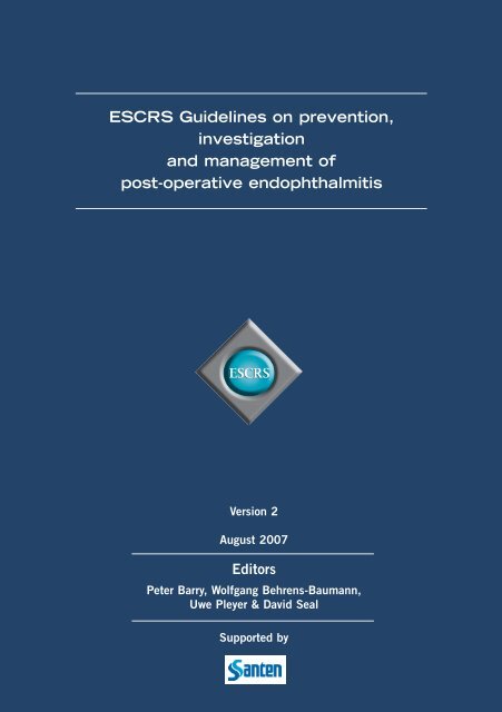
ESCRS Guidelines on prevention, investigation and management of
ESCRS Guidelines on prevention, investigation and management of
ESCRS Guidelines on prevention, investigation and management of

Create successful ePaper yourself
Turn your PDF publications into a flip-book with our unique Google optimized e-Paper software.
<str<strong>on</strong>g>ESCRS</str<strong>on</strong>g> <str<strong>on</strong>g>Guidelines</str<strong>on</strong>g> <strong>on</strong> preventi<strong>on</strong>,<br />
investigati<strong>on</strong><br />
<strong>and</strong> <strong>management</strong> <strong>of</strong><br />
post-operative endophthalmitis<br />
Versi<strong>on</strong> 2<br />
August 2007<br />
Editors<br />
Peter Barry, Wolfgang Behrens-Baumann,<br />
Uwe Pleyer & David Seal<br />
Supported by
C<strong>on</strong>tents<br />
1. INTRODUCTION 1<br />
1.1 Endophthalmitis 1<br />
1.2 Pathophysiology 1<br />
1.3 Microbial spectrum 2<br />
1.4 Incidence <strong>of</strong> endophthalmitis after different types <strong>of</strong> surgery 3<br />
2. PROPHYLAXIS 8<br />
2.1 Operating theatre 8<br />
2.2 Antisepsis 8<br />
2.3 Antibiotics 9<br />
2.4 Limitati<strong>on</strong>s <strong>of</strong> the <str<strong>on</strong>g>ESCRS</str<strong>on</strong>g> Study 12<br />
2.5 FLOW CHART – PROPHYLAXIS GUIDELINES 14<br />
3. DIAGNOSIS 15<br />
3.1 Commencement <strong>and</strong> symptoms 15<br />
3.2 FLOW CHART – DIAGNOSTIC GUIDELINES FOR ACUTE<br />
VIRULENT ENDOPHTHALMITIS 16<br />
3.3 FLOW CHART – DIAGNOSTIC GUIDELINES FOR CHRONIC ENDOPHTHALMITIS 17<br />
3.4 Differential diagnosis – the toxic anterior segment syndrome 18<br />
4. INVESTIGATION 19<br />
4.1 AC tap, vitreous tap, vitrectomy for Gram stain, culture <strong>and</strong> PCR tests 19<br />
4.2 Epidemiology 19<br />
5. TREATMENT 20<br />
5.1 Surgical <strong>management</strong> <strong>of</strong> endophthalmitis. Diagnostic <strong>and</strong> therapeutic vitrectomy 20<br />
5.2 Anti-microbial therapy 22<br />
5.3 Anti-inflammatory therapy 25<br />
5.4 Other types <strong>of</strong> post-operative endophthalmitis 25<br />
5.5 Limitati<strong>on</strong>s <strong>of</strong> EVS study 26<br />
5.6 FLOW CHART – TREATMENT GUIDELINES FOR<br />
ACUTE VIRULENT ENDOPHTHALMITIS 27<br />
5.7 FLOW CHART – TREATMENT GUIDELINES FOR<br />
CHRONIC ENDOPHTHALMITIS 28<br />
6. REFERENCES 29<br />
7. APPENDIX (Addresses <strong>of</strong> C<strong>on</strong>tributors) 36
1. Introducti<strong>on</strong><br />
1.1 Endophthalmitis<br />
Endophthalmitis is an inflammatory reacti<strong>on</strong> occurring as a result <strong>of</strong> intraocular col<strong>on</strong>isati<strong>on</strong> by bacteria,<br />
fungi or rarely parasites. It can be exogenous (post-operative, post-traumatic) due to microbial c<strong>on</strong>taminati<strong>on</strong><br />
spreading from the ocular surface or open incisi<strong>on</strong> (wound) or c<strong>on</strong>taminated instruments, intraocular lenses<br />
(IOLs) or intraocular foreign bodies or endogenous (septicaemia) in origin.<br />
These guidelines <strong>on</strong> the prophylaxis <strong>and</strong> treatment <strong>of</strong> post-operative endophthalmitis are supported in detail<br />
with literature references, which were classified according to the criteria <strong>of</strong> the Arbeitsgemeinschaft der<br />
Wissenschaftlichen Medizinischen Fachgesellschaften (AWMF) [Associati<strong>on</strong> <strong>of</strong> the Scientific Medical<br />
Societies in Germany] <strong>and</strong> the Ärztliches Zentrum für Qualität [Agency for Quality in Medicine] for evidencebased<br />
medicine (EBM) (Table 1.1). This enables the reader to form an accurate opini<strong>on</strong> <strong>of</strong> the value <strong>of</strong> the<br />
individual views stated. At the same time, he or she is able to form an opini<strong>on</strong> him/herself from the<br />
extensive literature. Ultimately, it is apparent that there is a lack <strong>of</strong> well-founded prospective <strong>and</strong> c<strong>on</strong>trolled<br />
studies <strong>of</strong> many procedures - an important task for the future. Results <strong>of</strong> the recently completed <str<strong>on</strong>g>ESCRS</str<strong>on</strong>g><br />
Study <strong>on</strong> the Antibiotic Prophylaxis <strong>of</strong> Post-operative Endophthalmitis (<str<strong>on</strong>g>ESCRS</str<strong>on</strong>g> Study) have been included in<br />
these <str<strong>on</strong>g>Guidelines</str<strong>on</strong>g>.<br />
Table 1.1 Classificati<strong>on</strong> <strong>of</strong> evidence type <strong>of</strong> studies<br />
Stage Evidence based <strong>on</strong>:<br />
I a meta-analysis <strong>of</strong> r<strong>and</strong>omised c<strong>on</strong>trolled studies<br />
I b at least <strong>on</strong>e r<strong>and</strong>omised c<strong>on</strong>trolled study<br />
II a at least <strong>on</strong>e well-designed c<strong>on</strong>trolled study without r<strong>and</strong>omisati<strong>on</strong><br />
II b at least <strong>on</strong>e well-designed, quasi-experimental study<br />
III well-designed, n<strong>on</strong>-experimental descriptive studies<br />
(e.g. comparative studies, correlative studies, case-c<strong>on</strong>trol studies)<br />
IV reports/opini<strong>on</strong>s <strong>of</strong> expert circles, c<strong>on</strong>sensus c<strong>on</strong>ferences<br />
<strong>and</strong>/or clinical experience <strong>of</strong> acknowledged authorities<br />
1.2 Pathophysiology<br />
The occurrence, severity <strong>and</strong> clinical course <strong>of</strong> endophthalmitis depends <strong>on</strong> the route <strong>of</strong> infecti<strong>on</strong>, the<br />
virulence <strong>and</strong> number <strong>of</strong> inoculated pathogens as well as the patient's immune state <strong>and</strong> the time <strong>of</strong><br />
examinati<strong>on</strong> [205]. In 29 to 43 per cent <strong>of</strong> cataract operati<strong>on</strong>s, intraocular c<strong>on</strong>taminati<strong>on</strong> occurs with<br />
facultative pathogenic bacteria from the ocular surface without the development <strong>of</strong> endophthalmitis [41],<br />
[68], [106]. Protective mechanisms, which have been summarised as the “immune privilege <strong>of</strong> the eye“<br />
(anterior or posterior chamber-associated immune deviati<strong>on</strong>, ACAID or POCAID) [205], are particularly<br />
effective in the anterior part <strong>of</strong> the eye, act as a protective barrier <strong>and</strong> can limit the inflammatory reacti<strong>on</strong><br />
[191], [198]. If this privilege is compromised, e.g. by an intra-operative capsular defect with vitreous loss,<br />
the risk <strong>of</strong> endophthalmitis increases by a factor <strong>of</strong> 14 [98].<br />
In microbial endophthalmitis, three phases <strong>of</strong> infecti<strong>on</strong> can be observed: an incubati<strong>on</strong> phase, an<br />
accelerati<strong>on</strong> phase <strong>and</strong> a destructive phase [173]. A clinically inapparent incubati<strong>on</strong> phase is observed<br />
initially, which lasts at least 16 to 18 hours, even with virulent micro-organisms. Intraocular bacterial<br />
inoculati<strong>on</strong> above a critical level then leads to breakdown <strong>of</strong> the aqueous barrier with fibrin exudati<strong>on</strong> <strong>and</strong><br />
cellular infiltrati<strong>on</strong> by neutrophilic granulocytes [24]. The incubati<strong>on</strong> phase is determined mainly by the<br />
generati<strong>on</strong> time <strong>of</strong> the pathogen (e.g. Staphylococcus aureus <strong>and</strong> Pseudom<strong>on</strong>as aeruginosa up to 10 min,<br />
Propi<strong>on</strong>ibacterium sp. > 5 h) <strong>and</strong> the specific characteristics <strong>of</strong> the pathogen such as toxin producti<strong>on</strong>. With<br />
the comm<strong>on</strong>est pathogens, Staphylococcus epidermidis (CNS) <strong>and</strong> Staphylococcus aureus, the greatest<br />
infiltrati<strong>on</strong> is observed <strong>on</strong>ly three days after infecti<strong>on</strong> [24], [39].<br />
In the case <strong>of</strong> primary infecti<strong>on</strong> <strong>of</strong> the posterior part <strong>of</strong> the eye, inflammati<strong>on</strong> <strong>of</strong> the anterior chamber occurs<br />
initially <strong>and</strong> this is accompanied within seven days by a specific immune resp<strong>on</strong>se with macrophages <strong>and</strong><br />
lymphocytes in the vitreous cavity. Just three days after intraocular infecti<strong>on</strong>, pathogen-specific antibodies<br />
can be detected, which c<strong>on</strong>tribute to pathogen eliminati<strong>on</strong> by ops<strong>on</strong>isati<strong>on</strong> <strong>and</strong> phagocytosis within about<br />
1
10 days. This can produce negative laboratory culture results but severe inflammatory disease within the eye<br />
[39]. The inflammatory mediators <strong>of</strong> infiltrating cells, especially cytokines, not <strong>on</strong>ly recruit further leukocytes<br />
but can directly result in destructive effects, retinal injury <strong>and</strong> vitreoretinal proliferati<strong>on</strong> (destructive phase)<br />
[30], [205].<br />
It has been suggested that the use <strong>of</strong> phacoemulsificati<strong>on</strong> increases pressure within the eye forcing bacteria<br />
backwards into the vitreous, where early multiplicati<strong>on</strong> gives rise to the anterior vitritis characteristically seen<br />
behind the posterior capsule (refer to Investigati<strong>on</strong> Secti<strong>on</strong> below).<br />
Intraocular lenses are a potential vector for bacteria. There are differences in adherence to different lens<br />
materials. Staphylococcus epidermidis adheres more to polypropylene haptics than to polymethyl methacrylate,<br />
(PMMA) [69], [98], [190]. Hydrophilic heparin-coated lenses dem<strong>on</strong>strate lower adherence for staphylococci<br />
[52]. The clinical effects have been variously interpreted [106]. A recent retrospective study has suggested,<br />
but not proven, that use <strong>of</strong> foldable IOLs inserted via a sterile injector lowers the incidence <strong>of</strong> post-operative<br />
endophthalmitis [95].<br />
1.3 Microbial spectrum<br />
This is described in the table <strong>of</strong> post-operative endophthalmitis below. The spectrum is dependent <strong>on</strong> various<br />
factors including envir<strong>on</strong>mental, geographic <strong>and</strong> climatic c<strong>on</strong>diti<strong>on</strong>s <strong>and</strong> which type <strong>of</strong> surgery is performed.<br />
Table 1.2: Microbial spectrum <strong>of</strong> post-operative endophthalmitis<br />
In summary, the most important pathogens causing post-operative phacoemulsificati<strong>on</strong> endophthalmitis are:<br />
Post-operative (cataract surgery) endophthalmitis [46], [70], [81], [100], [102], [105], [134], [205], [207]<br />
33 - 77 % CNS (coagulase-negative staphylococci)<br />
10 - 21 % Staphylococcus aureus<br />
9 - 19 % BHS (ß-haemolytic streptococci), S. pneum<strong>on</strong>iae, ∂-haemolytic<br />
streptococci including S. mitis <strong>and</strong> S. salivarius<br />
6 - 22 % Gram-negative bacteria including Ps. aeruginosa (occurs rarely)<br />
up to 8 % Fungi (C<strong>and</strong>ida sp., Aspergillus sp., Fusarium sp.)<br />
Delayed post-operative saccular or capsule bag endophthalmitis with IOL implantati<strong>on</strong><br />
Propi<strong>on</strong>ibacterium acnes, corynebacteria including C. macginleyi [164], [175], [205] <strong>and</strong> fungi [205]<br />
Post-operative (glaucoma surgery) endophthalmitis [116], [124]<br />
up to 67 % CNS<br />
Delayed post-operative (glaucoma surgery) endophthalmitis [78], [142]<br />
Streptococci<br />
Gram-negative bacteria (especially Haemophilus influenzae)<br />
Post-traumatic endophthalmitis<br />
Single pathogen identified in 62–65%, mixed infecti<strong>on</strong> in 12–42% [57], [91], [125], [130], [205],<br />
[207], [208]<br />
16 - 44 % CNS<br />
17 - 32 % Bacillus sp.<br />
10.5 - 18 % Gram-negative bacteria<br />
8 - 21 % Streptococci<br />
4 - 14 % Fungi<br />
4 - 8 % Corynebacterium sp.<br />
1 - 2 % Clostridum perfringens <strong>and</strong> other soil bacteria<br />
2
In the <str<strong>on</strong>g>ESCRS</str<strong>on</strong>g> Study, 29 patients presented with presumed post-operative endophthalmitis, <strong>of</strong> whom 20 were<br />
classified as having proven infective endophthalmitis. The causative bacteria were identified by<br />
microbiological methods <strong>and</strong> polymerase chain reacti<strong>on</strong> (PCR) [11]. From 19 cases the following bacteria<br />
have been c<strong>on</strong>firmed:<br />
Staphylococcus epidermidis 6<br />
CNS 4<br />
Staphylococcus aureus 2<br />
other staphylococci 1<br />
Streptococcus pneum<strong>on</strong>iae 2<br />
other streptococci 6<br />
Propi<strong>on</strong>ibacterium acnes 1<br />
Gemella haemolysans 1<br />
In summary, the most important pathogens causing post-operative phacoemulsificati<strong>on</strong> endophthalmitis are:<br />
Acute<br />
■ BHS, S. mitis, S. pneum<strong>on</strong>iae, E. faecalis<br />
■ S. aureus, CNS, (MRSA, MRSE)<br />
■ Gram-negative rods (GNR) including Haemophilus influenzae <strong>and</strong> Pseudom<strong>on</strong>as aeruginosa<br />
Chr<strong>on</strong>ic<br />
■ P. acnes<br />
■ Diphtheroids<br />
■ CNS<br />
■ Fungi<br />
1.4 Incidence <strong>of</strong> endophthalmitis after different types <strong>of</strong> surgery<br />
Phacoemulsificati<strong>on</strong><br />
At the start <strong>of</strong> the 20th century (c1910), the incidence <strong>of</strong> endophthalmitis after cataract operati<strong>on</strong>s was 10<br />
per cent. In the period <strong>of</strong> ECCE via a scleral or limbal incisi<strong>on</strong> <strong>and</strong> improved hygiene c<strong>on</strong>diti<strong>on</strong>s (1970-<br />
1990), the infecti<strong>on</strong> rate fell to 0.12 per cent in Europe [90] <strong>and</strong> to 0.072 per cent in the US [88].<br />
However, since the introducti<strong>on</strong> <strong>of</strong> phacoemulsificati<strong>on</strong> <strong>and</strong> clear cornea incisi<strong>on</strong>s, the retrospective data<br />
with phacoemulsificati<strong>on</strong> are between 0.3 to 0.5 <strong>and</strong> 0.015 per cent (Table 1.3).<br />
Table 1.3: Reported incidence for endophthalmitis after cataract surgery<br />
Country [reference] Year <strong>of</strong> Publicati<strong>on</strong> Incidence (%) No. <strong>of</strong> Operati<strong>on</strong>s<br />
USA [98] 1991 0.22 24105<br />
USA [203] 1992 0.015 27181<br />
France [40] 1992 0.32 ~ 34690<br />
Germany [126] 1999 0.15 ~ 103090<br />
Netherl<strong>and</strong>s [135] 2000 0.10 ~ 25330<br />
Canada [64] 2000 0.01 to 0.18 13886<br />
Sweden [37] 2002 0.10 54666<br />
Australia [110] 2003 0.16 to 0.36 83677<br />
Japan [8] 2003 0.05 to 0.29 11595<br />
USA [86] 2005 0.29 9079<br />
Irel<strong>and</strong> [87] 2005 0.5 8763<br />
UK [105] 2007 0.099 101920<br />
Sweden [94] 2007 0.048 225471<br />
Europe [5] 2007 0.05 to 0.35 16211<br />
3
While in the majority <strong>of</strong> studies patients received various additi<strong>on</strong>al forms <strong>of</strong> prophylaxis, the higher<br />
incidence rate reported by the <str<strong>on</strong>g>ESCRS</str<strong>on</strong>g> [5] may be regarded as the true background rate when <strong>on</strong>ly povid<strong>on</strong>eiodine<br />
is administered pre-operatively.<br />
Risk factors for endophthalmitis following cataract surgery<br />
The clear cornea incisi<strong>on</strong> is thought to have c<strong>on</strong>tributed to the increase in the number <strong>of</strong> endophthalmitis<br />
cases following phacoemulsificati<strong>on</strong> surgery [8], [64], [197], [205]. Taban (2005) performed a metaanalysis<br />
<strong>of</strong> 215 studies that addressed post-cataract surgical endophthalmitis which met his selecti<strong>on</strong><br />
criteria [129]. A total <strong>of</strong> 3,140,650 cataract extracti<strong>on</strong>s were pooled from ECCE <strong>and</strong> phacoemulsificati<strong>on</strong><br />
surgery giving an overall incidence <strong>of</strong> 0.128 per cent for post-operative endophthalmitis. He found this<br />
incidence varied with time from 0.265 per cent in 2000/2003, 0.087 per cent in the 1990s, 0.158 per<br />
cent in the 1980s to 0.327 per cent in the 1970s. He found the clear corneal incisi<strong>on</strong> <strong>of</strong><br />
phacoemulsificati<strong>on</strong> to be a risk factor between 1992 <strong>and</strong> 2003 with an increased rate <strong>of</strong> 0.189 per cent<br />
compared to 0.074 per cent for scleral tunnel incisi<strong>on</strong>. However, Taban reviewed the limitati<strong>on</strong>s <strong>of</strong> his metaanalysis<br />
study depending mostly <strong>on</strong> retrospective studies with limited statistical power. He commented <strong>on</strong><br />
the paucity <strong>of</strong> prospective r<strong>and</strong>omised case-c<strong>on</strong>trolled studies.<br />
Wallin et al. identified potential risk factors in a study <strong>of</strong> 27 endophthalmitis cases compared with 1525<br />
patients in the cohort c<strong>on</strong>trol. Main factors found to be statistically associated with endophthalmitis<br />
included i) wound leak <strong>on</strong> the first post-operative day (p
In a case-c<strong>on</strong>trol study <strong>of</strong> post-operative endophthalmitis cases in Sweden between 1994 <strong>and</strong> 2000, Wejde et al. found<br />
that silic<strong>on</strong>e intraocular lenses carried a higher risk than heparin surface modified PMMA implants [138]. Likewise, in<br />
the prospective <str<strong>on</strong>g>ESCRS</str<strong>on</strong>g> study, the type <strong>of</strong> IOL material was found to be a risk factor which was significantly associated<br />
with endophthalmitis. Patients receiving a silic<strong>on</strong>e intraocular lens were 3.13 times more likely to experience<br />
endophthalmitis than patients receiving an acrylic (or other material) lens. The hydrophobic nature <strong>of</strong> silic<strong>on</strong>e may not<br />
be the main characteristic explaining the apparent increased risk; the explanati<strong>on</strong> is likely to be more subtle involving<br />
an underst<strong>and</strong>ing <strong>of</strong> how differing bi<strong>of</strong>ilms are formed based <strong>on</strong> the surface properties <strong>of</strong> varying types <strong>of</strong> IOLs [5].<br />
Finally, the <str<strong>on</strong>g>ESCRS</str<strong>on</strong>g> study dem<strong>on</strong>strated that surgical complicati<strong>on</strong>s c<strong>on</strong>tribute to a higher incidence <strong>of</strong> c<strong>on</strong>tracting<br />
endophthalmitis following phacoemulsificati<strong>on</strong> cataract surgery. Patients experiencing complicati<strong>on</strong>s at the time <strong>of</strong><br />
surgery had a 4.95 times higher risk <strong>of</strong> infecti<strong>on</strong> [5].<br />
There are no definite data with regard to other factors such as durati<strong>on</strong> <strong>of</strong> operati<strong>on</strong>, tissue trauma, <strong>and</strong> choice <strong>of</strong><br />
viscoelastic <strong>and</strong> irrigati<strong>on</strong> soluti<strong>on</strong> [127], while there is limited retrospective data for use <strong>of</strong> injectors for lens (IOL)<br />
implantati<strong>on</strong>, suggesting they reduce the infecti<strong>on</strong> rate [95], <strong>and</strong> operative experience, suggesting a higher complicati<strong>on</strong><br />
rate by junior staff. All these factors have been assessed prospectively in the multi-centre <str<strong>on</strong>g>ESCRS</str<strong>on</strong>g> study (Table 1.4).<br />
Because <strong>of</strong> the low incidence <strong>of</strong> childhood cataract, an exact estimate <strong>of</strong> the endophthalmitis risk in this patient<br />
populati<strong>on</strong> is not possible. In 1990, Good et al. found three cases <strong>of</strong> endophthalmitis after 671 operati<strong>on</strong>s for<br />
paediatric or c<strong>on</strong>genital cataract (0.45 per cent). Two <strong>of</strong> the three cases occurred within the first 24 hours <strong>and</strong> Grampositive<br />
bacteria were isolated as the cause (S. aureus, S. epidermidis, Strep. pneum<strong>on</strong>iae) [76]. Wheeler et al. reported<br />
11 cases <strong>of</strong> endophthalmitis after cataract surgery out <strong>of</strong> 24,000 cataract or glaucoma operati<strong>on</strong>s in children [139].<br />
Table 1.4: Risk factors for endophthalmitis following phacoemulsificati<strong>on</strong> surgery being<br />
investigated in the <str<strong>on</strong>g>ESCRS</str<strong>on</strong>g> multi-centre study<br />
Risk Factor Odds Ratio<br />
Intra-cameral injecti<strong>on</strong> <strong>of</strong> cefuroxime – given or not given 4.92<br />
Clear cornea (<strong>and</strong> positi<strong>on</strong>) versus scleral tunnel incisi<strong>on</strong> 5.88<br />
Type <strong>of</strong> wound closure – suture or sutureless no evidence found<br />
Inserti<strong>on</strong> <strong>of</strong> IOL – injector or forceps not retained as a risk factor<br />
Type <strong>of</strong> IOL material 3.13<br />
Diabetic or n<strong>on</strong>-diabetic no evidence found<br />
Immuno-suppressi<strong>on</strong> or not no evidence found<br />
Equipment sterilisati<strong>on</strong> – disposable vs reusable no evidence found<br />
Complicati<strong>on</strong>s <strong>of</strong> surgery 4.95<br />
Glaucoma surgery<br />
Early post-operative endophthalmitis following glaucoma surgery has an incidence <strong>of</strong> about 0.1 per cent [89], [142].<br />
However, the majority <strong>of</strong> cases <strong>of</strong> endophthalmitis after glaucoma surgery occur after m<strong>on</strong>ths or years; the incidence is<br />
about 0.2 per cent to 0.7 per cent [89], [142]. The risk <strong>of</strong> endophthalmitis when using anti-metabolites depends,<br />
am<strong>on</strong>g other things, <strong>on</strong> the locati<strong>on</strong> <strong>of</strong> the filter bleb, where the inferior positi<strong>on</strong> has a markedly higher risk (Wolner:<br />
three per cent with superior vs. 9.4 per cent with inferior positi<strong>on</strong>, Greenfield: 1.3 per cent with superior vs. 7.8 per<br />
cent with inferior positi<strong>on</strong>, Car<strong>on</strong>ia: 11.9 per cent with inferior positi<strong>on</strong>, Higginbotham: 1.1 per cent with superior vs. 8<br />
per cent with inferior positi<strong>on</strong>) [59], [78], [82], [142]. After 5-FU the incidence <strong>of</strong> endophthalmitis is 5.7 per cent<br />
[142].<br />
In children, six cases <strong>of</strong> endophthalmitis after glaucoma surgery were reported after 24,000 cataract <strong>and</strong> glaucoma<br />
filtering operati<strong>on</strong>s [139]. However, it is not reported how many <strong>of</strong> the 24,000 operati<strong>on</strong>s were glaucoma surgeries<br />
al<strong>on</strong>e.<br />
Endophthalmitis after filter bleb operati<strong>on</strong> commences within four weeks in about 19 per cent, so the majority <strong>of</strong> cases<br />
occur later [92], [123]. In about half <strong>of</strong> the cases, the infecti<strong>on</strong> is due to streptococci <strong>and</strong> Gram-negative bacteria<br />
including Moraxella sp. [55], [60], [181]. The endophthalmitis is sometimes preceded for days or weeks by eyebrow<br />
pain, headache, blepharitis <strong>and</strong> c<strong>on</strong>junctivitis [123]. Filter bleb infecti<strong>on</strong> can still occur after many years [60], [78],<br />
[142].<br />
5
Penetrating keratoplasty<br />
The incidence <strong>of</strong> post-operative endophthalmitis after penetrating keratoplasty reported in the literature is<br />
between 0.08 per cent <strong>and</strong> 0.2 per cent (Eifrig: 0.08 per cent, Kattan: 0.11 per cent, Somani: 0.2 per<br />
cent) [71], [88], [127]. C<strong>on</strong>taminati<strong>on</strong> <strong>of</strong> the d<strong>on</strong>or cornea appears to be an important risk factor [152].<br />
Fungal endophthalmitis after keratoplasty tends to be rare [99], [151], [189].<br />
Pars plana vitrectomy<br />
The incidence <strong>of</strong> post-operative endophthalmitis reported in the literature after pars plana vitrectomy is<br />
between 0.05 per cent <strong>and</strong> 0.14 per cent [83], [88]. A few authors assumed that there was an increased<br />
incidence <strong>of</strong> endophthalmitis after pars plana vitrectomy, as the patient <strong>of</strong>ten suffers at the same time from,<br />
for example, diabetes mellitus. However, this assumpti<strong>on</strong> has not been c<strong>on</strong>firmed.<br />
The largest sample size comes from Cohen et al. in 1995 with 12,216 vitrectomies in eight centres; nine<br />
cases <strong>of</strong> endophthalmitis were reported with an incidence <strong>of</strong> 0.07 per cent [63]. Since then, Jager et al.<br />
have presented a retrospective review for 1972-2002 with 10,563 intravitreal taps in 1326 eyes from 42<br />
published reports [171]; the overall incidence <strong>of</strong> endophthalmitis was 0.17 per cent (18 cases). For those<br />
vitrectomies performed in the operating theatre, the rate was 0.11 per cent (4/3720) while for those<br />
performed in out-patients, the rate was 0.17 per cent (5/2965) which was not statistically significant.<br />
However, when there was use <strong>of</strong> topical povid<strong>on</strong>e iodine prior to the tap, the incidence rate was 0.14 per<br />
cent (9/6314), which increased to 0.69 per cent (6/869) when iodine was not used - this is statistically<br />
significant at p = 0.0009, but relates to multiple taps in n<strong>on</strong>-inflamed eyes.<br />
Post-traumatic endophthalmitis<br />
Post-traumatic endophthalmitis, al<strong>on</strong>g with post-operative endophthalmitis, is the sec<strong>on</strong>d comm<strong>on</strong>est form<br />
<strong>of</strong> endophthalmitis. The incidence <strong>of</strong> endophthalmitis after perforating injury is between two per cent <strong>and</strong><br />
17 per cent [75], [154].<br />
Trauma due to an intraocular foreign body involves a greater risk <strong>of</strong> endophthalmitis than trauma without a<br />
foreign body [131]. The signs <strong>of</strong> infecti<strong>on</strong> usually occur early, but are <strong>of</strong>ten masked by the post-traumatic<br />
reacti<strong>on</strong>s <strong>of</strong> the injured tissue.<br />
An exact history (e.g. “did the accident happen in the country or in the city?”, type <strong>of</strong> foreign body,<br />
symptoms) enables an early diagnosis to be made. In rural districts, the occurrence <strong>of</strong> post-traumatic<br />
endophthalmitis was reported in 30 per cent <strong>of</strong> 80 patients after an injury. In c<strong>on</strong>trast, post-traumatic<br />
endophthalmitis occurred in 11 per cent <strong>of</strong> 204 patients in n<strong>on</strong>-rural districts [154].<br />
The start, course <strong>and</strong> symptoms <strong>of</strong> endophthalmitis after trauma are very varied, corresp<strong>on</strong>ding to the<br />
causative organisms. The initial symptoms are usually pain, intraocular inflammati<strong>on</strong>, hypopy<strong>on</strong> <strong>and</strong> vitreous<br />
clouding. Similar to post-operative endophthalmitis, two thirds <strong>of</strong> the bacteria in post-traumatic<br />
endophthalmitis are Gram-positive <strong>and</strong> 10 to15 per cent are Gram-negative [25]. In c<strong>on</strong>trast to postoperative<br />
endophthalmitis, virulent Bacillus species are the comm<strong>on</strong>est pathogens in post-traumatic<br />
endophthalmitis. They were isolated in 20 per cent <strong>of</strong> all cases <strong>of</strong> post-traumatic endophthalmitis. In the<br />
rural populati<strong>on</strong>, they are found in 42 per cent <strong>of</strong> cases <strong>of</strong> post-traumatic endophthalmitis. They are the<br />
sec<strong>on</strong>d comm<strong>on</strong>est cause <strong>of</strong> all cases <strong>of</strong> endophthalmitis. Most Bacillus infecti<strong>on</strong>s are associated with<br />
intraocular foreign bodies [154]. Infecti<strong>on</strong>s that are caused by Bacillus species usually commence with rapid<br />
loss <strong>of</strong> visi<strong>on</strong> together with severe pain.<br />
Fungi are the causative organisms in 10 to 15 per cent <strong>of</strong> cases <strong>of</strong> endophthalmitis after trauma. Fungal<br />
endophthalmitis usually commences <strong>on</strong>ly weeks to m<strong>on</strong>ths after the injury. While mixed infecti<strong>on</strong>s tend to be<br />
rarer in post-operative endophthalmitis, they were isolated in 42 per cent <strong>of</strong> the trauma-associated cases <strong>of</strong><br />
endophthalmitis [154].<br />
Compared to post-operative endophthalmitis, the prognosis <strong>of</strong> post-traumatic endophthalmitis is usually<br />
poor. This is due to a spectrum <strong>of</strong> more virulent pathogens, to mixed infecti<strong>on</strong>s, to the degree <strong>of</strong> tissue<br />
injury caused by the preceding trauma <strong>and</strong> to the failure to instil prophylactic intravitreal antibiotics at the<br />
time <strong>of</strong> surgery. While final visi<strong>on</strong> <strong>of</strong> 20/400 or better occurs in 85 per cent <strong>of</strong> cases <strong>of</strong> post-operative<br />
endophthalmitis, patients with post-traumatic endophthalmitis achieve a final visi<strong>on</strong> <strong>of</strong> 20/400 or better in<br />
<strong>on</strong>ly 26 to 54 per cent with the remaining losing all visi<strong>on</strong> [49], [57], [114], [131].<br />
6
Medical c<strong>on</strong>diti<strong>on</strong>s<br />
Diabetes mellitus<br />
About 14 to 21 per cent <strong>of</strong> all patients who develop post-operative endophthalmitis after cataract<br />
operati<strong>on</strong>s are diabetic [67], [122]. However, pre-existing diabetes mellitus has not been c<strong>on</strong>firmed as an<br />
isolated risk factor for post-operative endophthalmitis after cataract extracti<strong>on</strong> [4], [5]. If endophthalmitis<br />
occurs in diabetics after cataract extracti<strong>on</strong>, however, the functi<strong>on</strong>al prognosis must be regarded as poorer<br />
especially if diabetic retinopathy is present pre-operatively [67]. Endophthalmitis in diabetics is caused<br />
more <strong>of</strong>ten by Gram-negative bacteria than in n<strong>on</strong>-diabetics [122]. In the EVS, endophthalmitis patients<br />
with diabetes mellitus benefited particularly from vitrectomy even when their initial visi<strong>on</strong> was better than<br />
light percepti<strong>on</strong> [4].<br />
Immune-suppressi<strong>on</strong><br />
Patients <strong>on</strong> topically or systemically administered immuno-suppressant therapy (corticosteroids, antimetabolites)<br />
at the time <strong>of</strong> intraoperative procedures have a significantly higher risk <strong>of</strong> endophthalmitis<br />
[106]. A change in the local pre-operative flora was not c<strong>on</strong>firmed in patients <strong>on</strong> immuno-suppressant<br />
therapy, nor was there an alterati<strong>on</strong> in the spectrum <strong>of</strong> the organisms causing the endophthalmitis [185].<br />
Altered bacterial flora<br />
Atopic patients <strong>and</strong> those with rosacea have altered c<strong>on</strong>junctival <strong>and</strong> lid bacterial flora with a prep<strong>on</strong>derance<br />
<strong>of</strong> Staphylococcus aureus [12]; in additi<strong>on</strong> rosacea patients have enhanced systemic cell-mediated immunity<br />
to S. aureus which is thought to c<strong>on</strong>tribute to their blepharitis <strong>and</strong> keratitis [12]. While no trial data exists<br />
for an increased incidence <strong>of</strong> endophthalmitis after cataract surgery in these patients, anecdotal evidence<br />
does exist <strong>and</strong> it is prudent to give them anti-staphylococcal prophylaxis prior to <strong>and</strong> after surgery [207].<br />
7
2. Prophylaxis<br />
2.1 Operating theatre<br />
Air flow design<br />
There are no current guidelines or data for the type <strong>of</strong> airflow best required to prevent post-operative<br />
endophthalmitis after phacoemulsificati<strong>on</strong>. For ECCE operati<strong>on</strong>s, it has been shown in the past that 85 per<br />
cent <strong>of</strong> endophthalmitis cases could be traced to the patient by comparing DNA pr<strong>of</strong>iles <strong>of</strong> vitreous isolates<br />
<strong>of</strong> bacteria with those collected from the lid <strong>and</strong> skin flora <strong>of</strong> the patient [42]. Nevertheless, a risk remains<br />
<strong>of</strong> infecting the eye with bacterial flora from the surgical team by the airborne route.<br />
Old data <strong>on</strong> aerobiology has suggested that a hospital operating theatre should have a minimum <strong>of</strong> 20 air<br />
changes per hour to reduce the airborne bacterial count, but this is arbitrary as all the airborne bacteria,<br />
attached to skin scales, will settle to the floor in still air after 30 minutes. Research <strong>on</strong> ultra-clean air for<br />
hip surgery has shown that a fast laminar flow <strong>of</strong> air in an operating theatre can remove airborne bacteria<br />
within sec<strong>on</strong>ds, rather than minutes with traditi<strong>on</strong>al airflow systems at 20 air changes per hour, but is this<br />
required for phacoemulsificati<strong>on</strong> surgery through a very small incisi<strong>on</strong>? This questi<strong>on</strong> has been investigated<br />
by the <str<strong>on</strong>g>ESCRS</str<strong>on</strong>g> multi-centre study <strong>of</strong> endophthalmitis after phacoemulsificati<strong>on</strong> surgery, as some clinical<br />
partners operate with minimal airflow, others 20 air changes per hour <strong>and</strong> others have ultraclean air systems<br />
with either horiz<strong>on</strong>tal or vertical laminar flow. However, the result <strong>of</strong> this investigati<strong>on</strong> was inc<strong>on</strong>clusive, a<br />
relati<strong>on</strong>ship between air changes per hour <strong>and</strong> incidence <strong>of</strong> endophthalmitis has not been established [5].<br />
Equipment – sterilisati<strong>on</strong> <strong>and</strong> single-use<br />
All instruments for surgery should be sterile. Single-use is even more robust, as there have been occasi<strong>on</strong>s<br />
recently when instruments have not been washed properly prior to sterilising which itself may have been<br />
faulty. Care is required with both washing the instruments <strong>and</strong> autoclaving them, as the latter is never<br />
absolute nor an exact science! Both matters should be investigated if there is an <strong>on</strong>going 'epidemic' <strong>of</strong> postoperative<br />
endophthalmitis with different types <strong>of</strong> skin bacteria viz. coagulase-negative staphylococci within a<br />
surgical unit for no obvious reas<strong>on</strong>.<br />
Single-use <strong>of</strong> tubing <strong>and</strong> other equipment that becomes wet within the operative procedure is always<br />
preferable, if cost allows. Tubing is not easy to effectively sterilise unless an ethylene oxide gas steriliser is<br />
available. Bottles <strong>of</strong> soluti<strong>on</strong> c<strong>on</strong>taining BSS (balanced salt soluti<strong>on</strong>) etc. should never be kept or used for<br />
more than <strong>on</strong>e operating sessi<strong>on</strong>. Any air vent applied to these bottles should be protected by a bacterial<br />
filter. Wet areas are easily c<strong>on</strong>taminated with Pseudom<strong>on</strong>as aeruginosa, which can then cause devastating<br />
endophthalmitis.<br />
2.2 Antisepsis<br />
The goal <strong>of</strong> pre-operative antisepsis is to reduce the likelihood <strong>of</strong> a wound infecti<strong>on</strong> by reducing the total<br />
bacterial count in the wound area <strong>and</strong> immediate wound envir<strong>on</strong>ment. In view <strong>of</strong> the low incidence <strong>of</strong><br />
endophthalmitis, c<strong>on</strong>trolled studies comparing the effectiveness <strong>of</strong> different antiseptics are hardly feasible<br />
because <strong>of</strong> the r<strong>and</strong>om sample sizes required.<br />
For peri-orbital skin antisepsis, a five to 10 per cent povid<strong>on</strong>e-iodine soluti<strong>on</strong> is recommended which should<br />
be allowed to act for a minimum <strong>of</strong> 3 min, as the skin c<strong>on</strong>tains many sebaceous gl<strong>and</strong>s. If this is<br />
c<strong>on</strong>traindicated (allergy or hyperthyroidism), an aqueous soluti<strong>on</strong> <strong>of</strong> chlorhexidine (0.05 per cent) should be<br />
used instead [212]. Single use <strong>of</strong> chlorhexidine is n<strong>on</strong>-toxic at this c<strong>on</strong>centrati<strong>on</strong>.<br />
For antisepsis <strong>of</strong> the c<strong>on</strong>junctiva <strong>and</strong> cornea, povid<strong>on</strong>e-iodine is the chemical preparati<strong>on</strong> <strong>of</strong> choice. The<br />
numbers <strong>of</strong> bacteria <strong>on</strong> the c<strong>on</strong>junctiva <strong>and</strong> cornea can be reduced by 1 log10 count (10 fold) to a<br />
maximum <strong>of</strong> 2 log10 count (100 fold) in the pre-operative phase by a five per cent povid<strong>on</strong>e-iodine soluti<strong>on</strong><br />
left in place for a minimum <strong>of</strong> three minutes [22], [23], [27], [28], [85]; a 10 per cent soluti<strong>on</strong> <strong>of</strong><br />
povid<strong>on</strong>e-iodine can be diluted 1:1 with BSS or isot<strong>on</strong>ic saline. Post-operatively, a significant reducti<strong>on</strong> in<br />
bacterial count in the c<strong>on</strong>junctival sac can also be achieved by antisepsis with a 1.25 per cent povid<strong>on</strong>eiodine<br />
soluti<strong>on</strong> [28].<br />
Schmitz et al. in their questi<strong>on</strong>naire survey <strong>of</strong> 469 ophthalmic surgical instituti<strong>on</strong>s in Germany, came to the<br />
c<strong>on</strong>clusi<strong>on</strong> that the pre-operative use <strong>of</strong> povid<strong>on</strong>e-iodine <strong>on</strong> the c<strong>on</strong>junctiva significantly reduced the risk <strong>of</strong><br />
endophthalmitis [126]. However, the survey gave no indicati<strong>on</strong> <strong>of</strong> the method <strong>of</strong> applicati<strong>on</strong>, c<strong>on</strong>tact time,<br />
or c<strong>on</strong>centrati<strong>on</strong> <strong>of</strong> the povid<strong>on</strong>e-iodine soluti<strong>on</strong> employed, so the c<strong>on</strong>clusi<strong>on</strong> remains unproven. In an<br />
analysis <strong>of</strong> the literature from 1966 to 2000 listed in Medline, the benefit <strong>of</strong> povid<strong>on</strong>e-iodine antisepsis is<br />
also c<strong>on</strong>firmed, even if <strong>on</strong>ly with category EbM III (= moderately important to clinical outcome) [61].<br />
8
In 1991, Bohigian was able to show in a retrospective analysis <strong>of</strong> a total <strong>of</strong> 19,269 cataract extracti<strong>on</strong>s that<br />
the incidence <strong>of</strong> endophthalmitis fell from 0.08 per cent to 0.03 per cent following the introducti<strong>on</strong> <strong>of</strong><br />
antisepsis with five per cent povid<strong>on</strong>e-iodine. This difference indicates a benefit but was not statistically<br />
significant <strong>and</strong> it is doubtful whether it can be attributed exclusively to povid<strong>on</strong>e-iodine antisepsis [56]. In<br />
the same year, Speaker <strong>and</strong> Menik<strong>of</strong>f in their study <strong>of</strong> 8,083 patients showed a significant difference, at the<br />
0.05 level, between the incidence <strong>of</strong> endophthalmitis with antisepsis using five per cent povid<strong>on</strong>e-iodine<br />
(0.06 per cent) <strong>and</strong> the c<strong>on</strong>trol group in which silver-protein soluti<strong>on</strong> was used (0.24 per cent) [18].<br />
The optimum c<strong>on</strong>centrati<strong>on</strong> <strong>of</strong> povid<strong>on</strong>e-iodine for use in pre-operative eye antisepsis has not been<br />
established at present. From the aspect <strong>of</strong> tolerability, there is evidence that even a 10 per cent povid<strong>on</strong>eiodine<br />
soluti<strong>on</strong> (without the additi<strong>on</strong> <strong>of</strong> detergent) is associated with low external corneal toxicity [177]. On<br />
the other h<strong>and</strong>, c<strong>on</strong>siderable side effects can be expected if even a five per cent povid<strong>on</strong>e-iodine soluti<strong>on</strong><br />
gets into the anterior chamber [145]. In animal experiments, the healing <strong>of</strong> skin wounds is significantly<br />
delayed even by two per cent povid<strong>on</strong>e-iodine [211], while it is tolerated by the sensitive nasociliary<br />
epithelium in a c<strong>on</strong>centrati<strong>on</strong> <strong>of</strong> 1.25 per cent [17] <strong>and</strong> by adult cartilage at <strong>on</strong>e per cent [16] without the<br />
efficacy in vitro being restricted at these diluti<strong>on</strong>s even with a large protein <strong>and</strong> blood load [38].<br />
Studies <strong>of</strong> the use <strong>of</strong> chlorhexidine digluc<strong>on</strong>ate <strong>on</strong> the c<strong>on</strong>junctiva or cornea give c<strong>on</strong>flicting results. A four<br />
per cent chlorhexidine soluti<strong>on</strong> induces corneal damage <strong>and</strong> should NOT be used [177], [200].<br />
Complicati<strong>on</strong>s have also been described for the 0.02 per cent chlorhexidine soluti<strong>on</strong> [188], but when used<br />
as l<strong>on</strong>g-term therapy (four times daily for eight weeks in the presence <strong>of</strong> a corneal ulcer presumed due to<br />
Acanthamoeba) rather than as single-use prophylaxis <strong>on</strong> an intact epithelium prior to surgery. On the other<br />
h<strong>and</strong>, pre-operative c<strong>on</strong>junctival irrigati<strong>on</strong> with 0.05 per cent chlorhexidine diacetate soluti<strong>on</strong> [107], as used<br />
by Per M<strong>on</strong>tan <strong>and</strong> all colleagues in Sweden, is found satisfactory.<br />
Applicati<strong>on</strong> <strong>of</strong> 10ml <strong>of</strong> five per cent povid<strong>on</strong>e-iodine <strong>on</strong>to a sp<strong>on</strong>ge pad <strong>on</strong>e hour prior to surgery clamped<br />
against the closed lids was associated with significantly fewer positive c<strong>on</strong>junctival cultures immediately<br />
prior to surgery (p = 0.02) <strong>and</strong> at the c<strong>on</strong>clusi<strong>on</strong> <strong>of</strong> the surgery (p = 0.05) compared with the applicati<strong>on</strong> <strong>of</strong><br />
two drops <strong>of</strong> five per cent povid<strong>on</strong>e-iodine [35].<br />
Clinicians should avoid use <strong>of</strong> large bottles <strong>of</strong> readily diluted povid<strong>on</strong>e-iodine or chlorhexidine whenever<br />
possible, <strong>and</strong> use single-use sachets or vials instead, as both antiseptics can become c<strong>on</strong>taminated with<br />
Ps. aeruginosa.<br />
In c<strong>on</strong>clusi<strong>on</strong>, it can be stated <strong>on</strong> the basis <strong>of</strong> the available clinical studies that <strong>on</strong>ly povid<strong>on</strong>e-iodine in a<br />
c<strong>on</strong>centrati<strong>on</strong> <strong>of</strong> five per cent in BSS or isot<strong>on</strong>ic saline can be recommended at present as the pre-operative<br />
antiseptic <strong>of</strong> choice. It has not been established whether similar results can be obtained with a lower<br />
c<strong>on</strong>centrati<strong>on</strong>, such as 2.5 or 1.25 per cent. However, a significant effect for a reducti<strong>on</strong> in the c<strong>on</strong>junctival<br />
bacterial count after surgery has been c<strong>on</strong>firmed when 1.25 per cent povid<strong>on</strong>e-iodine was used [28].<br />
Povid<strong>on</strong>e iodine soluti<strong>on</strong> c<strong>on</strong>taining a detergent must NOT be used as it coagulates the cornea irreversibly.<br />
2.3 Antibiotics<br />
Pre-operative prophylaxis<br />
Pre-operative topical antibiotic prophylaxis appears rati<strong>on</strong>al to reduce the number <strong>of</strong> bacteria in the<br />
c<strong>on</strong>junctival sac. Various antibiotics have been used in the past including fluoroquinol<strong>on</strong>es,<br />
chloramphenicol, aminoglycosides, fusidic acid <strong>and</strong> combinati<strong>on</strong> products <strong>of</strong> polymyxin / bacithracin /<br />
neomycin, but their use, including current use <strong>of</strong> topical fluoroquinol<strong>on</strong>es, is not yet scientifically proven to<br />
reduce the rate <strong>of</strong> post-operative endophthalmitis [53], [61], [77], [146], [178]. However, a recent<br />
retrospective analysis has suggested a lower endophthalmitis rate following use <strong>of</strong> topical <strong>of</strong>loxacin compared<br />
to topical cipr<strong>of</strong>loxacin [86].<br />
The r<strong>and</strong>omised, placebo-c<strong>on</strong>trolled prospective multi-centre <str<strong>on</strong>g>ESCRS</str<strong>on</strong>g> study has investigated if use <strong>of</strong> perioperative<br />
topical lev<strong>of</strong>loxacin, which reaches significantly higher c<strong>on</strong>centrati<strong>on</strong>s in the anterior chamber than<br />
<strong>of</strong>loxacin <strong>and</strong> cipr<strong>of</strong>loxacin [2], [3], [7], can prevent post-operative endophthalmitis. Patients were<br />
administered <strong>on</strong>e drop <strong>of</strong> lev<strong>of</strong>loxacin 0.5 per cent soluti<strong>on</strong> <strong>on</strong>e hour before surgery, <strong>on</strong>e drop 30 minutes<br />
before surgery <strong>and</strong> three drops at five-minute intervals immediately following surgery. Dosing was interrupted<br />
until the following day to study the effect <strong>of</strong> peri-operative prophylaxis <strong>on</strong>ly. Although there appeared to be<br />
some benefit from the use <strong>of</strong> peri-operative lev<strong>of</strong>loxacin, the effect was smaller than for intracameral<br />
injecti<strong>on</strong> <strong>of</strong> cefuroxime (see below) <strong>and</strong> did not reach statistical significance [5]. To maintain an adequate<br />
level <strong>of</strong> lev<strong>of</strong>loxacin in the anterior chamber it may be c<strong>on</strong>sidered c<strong>on</strong>tinuing to dose every two hours postoperatively<br />
<strong>on</strong> the day <strong>of</strong> surgery [43].<br />
9
In a retrospective analysis <strong>of</strong> 20,013 cataract surgeries, the effect <strong>of</strong> gatifloxacin 0.3 per cent <strong>and</strong> moxifloxacin<br />
0.5 per cent given three times within <strong>on</strong>e hour before surgery <strong>and</strong> four times daily for <strong>on</strong>e week after surgery<br />
was shown to be comparable with other prophylactic measures. Estimated endophthalmitis rates <strong>of</strong> 0.06 per<br />
cent for gatifloxacin <strong>and</strong> 0.1 per cent for moxifloxacin, respectively, were reported. The authors c<strong>on</strong>cluded that<br />
older fluoroquinol<strong>on</strong>es may be used instead <strong>and</strong> these newer <strong>on</strong>es be reserved for frank infecti<strong>on</strong>s [112].<br />
Combining prophylaxis for three days by the oral route with short-term prophylaxis by the topical route (1 h)<br />
provides higher antibiotic levels in the anterior chamber than either al<strong>on</strong>e [44]. However, a reducti<strong>on</strong> in<br />
intraocular bacterial c<strong>on</strong>taminati<strong>on</strong> using this approach has not been proven [74], [157]. From c<strong>on</strong>siderati<strong>on</strong>s<br />
<strong>of</strong> principle (development <strong>of</strong> resistance, allergies) <strong>and</strong> because <strong>of</strong> the higher efficacy <strong>of</strong> antiseptics in vitro,<br />
equivalent results can be expected with antiseptics (e.g. povid<strong>on</strong>e-iodine) <strong>on</strong> the ocular surface, but with the<br />
major disadvantage <strong>of</strong> a lack <strong>of</strong> an antibiotic effect within the anterior chamber.<br />
The effect <strong>of</strong> a combined antiseptic <strong>and</strong> antibiotic approach with cipr<strong>of</strong>loxacin, <strong>of</strong>loxacin <strong>and</strong> povid<strong>on</strong>e-iodine<br />
has been investigated in vitro [174], but large numbers <strong>of</strong> patients are needed for a prospective clinical trial.<br />
Ofloxacin <strong>and</strong> lev<strong>of</strong>loxacin are absorbed through the cornea into the anterior chamber to give an antibiotic<br />
effect which does not happen with povid<strong>on</strong>e iodine. Topical lev<strong>of</strong>loxacin four times <strong>on</strong> the day prior to surgery<br />
<strong>and</strong> three drops within <strong>on</strong>e hour before surgery in combinati<strong>on</strong> with povid<strong>on</strong>e-iodine irrigati<strong>on</strong> was shown to be<br />
highly effective in reducing the bacterial c<strong>on</strong>junctival flora compared to the use <strong>of</strong> povid<strong>on</strong>e-iodine prophylaxis<br />
al<strong>on</strong>e. This schedule may be prudent in reducing bacteria from the surgical field in order to minimise the risk<br />
<strong>of</strong> endophthalmitis [36].<br />
Vancomycin <strong>and</strong> other reserve antibiotics should not be used for prophylaxis [156].<br />
Systemic antibiotic prophylaxis<br />
Intravenous antibiotic prophylaxis is not used for c<strong>on</strong>venti<strong>on</strong>al intra- <strong>and</strong> extra-ocular procedures <strong>and</strong> is not<br />
proven to be <strong>of</strong> benefit against post-operative endophthalmitis. As low levels <strong>of</strong> antibiotic penetrate the globe in<br />
the n<strong>on</strong>-inflamed eye, this route is NOT recommended. Oral quinol<strong>on</strong>es, however, penetrate the globe to give<br />
c<strong>on</strong>centrati<strong>on</strong> levels (up to 1.3 µg/ml after three doses <strong>of</strong> 200mg lev<strong>of</strong>loxacin), which increase up to 1.86<br />
µg/ml with topical therapy pre-operatively [6]. However, routine cataract surgery does not require oral<br />
prophylaxis unless the patient has severe atopic disease when the lid margins are more frequently col<strong>on</strong>ised<br />
with S. aureus [133].<br />
After a penetrating injury, a prophylactic antibiotic is given systemically, as well as by the intravitreal route, <strong>and</strong><br />
the same drug should be used. This has been reviewed recently [150], [205]. Antibiotic cover must include<br />
Bacillus sp., with vancomycin, gentamicin or clindamycin, as well as CNS, S. aureus <strong>and</strong> Clostridium sp. The<br />
importance <strong>of</strong> intravitreal antibiotic was shown in two recent studies. In the first study, at the end <strong>of</strong> wound<br />
repair, vancomycin (1mg) plus ceftazidime (2.25mg) were injected intravitreally in 32 eyes, whereas 38 eyes<br />
remained without this prophylaxis. Seven <strong>of</strong> the latter 38 eyes (18.42 per cent) developed endophthalmitis<br />
compared to <strong>on</strong>ly two <strong>of</strong> the prophylaxis group (6.25 per cent), which had an initially undetected intraocular<br />
foreign body (cilia) in the vitreous cavity [9]. In the sec<strong>on</strong>d study, 179 eyes were r<strong>and</strong>omly assigned to either<br />
intracameral injecti<strong>on</strong>s <strong>of</strong> 40µg <strong>of</strong> gentamicin <strong>and</strong> 45µg <strong>of</strong> clindamycin or to injecti<strong>on</strong> <strong>of</strong> balanced salt soluti<strong>on</strong>.<br />
Endophthalmitis developed within two weeks postop in eight eyes (2.3 per cent) in the c<strong>on</strong>trol group, compared<br />
to <strong>on</strong>e eye (0.3 per cent) in the study group. [13]. A significant associati<strong>on</strong> was also noted between<br />
endophthalmitis <strong>and</strong> the presence <strong>of</strong> an intraocular foreign body (IOFB). Endophthalmitis developed in seven <strong>of</strong><br />
25 c<strong>on</strong>trol eyes with an IOFB, but in n<strong>on</strong>e <strong>of</strong> the 27 antibiotic-treated eyes with an IOFB.<br />
Irrigati<strong>on</strong> <strong>of</strong> the lacrimal passages<br />
Pre-operative irrigati<strong>on</strong> <strong>of</strong> the lacrimal passages has no significant effect <strong>on</strong> the c<strong>on</strong>taminati<strong>on</strong> <strong>of</strong> investigated<br />
aqueous aspirates. However, it should not be performed immediately before the operati<strong>on</strong>, as then even more<br />
bacteria are washed from the lacrimal sac into the c<strong>on</strong>junctival sac [51].<br />
Covering the periorbital area<br />
With regard to the endophthalmitis risk, there has been no r<strong>and</strong>omised c<strong>on</strong>trolled study <strong>of</strong> pre-operative cutting<br />
<strong>of</strong> the eyelashes.<br />
The informati<strong>on</strong> available in the literature suggests that cutting the lashes is not associated with a reducti<strong>on</strong> <strong>of</strong><br />
risk [126], <strong>and</strong> dem<strong>on</strong>strates that the periocular flora is not influenced <strong>on</strong> the day <strong>of</strong> the operati<strong>on</strong> or in the<br />
immediately succeeding days [194].<br />
Taping back the lashes with adhesive tape nevertheless appears recommendable after PVP treatment <strong>of</strong> the skin<br />
[53], as in this way a c<strong>on</strong>ceivable additi<strong>on</strong>al risk is eliminated without side effects, all the more so as the<br />
annoying presence <strong>of</strong> lashes in the operating area can be avoided.<br />
10
Intra-operative prophylaxis<br />
Additi<strong>on</strong> <strong>of</strong> antibiotic to irrigating soluti<strong>on</strong>s<br />
According to Questi<strong>on</strong>naire Reporting Surveys in various countries, antibiotics are used in the irrigating soluti<strong>on</strong><br />
by approximately 60 per cent <strong>of</strong> resp<strong>on</strong>ding cataract surge<strong>on</strong>s in Germany [126], 35 per cent in the US [182],<br />
16 per cent in New Zeal<strong>and</strong> [163], 8.5 per cent in Engl<strong>and</strong> [161] <strong>and</strong> 8 per cent in Australia [186]. However,<br />
while these surveys can be used for crude comparative rates, they are not accurate because <strong>of</strong> a limited<br />
resp<strong>on</strong>se rate around the 65 per cent limit [205]. Antibiotics added to the irrigating soluti<strong>on</strong> include<br />
vancomycin <strong>and</strong> gentamicin.<br />
While it is suggested in various surveys that the additi<strong>on</strong> <strong>of</strong> antibiotics to the irrigating soluti<strong>on</strong> has a protective<br />
effect, it has not been possible to reduce the incidence <strong>of</strong> endophthalmitis in any prospective scientific study.<br />
All informati<strong>on</strong> <strong>on</strong> the incidence <strong>of</strong> endophthalmitis comes either from retrospective studies or from studies <strong>of</strong><br />
antibiotic use where there was no c<strong>on</strong>trol group [108], [167], [169].<br />
Anterior chamber c<strong>on</strong>taminati<strong>on</strong> at the end <strong>of</strong> a cataract operati<strong>on</strong> varies between 0 per cent (0 <strong>of</strong> 98 eyes) <strong>and</strong><br />
an extreme figure <strong>of</strong> 43 per cent (13 <strong>of</strong> 30 eyes) [68], [93], but even with a greater number <strong>of</strong> investigated<br />
eyes it is between 0.18 per cent (1/552) <strong>and</strong> 13.7 per cent (98/700) [104], [187]. Whether the reducti<strong>on</strong> in<br />
the c<strong>on</strong>taminati<strong>on</strong> rate from 12/100 to 5/100 when vancomycin is used in the irrigati<strong>on</strong> soluti<strong>on</strong> [97] <strong>and</strong> from<br />
22/110 to 3/110 with vancomycin/gentamicin [54] is meaningful remains doubtful, especially as no difference<br />
was found in another corresp<strong>on</strong>ding study (8/190 c<strong>on</strong>trol, 9/182 vancomycin) [72].<br />
In any case, the <strong>on</strong>set <strong>of</strong> acti<strong>on</strong> <strong>of</strong> various antibiotics, but in particular vancomycin, in vitro is observed <strong>on</strong>ly<br />
after three to four hours <strong>and</strong> full activity <strong>on</strong>ly occurs after about <strong>on</strong>e day [31], [58], [79], [174]. This c<strong>on</strong>trasts<br />
with the half-life <strong>of</strong> the drug in the anterior chamber <strong>of</strong> three hours. In animal studies <strong>of</strong> pars plana vitrectomy,<br />
antibiotic prophylaxis can be established <strong>on</strong>ly for low but not for moderate numbers <strong>of</strong> bacteria [33].<br />
There is also the risk <strong>of</strong> overdose (aminoglycoside retinal toxicity) <strong>and</strong> the danger <strong>of</strong> developing resistance,<br />
which is disturbing particularly with the reserve antibiotic vancomycin. Relevant scientific organisati<strong>on</strong>s <strong>and</strong><br />
authors therefore advise against giving prophylactic antibiotics in the irrigating soluti<strong>on</strong>s or call this in questi<strong>on</strong>,<br />
especially as a benefit has not so far been proven (Center for Disease C<strong>on</strong>trol in 1995 regarding vancomycin<br />
[156], American Academy <strong>of</strong> Ophthalmology in 1999 regarding vancomycin [144] <strong>and</strong> May et al. in 2000<br />
regarding aminoglycosides [184]).<br />
Additi<strong>on</strong> <strong>of</strong> antibiotic as an intra-cameral injecti<strong>on</strong> in 0.1ml at the end <strong>of</strong> surgery<br />
All Swedish cataract surge<strong>on</strong>s now routinely give an intra-cameral injecti<strong>on</strong> <strong>of</strong> 1mg cefuroxime in 0.1ml at<br />
the end <strong>of</strong> phacoemulsificati<strong>on</strong> surgery before closing the wound [108], [109]. This technique has been<br />
developed in Sweden <strong>and</strong> data from over 400,000 Swedish patients both retrospective <strong>and</strong> prospective<br />
dem<strong>on</strong>strate the efficacy <strong>of</strong> intracameral cefuroxime [94][137]. The technique has now been proven by the<br />
results <strong>of</strong> the prospective, r<strong>and</strong>omised <strong>and</strong> c<strong>on</strong>trolled multi-centre <str<strong>on</strong>g>ESCRS</str<strong>on</strong>g> study [1], [5], [10]. Cefuroxime is<br />
very active against staphylococci <strong>and</strong> streptococci (except MRSA, MRSE <strong>and</strong> Enterococcus faecalis), Gramnegative<br />
bacteria (except Pseudom<strong>on</strong>as aeruginosa) <strong>and</strong> P. acnes.<br />
The study found that the risk for c<strong>on</strong>tracting endophthalmitis following phacoemulsificati<strong>on</strong> cataract surgery<br />
was significantly reduced (five fold) by an intracameral injecti<strong>on</strong> <strong>of</strong> cefuroxime at the end <strong>of</strong> surgery,<br />
p=0.001 for presumed endophthalmitis <strong>and</strong> p=0.005 for proven endophthalmitis [1], [5], [10]. The lowest<br />
observed incidence rates were in Group D <strong>of</strong> this four-arm study [10], which received both intracameral<br />
cefuroxime <strong>and</strong> peri-operative topical lev<strong>of</strong>loxacin. These rates were 0.049 per cent for presumed<br />
endophthalmitis <strong>and</strong> 0.025 per cent for proven endophthalmitis.<br />
It must be remembered that some patients still developed post-operative endophthalmitis even after they<br />
received intracameral cefuroxime in both Sweden <strong>and</strong> the <str<strong>on</strong>g>ESCRS</str<strong>on</strong>g> study. There were three isolates <strong>of</strong><br />
coagulase-negative staphylococci in the <str<strong>on</strong>g>ESCRS</str<strong>on</strong>g> study that were shown to be resistant to cefuroxime. On subanalysis,<br />
the evidence for benefit <strong>of</strong> cefuroxime against CNS endophthalmitis appears to be weaker than<br />
against streptococcal endophthalmitis, while intensive peri-operative antibiotic eye drops are likely to work<br />
synergistically with cefuroxime.<br />
The risk <strong>of</strong> an allergic reacti<strong>on</strong> to cefuroxime in patients with a known allergy to penicillin is present but<br />
small [195] <strong>and</strong> must be weighed up against the increased risk <strong>of</strong> endophthalmitis if the cefuroxime<br />
injecti<strong>on</strong> is withheld. In these patients, an antihistamine tablet 15 minutes before surgery may be<br />
c<strong>on</strong>sidered. There has been a case report <strong>of</strong> severe anaphylactic reacti<strong>on</strong> attributed to intracameral injecti<strong>on</strong><br />
<strong>of</strong> cefuroxime [199]. In patients with a known allergy to cephalosporins, cefuroxime should not be used <strong>and</strong><br />
11
as an excepti<strong>on</strong> an intracameral injecti<strong>on</strong> <strong>of</strong> vancomycin should be c<strong>on</strong>sidered instead. Intensive topical<br />
quinol<strong>on</strong>es, e.g. lev<strong>of</strong>loxacin, may be a useful adjunct for coverage <strong>of</strong> Gram-negative bacteria.<br />
Besides cefuroxime, other antibiotics that have been given intracamerally include vancomycin (effective<br />
against all Gram-positive bacteria <strong>on</strong>ly) [168], gentamicin (effective against staphylococci, but not<br />
streptococci <strong>and</strong> P. acnes, <strong>and</strong> effective against Gram-negative bacteria including Ps. aeruginosa) <strong>and</strong><br />
clindamycin in combinati<strong>on</strong> with gentamicin [13], [14]. Anecdotal evidence from those who use antibiotics<br />
in this way suggests that they prevent post-operative or post-traumatic endophthalmitis but no scientific<br />
study has been mounted in their support.<br />
The intracameral applicati<strong>on</strong> <strong>of</strong> antibiotics, including cefuroxime, vancomycin <strong>and</strong> aminoglycosides, is not<br />
licensed by regulatory authority <strong>and</strong> it is given at the surge<strong>on</strong>'s discreti<strong>on</strong>. Clinicians should be aware <strong>of</strong><br />
country specific implicati<strong>on</strong>s as regards liability, medical insurance <strong>and</strong> reimbursement.<br />
Subc<strong>on</strong>junctival antibiotic injecti<strong>on</strong> prophylaxis<br />
This technique has been much used over the last 30 years, especially in the UK, but probably has little<br />
prophylactic effect <strong>on</strong> the preventi<strong>on</strong> <strong>of</strong> endophthalmitis [61], [205]. While there has been no formal study<br />
to establish the technique, there have been plenty <strong>of</strong> studies, including the EVS, in which patients who<br />
develop endophthalmitis have received a prophylactic subc<strong>on</strong>junctival injecti<strong>on</strong>. One <strong>of</strong> the reas<strong>on</strong>s why this<br />
might have occurred is that many surge<strong>on</strong>s used gentamicin which has no antibiotic effect <strong>on</strong> streptococci<br />
<strong>and</strong> Propi<strong>on</strong>ibacterium acnes. Researchers have investigated the pharmacokinetics <strong>of</strong> cefuroxime when<br />
125mg given by the subc<strong>on</strong>junctival route gave levels <strong>of</strong> 20 µg/ml in the anterior chamber [29], which is<br />
far lower than that (3,000 µg/ml) which occurs when injected by the intracameral route. However, <strong>on</strong>e<br />
recent retrospective unc<strong>on</strong>trolled study from Canada found <strong>on</strong>e case <strong>of</strong> endophthalmitis in 8856 surgeries<br />
using subc<strong>on</strong>junctival antibiotics <strong>and</strong> nine cases <strong>of</strong> endophthalmitis out <strong>of</strong> 5030 surgeries not using them<br />
[64]. While this was statistically significant (p < 0.009), the study was open to bias as regards surge<strong>on</strong>,<br />
incisi<strong>on</strong> <strong>and</strong> type <strong>of</strong> patient; such data needs to be repeated in a c<strong>on</strong>trolled prospective study.<br />
Post-operative prophylaxis<br />
In order to minimise the risk <strong>of</strong> infecti<strong>on</strong>, particularly after clear cornea incisi<strong>on</strong>s (see above) until wound<br />
healing is secure, use <strong>of</strong> the pre-operatively applied topical antibiotic (a quinol<strong>on</strong>e or other drug - see below)<br />
is recommended for up to two weeks but is not proven. L<strong>on</strong>ger applicati<strong>on</strong> (more than two weeks) is<br />
discouraged unless there are other medical reas<strong>on</strong>s for it. Post-operative quinol<strong>on</strong>e drops should be applied<br />
every <strong>on</strong>e to two hours after surgery to sleeping <strong>and</strong> from the next day <strong>on</strong> four times daily, from first waking<br />
in the morning to just before sleeping, with <strong>on</strong>e drop well placed in the c<strong>on</strong>junctival sac [193]. Alternatively,<br />
topical chloramphenicol has been dem<strong>on</strong>strated to be safe for use for <strong>on</strong>e week, without the risk <strong>of</strong> causing<br />
aplastic anaemia [45], or a combinati<strong>on</strong> product such as polymyxin/bacithracin/neomycin may be used,<br />
which has broad spectrum, low resistance <strong>and</strong> is not used systemically.<br />
2.4 Limitati<strong>on</strong>s <strong>of</strong> the <str<strong>on</strong>g>ESCRS</str<strong>on</strong>g> Study<br />
One <strong>of</strong> the objectives <strong>of</strong> the study was to assess the true background rate <strong>of</strong> post-operative endophthalmitis<br />
in Europe when no antibiotics were given <strong>on</strong> the days before or at the time <strong>of</strong> surgery. Patients in Group A,<br />
the c<strong>on</strong>trol group, received instead peri-operative placebo eye drops without the cefuroxime injecti<strong>on</strong> [10].<br />
However, it was c<strong>on</strong>sidered unethical to withhold povid<strong>on</strong>e iodine at the time <strong>of</strong> study design as it was the<br />
<strong>on</strong>ly prophylaxis for which there was a true evidence base [18]. Hence, all patients received povid<strong>on</strong>e-iodine<br />
antisepsis following a st<strong>and</strong>ard protocol, which the background rate was established with. Topical<br />
lev<strong>of</strong>loxacin was given as st<strong>and</strong>ardised medicati<strong>on</strong> for six days beginning <strong>on</strong> the first post-operative day, to<br />
prevent wound infecti<strong>on</strong> [149], but this was c<strong>on</strong>sidered irrelevant for testing, whether or not an antibiotic<br />
administered at the time <strong>of</strong> surgery was effective.<br />
The study did not evaluate the role <strong>of</strong> pre-operative antibiotics, which are normally administered anywhere<br />
between <strong>on</strong>e hour to three days before surgery. Instead, it tested intensive peri-operative antibiotic drops<br />
versus intensive peri-operative placebo drops, commencing <strong>on</strong>e hour before <strong>and</strong> c<strong>on</strong>cluding 15 minutes after<br />
the surgery.<br />
Cefuroxime was chosen for the study, because its benefit had been dem<strong>on</strong>strated in more than 32,000<br />
procedures in Sweden by the time <strong>of</strong> study design [109], [149], <strong>and</strong> it is effective against the majority <strong>of</strong><br />
organisms that cause post-operative endophthalmitis [10]. The objective <strong>of</strong> the study was to prove the<br />
efficacy <strong>of</strong> the technique <strong>of</strong> intracameral cefuroxime in a prospective, r<strong>and</strong>omised c<strong>on</strong>trolled trial. Although<br />
other intracameral agents or alternative systems <strong>of</strong> drug delivery might be superior to cefuroxime, they would<br />
have required additi<strong>on</strong>al safety studies prior to their use in this trial.<br />
12
Lev<strong>of</strong>loxacin was chosen as a topical antibiotic for similar reas<strong>on</strong>s. It is well absorbed into the anterior<br />
chamber <strong>and</strong> has enhanced antibacterial activity compared to older fluoroquinol<strong>on</strong>es [10]. Whilst newer<br />
fluoroquinol<strong>on</strong>es, such as moxifloxacin <strong>and</strong> gatifloxacin may have a higher potency against Gram-positive<br />
bacteria <strong>and</strong> are widely used pre-operatively in the US, their use, like that <strong>of</strong> intracameral vancomycin,<br />
raises ethical questi<strong>on</strong>s about the utilisati<strong>on</strong> <strong>of</strong> reserve antibiotics for prophylaxis as opposed to treatment<br />
[149]. Furthermore, both antibiotics are not yet available for topical ophthalmic use in Europe.<br />
The applied dosing regimen <strong>of</strong> topical lev<strong>of</strong>loxacin was based up<strong>on</strong> studies investigating its ocular<br />
penetrati<strong>on</strong> with four or five drops pre-operatively [2], [3], [7]. There has been a lack <strong>of</strong> specific<br />
pharmacokinetic data <strong>of</strong> fluoroquinol<strong>on</strong>es in the anterior chamber <strong>and</strong> the PK study by Sundelin et al. [43]<br />
was c<strong>on</strong>ducted <strong>on</strong>ly after completi<strong>on</strong> <strong>of</strong> the <str<strong>on</strong>g>ESCRS</str<strong>on</strong>g> study. The results <strong>of</strong> this latter study suggest a<br />
c<strong>on</strong>tinued dosing <strong>on</strong> the day <strong>of</strong> surgery to maintain adequate drug levels in the aqueous humour. This is<br />
endorsed by Wallin et al., who showed that commencing post-operative topical antibiotics as late as <strong>on</strong>e day<br />
after surgery is associated with a higher risk <strong>of</strong> c<strong>on</strong>tracting endophthalmitis [136].<br />
Preparati<strong>on</strong> <strong>of</strong> cefuroxime soluti<strong>on</strong> for intra-cameral injecti<strong>on</strong><br />
The use <strong>of</strong> cefuroxime should be discussed with the hospital pharmacist <strong>and</strong>, in particular, the<br />
method agreed about how to make the diluti<strong>on</strong> to 10 mg/ml.<br />
It is recommended to use ZINACEF (GlaxoSmithKline) or an equivalent preparati<strong>on</strong> c<strong>on</strong>taining<br />
cefuroxime as cefuroxime sodium in powder form, without any excipients. It MUST be diluted in<br />
sterile 0.9 per cent sodium chloride soluti<strong>on</strong> (saline) <strong>and</strong> NOT sterile water for injecti<strong>on</strong> as stated in<br />
the product data sheet, to achieve the correct pH <strong>of</strong> approx. 7.4 <strong>and</strong> osmolality <strong>of</strong> approx. 310<br />
mOsm/kg for ocular injecti<strong>on</strong> [109]. 250mg cefuroxime are dissolved in 2.5ml <strong>of</strong> 0.9 per cent saline.<br />
1ml <strong>of</strong> this soluti<strong>on</strong> is mixed with 9ml <strong>of</strong> 0.9 per cent saline to obtain a cefuroxime c<strong>on</strong>centrati<strong>on</strong> <strong>of</strong><br />
10 mg/ml. 0.4ml <strong>of</strong> this soluti<strong>on</strong> are drawn up in a 1ml syringe <strong>and</strong> a fine metal cannula is attached<br />
to it. The first 0.1ml is displaced through the cannula for disposal. The cannula is inserted through<br />
the clear corneal wound or the corneo-scleral tunnel to the locati<strong>on</strong> <strong>of</strong> the intra-ocular lens <strong>and</strong> 0.1ml<br />
is injected intra-camerally.<br />
13
14<br />
2.5 FLOW CHART – PROPHYLAXIS GUIDELINES<br />
(Based <strong>on</strong> the results <strong>of</strong> <str<strong>on</strong>g>ESCRS</str<strong>on</strong>g> multi-centre study [1], [5], [10] as well as Healy et al. 2004 [26],<br />
Jensen et al. 2005 [86] <strong>and</strong> Peyman, Lee & Seal 2004 [205]<br />
C<strong>on</strong>sider use <strong>of</strong> topical quinol<strong>on</strong>e (lev<strong>of</strong>loxacin* or <strong>of</strong>loxacin <strong>on</strong>e drop four times daily) or a topical<br />
combinati<strong>on</strong> <strong>of</strong> polymyxin B/bacithracin/neomycin for 24 or 48 hours prior to surgery<br />
<strong>and</strong>/or Apply topical quinol<strong>on</strong>e (same type) to cornea <strong>and</strong> c<strong>on</strong>junctiva with <strong>on</strong>e drop<br />
<strong>on</strong>e hour prior to surgery <strong>and</strong> <strong>on</strong>e drop <strong>on</strong>e half-hour prior to surgery<br />
It is m<strong>and</strong>atory to apply <strong>on</strong>e drop povid<strong>on</strong>e iodine five per cent [61], or 10ml povid<strong>on</strong>e iodine five<br />
per cent <strong>on</strong> a sp<strong>on</strong>ge pad [35], or aq. chlorhexidine 0.05 per cent [107], to the cornea <strong>and</strong><br />
c<strong>on</strong>junctival sac for a minimum <strong>of</strong> three minutes prior to surgery – preferably this is d<strong>on</strong>e in the<br />
preparati<strong>on</strong> room prior to taking the patient into the operating theatre (OT)<br />
Apply 10 per cent povid<strong>on</strong>e iodine or 0.05 per cent chlorhexidine to the peri-orbital area in the OT<br />
as skin antisepsis, <strong>and</strong> allow to spill over into the c<strong>on</strong>junctival sac<br />
Surge<strong>on</strong> washes h<strong>and</strong>s with antiseptic soap soluti<strong>on</strong> (povid<strong>on</strong>e iodine or chlorhexidine), gowns up<br />
<strong>and</strong> wears sterile gloves <strong>and</strong> a mask. Check that theatre airflow is running <strong>and</strong> that doors are closed<br />
Apply surgical drapes <strong>and</strong> taping <strong>of</strong> eyelids to remove eye lashes<br />
from the surgical field (do not cut eyelashes)<br />
Perform phacoemulsificati<strong>on</strong> surgery. C<strong>on</strong>sider using foldable IOLs<br />
that can be inserted through a sterile injector<br />
Apply 1mg cefuroxime in 0.1ml saline (0.9 per cent) by intra-cameral injecti<strong>on</strong> [108], [109] at the<br />
end <strong>of</strong> surgery. This is an unlicensed use <strong>of</strong> cefuroxime given at the surge<strong>on</strong>'s discreti<strong>on</strong>.<br />
Use <strong>of</strong> vancomycin (or gentamicin) in the irrigati<strong>on</strong> fluid or by intra-cameral injecti<strong>on</strong><br />
is not proven <strong>and</strong> is not encouraged (refer to text)<br />
Re-apply topical quinol<strong>on</strong>e (same type) at the end <strong>of</strong> surgery as <strong>on</strong>e drop stat, <strong>on</strong>e drop<br />
five minutes later <strong>and</strong> <strong>on</strong>e drop five minutes later again, or reapply polymyxin B/<br />
bacithracin/neomycin or apply topical chloramphenicol (1 drop stat)<br />
Use <strong>of</strong> subc<strong>on</strong>junctival antibiotic (gentamicin, cefuroxime) is not thought to <strong>of</strong>fer effective<br />
prophylaxis (refer to text)<br />
Give post-operative topical prophylaxis with the same quinol<strong>on</strong>e as <strong>on</strong>e drop every <strong>on</strong>e to two hours<br />
<strong>on</strong> the day <strong>of</strong> surgery. From the next day, give four times daily (six hourly) for <strong>on</strong>e week if scleral<br />
tunnel or sutured clear cornea incisi<strong>on</strong> was used, <strong>and</strong> for two weeks if an unsutured clear cornea<br />
incisi<strong>on</strong> was used. If not using quinol<strong>on</strong>es, give topical chloramphenicol or polymyxin B/<br />
bacithracin/neomycin using the same regimen as with quinol<strong>on</strong>es.<br />
* gives three to four times higher levels <strong>of</strong> the active isomer in the<br />
anterior chamber than <strong>of</strong>loxacin [7], [26], [205]
3. Diagnosis<br />
3.1 Commencement <strong>and</strong> symptoms<br />
Acute early endophthalmitis after cataract operati<strong>on</strong>s commences between the first post-operative day <strong>and</strong><br />
approximately two weeks after the operati<strong>on</strong>. It is associated in 74 to 85 per cent with ocular pain <strong>and</strong> in ><br />
90 per cent with reduced visi<strong>on</strong> [15], [122]. It is characterised in 75 to 86 per cent by hypopy<strong>on</strong>, in > 80<br />
per cent by a red eye <strong>and</strong> in 35 per cent by lid swelling [141]. Additi<strong>on</strong>ally, there can be corneal oedema<br />
<strong>and</strong> involvement <strong>of</strong> the posterior segment (refer to chart below) [205].<br />
Chr<strong>on</strong>ic late endophthalmitis after cataract operati<strong>on</strong>s commences <strong>on</strong>ly after two weeks but may also take<br />
many m<strong>on</strong>ths to appear [50], [96], [113], [117]. It is usually caused by Propi<strong>on</strong>ibacterium acnes, S.<br />
epidermidis (CNS), diphtheroids <strong>and</strong> fungi [15], [122]. In P. acnes endophthalmitis, whitish plaques are<br />
found in the capsular sac in 40 to 89 per cent [50], [62], [205]. Hypopy<strong>on</strong> is found in 67 per cent, corneal<br />
oedema in 48 per cent <strong>and</strong> keratitis in 26 per cent <strong>of</strong> cases <strong>of</strong> fungal endophthalmitis [113], [124];<br />
pyramid-shaped hypopy<strong>on</strong> is typical <strong>of</strong> a mycotic cause [204], [210]. Refer to chart below.<br />
The course <strong>and</strong> final outcome depend <strong>on</strong> the type <strong>and</strong> number <strong>of</strong> pathogens. In 44 to 53 per cent there is<br />
an achievement <strong>of</strong> <strong>on</strong>ly <strong>on</strong>e metre visi<strong>on</strong> with bacterial infecti<strong>on</strong> [15], [122], <strong>and</strong> similar for fungal infecti<strong>on</strong><br />
in 41 to 70 per cent <strong>of</strong> patients [113], [151]. Endophthalmitis after pars plana vitrectomy has a poorer<br />
prognosis than after cataract or glaucoma surgery [46], [63].<br />
The diagnosis <strong>of</strong> acute bacterial endophthalmitis is a medical emergency requiring an immediate vitreal tap<br />
<strong>and</strong> instillati<strong>on</strong> <strong>of</strong> intravitreal antibiotics <strong>and</strong> corticosteroid.<br />
Figure 3.1 Early presentati<strong>on</strong> <strong>of</strong> acute endophthalmitis<br />
due to Streptococcus mitis<br />
(Courtesy <strong>of</strong> Suleyman Kaynak)<br />
Figure 3.2 Presentati<strong>on</strong> <strong>of</strong> acute endophthalmitis<br />
due to Staphylococcus aureus<br />
(Courtesy <strong>of</strong> Uwe Pleyer)<br />
15
16<br />
3.2 FLOW CHART – DIAGNOSTIC GUIDELINES FOR<br />
ACUTE VIRULENT ENDOPHTHALMITIS [205]<br />
Observe the patient for:<br />
■ pain<br />
■ blurring or loss <strong>of</strong> visi<strong>on</strong>, which may appear as a darkened image<br />
due to developing vitritis, down to percepti<strong>on</strong> <strong>of</strong> light<br />
■ swollen lids<br />
■ inflamed or oedematous c<strong>on</strong>junctiva<br />
■ discharge into c<strong>on</strong>junctiva<br />
■ corneal oedema sometimes with an infiltrate or ring abscess<br />
■ cloudy anterior chamber with cells, hypopy<strong>on</strong> or fibrin clot<br />
■ afferent pupillary defect<br />
■ vitreous clouding (vitritis) from inflammati<strong>on</strong> precluding a view <strong>of</strong> the retinal vessels<br />
■ involvement <strong>of</strong> posterior segment with retinitis, <strong>and</strong>/or retinal periphlebitis, retinal oedema <strong>and</strong><br />
papillary oedema<br />
■ absent red reflex when the vitreous is viewed through the pupil may be a poor guide to the state <strong>of</strong><br />
the vitreous, which may be most opaque anteriorly where the inflammatory process has begun. If<br />
the pupil is observed while transilluminating the sclera, the red reflex may become apparent <strong>and</strong><br />
can then form a better guide to c<strong>on</strong>trol <strong>of</strong> the disease.<br />
Check B-scan ultras<strong>on</strong>ography for vitritis <strong>and</strong> retinal detachment – this is a useful adjunct<br />
for the clinical evaluati<strong>on</strong> <strong>of</strong> infectious endophthalmitis especially in an eye with opaque media.<br />
MAKE A CLINICAL DIAGNOSIS OF ENDOPHTHALMITIS (with photography if possible)<br />
BEWARE OF DELAYING THE DIAGNOSIS WITH A TRIAL OF CORTICOSTEROID DROPS<br />
(refer to Secti<strong>on</strong> 5.3)<br />
THIS IS A MEDICAL EMERGENCY!<br />
PERFORM AN INTRAVITREAL TAP WITHIN ONE HOUR!<br />
■ Perform a vitreous tap in the OT using a vitrector or phaco/vitrector or use a portable vitrector<br />
(BD Visitrec 5100) in the outpatient clinic (refer to text)<br />
■ Also, perform an anterior chamber tap for microbiology<br />
■ Collect samples <strong>of</strong> vitreous <strong>and</strong> aqueous for microbiology (Gram stain, culture & PCR*)<br />
■ Inject empirical choice <strong>of</strong> antibiotics** (refer to text <strong>and</strong> flow diagram below) AND dexamethas<strong>on</strong>e<br />
(400 µg unpreserved drug in 0.1ml) into vitreous with separate syringes <strong>and</strong> needles<br />
* Expert PCR (polymerase chain reacti<strong>on</strong>) for bacteria <strong>and</strong> fungi causing acute <strong>and</strong> chr<strong>on</strong>ic endophthalmitis is<br />
available from Dr Udo Reischl, Institute for Medical Microbiology <strong>and</strong> Hygiene, University Hospital, 93053<br />
Regensburg, Germany (udo.reischl@klinik.uni-regensburg.de; Tel: +49-941-944-6450, Fax: -6402) or from Pr<strong>of</strong><br />
Jorge Alio & Dr C<strong>on</strong>suelo Ferrer, VISSUM-Instituto Oftalmologico de Alicante, 03016 Alicante, Spain<br />
(cferrer@vissum.com; Tel: + 34-9651-50025, Fax: -60468) at a cost <strong>of</strong> 250 EUR per sample. Collect samples<br />
<strong>of</strong> <strong>on</strong>e drop <strong>of</strong> aqueous <strong>and</strong> <strong>on</strong>e drop <strong>of</strong> vitreous, each placed in a separate sterile Eppendorf plastic tube or<br />
other sealable tube. Specimens should be stored at + 4°C for up to 24 hours <strong>and</strong> sent by next-day courier<br />
service to the laboratory or stored at -20°C for l<strong>on</strong>ger periods. Do not send by courier over a weekend. It is<br />
advisable to c<strong>on</strong>tact the units by teleph<strong>on</strong>e, email or fax before sending any samples.<br />
** ALWAYS have a chosen empirical regime <strong>of</strong> antibiotics ready in advance for intravitreal use in a clinic or OT<br />
setting. Have instructi<strong>on</strong>s prepared for making-up correct diluti<strong>on</strong>s <strong>and</strong> have necessary sterile equipment (bottles<br />
<strong>and</strong> syringes) available in an 'endophthalmitis pack' within the operating theatre.
3.3 FLOW CHART – DIAGNOSTIC GUIDELINES FOR<br />
CHRONIC ENDOPHTHALMITIS [205]<br />
Observe the patient for:<br />
■ pain<br />
■ blurring or loss <strong>of</strong> visi<strong>on</strong><br />
■ cloudy anterior chamber with cells<br />
■ recurrent hypopy<strong>on</strong> uveitis that fails to resp<strong>on</strong>d to corticosteroids<br />
■ plaque in the capsular bag (= saccular or granulomatous endophthalmitis)<br />
■ vitreous clouding (vitritis) from chr<strong>on</strong>ic inflammati<strong>on</strong> reducing a view <strong>of</strong> the retinal vessels<br />
Check B-scan ultras<strong>on</strong>ography for vitritis <strong>and</strong> retinal detachment<br />
MAKE A CLINICAL DIAGNOSIS OF CHRONIC ENDOPHTHALMITIS<br />
INVESTIGATE FOR A MICROBIAL SOURCE<br />
■ perform an anterior chamber tap for microbiology (Gram stain, culture & PCR) (refer above for<br />
details) – PCR is <strong>of</strong>ten positive here when culture is negative as bacteria are intra-cellular<br />
■ perform a vitreous tap, if vitritis is present, for microbiology (Gram stain, culture & PCR)<br />
■ if a decisi<strong>on</strong> is made to remove the IOL, then collect <strong>and</strong> send the capsule fragments to the<br />
microbiologist <strong>and</strong> to the histopathologist for paraffin-secti<strong>on</strong> based Gram stained films which<br />
will reveal the presence <strong>of</strong> intra-cellular Gram-positive bacteria within macrophages lining the<br />
capsule [47], [205]. Also collect a sample into glutaraldehyde to perform electi<strong>on</strong> microscopy,<br />
which can identify the intra-cellular bacteria [205].<br />
Figure 3.3 Gram stain <strong>of</strong> vitreous tap from acute endophthalmitis<br />
due to Streptococcus salivarius<br />
(Courtesy <strong>of</strong> Rol<strong>and</strong> Koerner)<br />
17
3.4 Differential diagnosis – the toxic anterior segment syndrome<br />
The toxic anterior segment syndrome (TASS) is an acute inflammati<strong>on</strong> <strong>of</strong> the anterior chamber <strong>of</strong> the eye.<br />
TASS may be related to any <strong>of</strong> the irrigating soluti<strong>on</strong>s, medicati<strong>on</strong>s, or materials that gain access to the eye<br />
during anterior segment surgery. In additi<strong>on</strong>, factors related to the cleaning <strong>and</strong> sterilisati<strong>on</strong> <strong>of</strong> instruments<br />
may be a major problem causing TASS. Some cases have been related to heat stable endotoxins from<br />
overgrowth <strong>of</strong> Gram-negative bacilli in water baths <strong>of</strong> ultras<strong>on</strong>ic cleaners.<br />
TASS rarely occurs in <strong>on</strong>e patient <strong>on</strong>ly, but usually in three or more, because most or all the patients have<br />
been exposed to the incriminating toxin during <strong>on</strong>e or two operating sessi<strong>on</strong>s. If an outbreak occurs, then<br />
the surge<strong>on</strong> must stop operating <strong>and</strong> investigate for the source <strong>of</strong> the problem.<br />
The comm<strong>on</strong> signs noted in the patients in a recent outbreak <strong>of</strong> TASS in the US included blurred visi<strong>on</strong>,<br />
marked increase in anterior segment inflammati<strong>on</strong>, including hypopy<strong>on</strong> formati<strong>on</strong> as well as fibrin in the<br />
anterior chamber <strong>of</strong> the eye. It is always Gram stain <strong>and</strong> culture negative. There may be diffuse corneal<br />
oedema, classically from limbus to limbus, with endothelial cell damage. Patients present within 12 to 48<br />
hours <strong>of</strong> cataract surgery <strong>and</strong> seem to resp<strong>on</strong>d well to intense topical corticosteroid treatment [147],<br />
[148], [179].<br />
18
4. Investigati<strong>on</strong><br />
4.1 AC tap, vitreous tap, vitrectomy for Gram stain, culture <strong>and</strong> PCR tests<br />
The previous secti<strong>on</strong> describes diagnostic guidelines. A medical emergency exists as so<strong>on</strong> as the clinical<br />
diagnosis is made <strong>of</strong> acute bacterial endophthalmitis. There is a need to perform an anterior chamber tap <strong>and</strong> a<br />
vitreous tap within ONE hour <strong>of</strong> clinical diagnosis <strong>and</strong> to instil intravitreal antibiotics. The acute bacterial<br />
process is very 'unforgiving', giving rise to massive inflammati<strong>on</strong> within the posterior segment with a breakdown<br />
<strong>of</strong> the blood-ocular barrier. Hence, to save visi<strong>on</strong> there is need to stop the acute bacterial process but also to<br />
minimise the acute inflammati<strong>on</strong> that it has caused, hence the current opini<strong>on</strong> that unpreserved dexamethas<strong>on</strong>e<br />
400 µg in 0.1ml should be injected intravitreally at the same time as the antibiotics. The full arguments for <strong>and</strong><br />
against this approach, based <strong>on</strong> a scientific review, are c<strong>on</strong>sidered by Peyman, Lee & Seal [205].<br />
The questi<strong>on</strong> arises how to carry out the urgent taps <strong>and</strong> antibiotic instillati<strong>on</strong>. In some hospitals it is not<br />
possible to take the patient to the operating theatre (OT) at any given time because <strong>of</strong> use <strong>of</strong> the OT by others. It<br />
is not advisable to keep the patient waiting for more than three hours, because the chances <strong>of</strong> retaining<br />
reas<strong>on</strong>able visi<strong>on</strong> are diminishing fast. For this reas<strong>on</strong>, a portable vitrector has been developed by Peyman <strong>and</strong><br />
Visitec (Figure 5.1) to make an intravitreal tap within the outpatient clinic <strong>and</strong> to instil intravitreal antibiotics<br />
<strong>and</strong> dexamethas<strong>on</strong>e at the same time in the clinic (refer to surgical secti<strong>on</strong> below). Jager et al. [171] have found<br />
that it is safe to perform vitrectomy in the outpatient (OP) clinic providing that antiseptic prophylaxis is used<br />
with povid<strong>on</strong>e iodine; they found no statistical difference in acquired endophthalmitis rates after vitrectomy<br />
between the two sites – OT <strong>and</strong> OP (refer to incidence secti<strong>on</strong> above).<br />
Use <strong>of</strong> the phaco/vitrector equipment, for a partial or full vitrectomy, is c<strong>on</strong>sidered below in the treatment<br />
(surgical) secti<strong>on</strong>. It can be used for both sample collecti<strong>on</strong> <strong>and</strong> a full vitrectomy.<br />
Antibiotics (refer below) are instilled with separate syringes <strong>and</strong> 25 or 30 G needles for each drug, either<br />
injecting directly through the pars plana or by injecting through the sclerotomy wound if present. The scleral<br />
wound is usually self-sealing <strong>and</strong> does not need to be sutured.<br />
Identificati<strong>on</strong> <strong>of</strong> the pathogen in infectious endophthalmitis is rati<strong>on</strong>al as this allows targeted antibiotic therapy.<br />
The sampling should be d<strong>on</strong>e as so<strong>on</strong> as possible, <strong>and</strong> within ONE hour, after the diagnosis is made. Enlist the<br />
help <strong>of</strong> the microbiologist.<br />
Microscopy <strong>and</strong> Gram stain results are available after <strong>on</strong>e hour, pathogen culture results after 24 h, <strong>and</strong><br />
antibiotic sensitivity testing results after six to 10 hours when the RAST method is used, or after 24 to 48 h<br />
with c<strong>on</strong>venti<strong>on</strong>al methods [103].<br />
The highest rate <strong>of</strong> pathogen identificati<strong>on</strong> is obtained with microscopic <strong>and</strong> microbiological processing <strong>of</strong><br />
vitreous material, obtained either using the vitrectomy cutter before switching <strong>on</strong> the irrigati<strong>on</strong> or as an aspirate,<br />
or using the portable vitrector. Use <strong>of</strong> a syringe <strong>and</strong> needle gives an unreliable sample that is <strong>of</strong>ten dry <strong>and</strong><br />
culture-negative.<br />
Pathogen identificati<strong>on</strong> from the anterior chamber is less successful <strong>and</strong> also potentially c<strong>on</strong>taminated by the<br />
vitrectomy cassette [20], [101]. C<strong>on</strong>junctival <strong>and</strong> corneal swabs are pointless, as the correlati<strong>on</strong> with the microorganisms<br />
isolated is too low [42]. The culture media should be inoculated directly in the operating theatre<br />
[189]. Transportati<strong>on</strong> <strong>of</strong> material <strong>on</strong> cott<strong>on</strong> buds produces a high loss <strong>of</strong> pathogens <strong>and</strong> reduces the detecti<strong>on</strong><br />
rate, which ideally is about 90 per cent [102].<br />
The polymerase chain reacti<strong>on</strong> (PCR) method <strong>of</strong>fers much improved pathogen detecti<strong>on</strong> especially in the case <strong>of</strong><br />
chr<strong>on</strong>ic endophthalmitis with low pathogen counts [34], [205]. However, the increased risk <strong>of</strong> c<strong>on</strong>taminati<strong>on</strong><br />
because <strong>of</strong> the high sensitivity <strong>of</strong> the method, the lack <strong>of</strong> antibiotic sensitivity testing <strong>and</strong> the partial lack <strong>of</strong><br />
quality c<strong>on</strong>trol st<strong>and</strong>ards in routine diagnostic laboratories have limited its routine use so far. PCR has been<br />
evaluated in depth in the multi-centre <str<strong>on</strong>g>ESCRS</str<strong>on</strong>g> study by the two expert laboratories menti<strong>on</strong>ed above. Results <strong>of</strong><br />
this cross-validati<strong>on</strong> study are not yet published [11].<br />
4.2 Epidemiology<br />
For detailed discussi<strong>on</strong> <strong>on</strong> how to investigate an outbreak <strong>of</strong> cataract-surgery-associated post-operative<br />
endophthalmitis, the reader is referred to Chapter 4 <strong>of</strong> Peyman, Lee & Seal's book [205]. This chapter<br />
c<strong>on</strong>siders the epidemiology <strong>of</strong> post-operative endophthalmitis in detail <strong>and</strong> has tables from which actual<br />
numbers <strong>of</strong> cases <strong>of</strong> endophthalmitis can be compared with predicted numbers for different incidence rates<br />
<strong>of</strong> infecti<strong>on</strong>, <strong>and</strong> thus determine whether the number <strong>of</strong> cases is greater-than-expected statistically, requiring<br />
the unit to close for detailed investigati<strong>on</strong>s. One <strong>of</strong> the most important aspects to then determine is whether<br />
there is a comm<strong>on</strong> source outbreak or whether it is due to a selecti<strong>on</strong> <strong>of</strong> different types <strong>of</strong> bacteria out <strong>of</strong><br />
those normally found <strong>on</strong> the lid margin or in the c<strong>on</strong>junctival sac [214]. The former is <strong>of</strong>ten associated with<br />
a staff carrier for Staphylococcus aureus or Pseudom<strong>on</strong>as aeruginosa if c<strong>on</strong>taminated soluti<strong>on</strong>s are involved,<br />
while the latter is more <strong>of</strong>ten associated with failures <strong>of</strong> either washing the instruments or with their<br />
sterilisati<strong>on</strong> or with a failure <strong>of</strong> the air c<strong>on</strong>diti<strong>on</strong>ing system in summer, causing the surge<strong>on</strong>s to shed<br />
excessive numbers <strong>of</strong> bacteria. It may be necessary to introduce an intra-cameral injecti<strong>on</strong> <strong>of</strong> antibiotic when<br />
the unit reopens if the source cannot be found.<br />
19
5. Treatment<br />
5.1 Surgical <strong>management</strong> <strong>of</strong> endophthalmitis.<br />
Diagnostic <strong>and</strong> therapeutic vitrectomy<br />
The GOLD STANDARD <strong>of</strong> treatment <strong>of</strong> acute post-operative endophthalmitis is immediate (within a few<br />
hours) "complete" three port pars plana vitrectomy by a vitreoretinal surge<strong>on</strong>. First, the infusi<strong>on</strong> port is<br />
inserted through the pars plana, 3.5mm from the limbus, but is NOT TURNED ON. The vitreous cutter is<br />
inserted through a separate 3.5mm sclerotomy <strong>and</strong> directly visualised through the pupil. A h<strong>and</strong>-held syringe<br />
is attached to the aspirating line <strong>and</strong> the surgical assistant aspirates whilst the surge<strong>on</strong> activates the cutter<br />
until the eye visibly s<strong>of</strong>tens <strong>and</strong> the cutter is disappearing from view. The infusi<strong>on</strong> is turned <strong>on</strong> to reform the<br />
globe <strong>and</strong> the cutter removed. The syringe <strong>and</strong> the tubing now c<strong>on</strong>tain 1-2ml <strong>of</strong> infected but undiluted<br />
vitreous <strong>and</strong> the two together are promptly sent to the laboratory for immediate Gram stain, culture <strong>and</strong> PCR<br />
analysis.<br />
The microbiologist has previously been informed the sample is en route.<br />
The vitreous cutter is now c<strong>on</strong>nected to the machine for aspirati<strong>on</strong> c<strong>on</strong>trol <strong>and</strong> a light pipe is inserted<br />
through the pars plana. A st<strong>and</strong>ard three-port vitrectomy is performed within the limits <strong>of</strong> visualisati<strong>on</strong>. It is<br />
useful to perform a posterior capsulotomy with the cutter <strong>and</strong> aspirate the fibrin <strong>and</strong> pus from the anterior<br />
chamber <strong>and</strong> intraocular lens surface. This procedure not <strong>on</strong>ly improves visualisati<strong>on</strong> but permits flow<br />
through the entire eye <strong>and</strong> facilitates recovery.<br />
Cauti<strong>on</strong> must be exercised against too aggressive surgery. These eyes have c<strong>on</strong>comitant retinal vasculitis <strong>and</strong><br />
retinal oedema <strong>and</strong> the inadvertent creati<strong>on</strong> <strong>of</strong> a retinal break can be catastrophic. Iatrogenic retinal<br />
detachment in eyes with acute endophthalmitis is comparable to that <strong>of</strong> AIDS.<br />
Once the vitrectomy is as complete as possible, the intravitreal antibiotics (see text) are injected. Note that<br />
the dose is reduced by 50 per cent if a full vitrectomy has been performed. This injecti<strong>on</strong> should be given<br />
SLOWLY, over minutes, <strong>and</strong> the needle pointed away from the macula. Separate syringes <strong>and</strong> separate<br />
needles are used through an existing entry site. Intravitreal dexamethas<strong>on</strong>e (preservative free) is then<br />
injected.<br />
The procedure is performed under general, peribulbar or retrobulbar anaesthesia but not topical.<br />
The authors favour general anaesthesia as the patients are usually old, frail, sick, <strong>and</strong> in pain. Furthermore,<br />
the eyes are inflamed, hyperaemic <strong>and</strong> bleed.<br />
While the above GOLD STANDARD achieves immediate diagnostic <strong>and</strong> therapeutic vitrectomy <strong>and</strong> reduces<br />
the need for re-operati<strong>on</strong>, it is <strong>of</strong>ten not possible due to the lack <strong>of</strong> a vitreoretinal surge<strong>on</strong> <strong>and</strong> a vitreoretinal<br />
operating room. The duty surge<strong>on</strong> frequently does not have the required skill.<br />
As TIME is <strong>of</strong> the essence, a SILVER STANDARD may be the practical opti<strong>on</strong>. The basic fundamental<br />
requirement is the intravitreal injecti<strong>on</strong> <strong>of</strong> the antibiotics. This should be preceded by vitreous biopsy.<br />
Simple aspirati<strong>on</strong> with a needle is frequently unsuccessful unless the needle fortuitously enters the syneretic<br />
vitreous cavity; infected vitreous cannot be aspirated with a syringe <strong>and</strong> needle. Every duty surge<strong>on</strong> must be<br />
taught to perform a vitreous biopsy with a vitreous cutter. The simplest technique is with the portable<br />
vitrector with a microvitreoretinal (MVR) blade at its tip as marketed by Visitec (Bect<strong>on</strong> Dickins<strong>on</strong> Visitrec<br />
Vitrectomy System 5100 Visitrec Surgical Vitrectomy Unit) <strong>and</strong> Insight Instruments, Inc. (The Intrector®<br />
Portable Office-based Drug Injecti<strong>on</strong>/Vitrectomy System). Following the sampling, antibiotics <strong>and</strong><br />
corticosteroids are injected through the sclerotomy as above. The incisi<strong>on</strong> does not require suture closure<br />
<strong>and</strong> no c<strong>on</strong>junctival incisi<strong>on</strong> is necessary.<br />
This SILVER STANDARD has the advantage <strong>of</strong> time over completeness. While it ignores the fundamental<br />
surgical principle <strong>of</strong> "Ubi pus, ibi evacuat" (where there is pus, let it out) <strong>and</strong> it provides a smaller sample, it<br />
permits the earlier injecti<strong>on</strong> <strong>of</strong> intravitreal antibiotics <strong>and</strong> earlier microbiology. It also buys time pending the<br />
availability <strong>of</strong> a vitreoretinal surge<strong>on</strong> <strong>and</strong> vitreoretinal operating room <strong>and</strong> the technique should be<br />
m<strong>and</strong>atory for all cataract surge<strong>on</strong>s.<br />
The portable vitrector c<strong>on</strong>sists <strong>of</strong> a battery box, with '<strong>of</strong>f/<strong>on</strong>' <strong>and</strong> cutting speed switches, <strong>and</strong> a h<strong>and</strong>held<br />
vitrector (Figure 5.1). The vitrector has a 23 G guillotine probe, for use through a sclerotomy puncture site.<br />
In additi<strong>on</strong>, a 20 G bevel needle can be placed <strong>on</strong> it, to allow direct puncture <strong>and</strong> use at the pars plana.<br />
The micro-guillotine cutter is c<strong>on</strong>trolled with a switch <strong>on</strong> the h<strong>and</strong>-piece. There is also an irrigating <strong>and</strong><br />
aspirati<strong>on</strong> module, to allow collecti<strong>on</strong> <strong>of</strong> the vitreous sample (Figure 5.1). The portable vitrector is designed<br />
for sample collecti<strong>on</strong> <strong>on</strong>ly <strong>and</strong> not for use for a therapeutic vitrectomy. These techniques are also described<br />
by Peyman et al. [206].<br />
20
Figure 5.1 Portable Vitrector* – Bect<strong>on</strong> Dickins<strong>on</strong> Visitrec Vitrectomy<br />
System 5100 Visitrec<br />
* PORTABLE VITRECTOR – Bect<strong>on</strong> Dickins<strong>on</strong> Visitrec Vitrectomy System 5100 Visitrec TM Surgical<br />
Vitrectomy Unit marketed by Visitec, Waterloo Industrial Estate, Bidford-<strong>on</strong>-Av<strong>on</strong>, Warwickshire B50 4JH, UK.<br />
Tel: +44-1789-490909 Fax: +44-1789-490511<br />
Figure 5.2 Site <strong>of</strong> inserti<strong>on</strong> <strong>of</strong> vitrector guillotine<br />
21
The patient is supine (lying flat). The eye is prepared with povid<strong>on</strong>e iodine <strong>and</strong> then anaesthetised by<br />
topical, peri- or retro-bulbar anaesthetic. A lid speculum is inserted <strong>and</strong> an anterior chamber tap is made for<br />
Gram stain, culture <strong>and</strong> PCR.<br />
For the portable vitrector, the biopsy probe is inserted through the pars plana transc<strong>on</strong>junctivally. The probe<br />
has a MVR blade at its tip so no incisi<strong>on</strong> is necessary. The single instrument provides <strong>on</strong>e-step sclerotomy,<br />
cutting <strong>and</strong> aspirati<strong>on</strong>. A mini-core vitrectomy can also be performed with slow manual aspirati<strong>on</strong> <strong>of</strong> up to<br />
1ml vitreous for microbiology <strong>and</strong> PCR.<br />
The cutting probe is removed from the eye. Antibiotics <strong>and</strong> corticosteroids are injected with a 30mm (30 G)<br />
needle through the same incisi<strong>on</strong> site with separate syringes <strong>and</strong> needles. The incisi<strong>on</strong> is small enough not<br />
to need suture closure.<br />
5.2 Anti-microbial therapy<br />
An antibiotic combinati<strong>on</strong> is injected intravitreally <strong>and</strong> repeated as necessary according to the clinical<br />
resp<strong>on</strong>se at intervals <strong>of</strong> 48 to 72 hours, depending <strong>on</strong> the persistence <strong>of</strong> the drugs selected within the eye.<br />
Intravitreal antibiotic doses are scaled down to avoid retinal toxicity but the margin for error between<br />
chemotherapy <strong>and</strong> toxicity is narrow for the aminoglycosides (for gentamicin, 200 µg is effective but 400 µg<br />
can be toxic, causing macular infarcti<strong>on</strong>); thus, the total dose injected must be highly accurate.<br />
A combinati<strong>on</strong> <strong>of</strong> intravitreal antibiotics should be instilled as vancomycin (1mg) plus ceftazidime (2mg)<br />
(first choice) [32] (cave: physical incompatibility <strong>of</strong> vancomycin <strong>and</strong> ceftazidime [165], [176]) or amikacin<br />
(0.4mg) plus vancomycin (2mg) (sec<strong>on</strong>d choice); 0.1ml <strong>of</strong> each <strong>of</strong> the two chosen antibiotics should be<br />
injected separately intravitreally after the tap or vitrectomy has been performed. An intravitreal injecti<strong>on</strong> <strong>of</strong><br />
dexamethas<strong>on</strong>e 0.4mg in 0.1ml should be given at the same time in most circumstances, again by a<br />
separate syringe <strong>and</strong> needle.<br />
Antibiotics that have been used relatively safely for intravitreal use are shown in Table 5.1 below. The table<br />
lists the n<strong>on</strong>-toxic doses <strong>of</strong> antibiotics, but the dose should be reduced by at least 50 per cent if given with<br />
a full vitrectomy, since the vitreous prevents rapid diffusi<strong>on</strong> <strong>of</strong> antibiotics towards the retina [205]; some<br />
clinicians reduce the dose even by 90 per cent.<br />
Table 5.1 - Doses <strong>of</strong> antibiotics for intra-vitreal treatment <strong>of</strong> acute<br />
post-operative endophthalmitis<br />
Antibiotic injecti<strong>on</strong> intravitreal dose* (micrograms) Durati<strong>on</strong> (h)<br />
Amikacin 400 24-48<br />
Ampicillin 2000 24<br />
Amphotericin 5 or 10 24-48<br />
Cefazolin 2000 16<br />
Ceftazidime 2000 16-24<br />
Cefuroxime 2000 16-24<br />
Clindamycin 1000 16-24<br />
Erythromycin 500 24<br />
Gentamicin 200 48<br />
Methicillin 2000 16-24<br />
Mic<strong>on</strong>azole 5 or 10 24-48<br />
Oxacillin 500 24<br />
Vancomycin 1000 48-72<br />
* maximum intravitreal injecti<strong>on</strong> volume is usually 0.3ml but give each drug separately in 0.1ml amounts (use the<br />
intrathecal preparati<strong>on</strong> when available because they are preservative free); intravitreal cipr<strong>of</strong>loxacin has been used<br />
[205], while the use <strong>of</strong> intravitreal lev<strong>of</strong>loxacin is still experimental [19]. Intravitreal c<strong>on</strong>centrati<strong>on</strong>s <strong>of</strong> 625µg or<br />
less <strong>of</strong> lev<strong>of</strong>loxacin <strong>and</strong> 400µg or less <strong>of</strong> gatifloxacin appeared to be n<strong>on</strong>-toxic in rabbit eyes [172].<br />
22
How To Make Up The Antibiotics<br />
The antibiotics should be supplied freshly diluted by the hospital pharmacy department. However, for<br />
emergency cases, a method for diluting the drugs in the operating theatre is given below. The procedure<br />
must use sterile equipment <strong>and</strong> be undertaken <strong>on</strong> a sterile surface; ideally, the hospital makes up sterile<br />
packs with drugs, bottles for diluti<strong>on</strong> <strong>and</strong> instructi<strong>on</strong>s in advance for this purpose. All drugs should be mixed<br />
by inverting or rolling the bottle 25 times, avoiding frothing.<br />
Some important “dos” <strong>and</strong> “d<strong>on</strong>’ts” are:<br />
■ Never return diluted drugs to the same or original vial for further diluti<strong>on</strong><br />
■ Never dilute at greater than 1 in 10<br />
■ Do not use syringes more than <strong>on</strong>ce<br />
■ Do not reuse bottles<br />
■ Avoid use <strong>of</strong> drugs with preservatives if possible<br />
■ Do inject the drugs slowly over 1 to 2 minutes<br />
Prior to preparing the diluti<strong>on</strong>s it is m<strong>and</strong>atory to check the amount <strong>of</strong> the antibiotic in the vial as the same<br />
antibiotic may be sold in different strengths in each EU country.<br />
Vancomycin Dose for use = 1000µg. Rec<strong>on</strong>stitute <strong>on</strong>e vial <strong>of</strong> 250mg <strong>and</strong> make up to 10ml with sterile<br />
normal (0.9 per cent) saline (SNS) in a sterile bottle with lid. Mix well. Withdraw 2ml accurately <strong>and</strong> add to<br />
3ml <strong>of</strong> SNS in a sterile bottle with lid. Mix well (= 10mg/ml). Use 0.1ml = 1000µg.<br />
Ceftazidime (or other cephalosporin) Dose for use = 2000µg. Rec<strong>on</strong>stitute <strong>on</strong>e vial <strong>of</strong> 500mg <strong>and</strong> make up<br />
to 10ml with SNS in a sterile bottle with lid. Mix well. Withdraw 2ml accurately <strong>and</strong> add to 3ml <strong>of</strong> SNS in a<br />
sterile bottle with lid. Mix well (= 20mg/ml). Use 0.1ml = 2000µg.<br />
Note: the percentage <strong>of</strong> drug precipitati<strong>on</strong> is less when using SNS instead <strong>of</strong> BSS [32].<br />
Amikacin Dose for use = 400µg. Rec<strong>on</strong>stitute <strong>on</strong>e vial <strong>of</strong> 500mg <strong>and</strong> make up to 10ml with SNS or<br />
balanced salt soluti<strong>on</strong> (BSS) in a sterile bottle with lid. Mix well. Withdraw 0.8ml, using a 1ml syringe, <strong>and</strong><br />
add to 9.2ml <strong>of</strong> SNS or BSS in a sterile bottle with lid. Mix well (= 4.0mg/ml). Use 0.1ml = 400µg.<br />
Gentamicin Dose for use = 200µg. Method 1: Use a 'Minim' which c<strong>on</strong>tains 3000µg/ml. Dilute to<br />
2000µg/ml by adding 2ml <strong>of</strong> the Minim formulati<strong>on</strong> to 1ml SNS in a sterile bottle with lid. Mix well. Use<br />
0.1ml = 200µg for injecti<strong>on</strong>. Method 2: Remove 0.5ml, using a 1ml syringe, from a vial c<strong>on</strong>taining<br />
40mg/ml unpreserved gentamicin <strong>and</strong> place in a sterile bottle with lid. Add 9.5ml <strong>of</strong> SNS or BSS <strong>and</strong> mix<br />
well (= 2.0mg/ml). Use 0.1ml = 200µg.<br />
Clindamycin Dose for use = 1000µg. Transfer the c<strong>on</strong>tents <strong>of</strong> a 2ml ampule c<strong>on</strong>taining 300mg to a sterile<br />
bottle <strong>and</strong> add 1ml SNS or BSS, replace lid, <strong>and</strong> mix well. Withdraw 1ml, using a 1ml syringe, <strong>and</strong> add to<br />
9ml <strong>of</strong> SNS or BSS in a sterile bottle with lid. Mix well (=10mg/ml). Use 0.1ml = 1000µg.<br />
Amphotericin Dose for use = 5µg. Rec<strong>on</strong>stitute a 50mg vial with 10ml water for injecti<strong>on</strong>. Withdraw 1ml,<br />
using a 1ml syringe, <strong>and</strong> add to 9ml <strong>of</strong> water in a sterile bottle with lid for injecti<strong>on</strong>. Mix well. Withdraw 1ml<br />
<strong>of</strong> this diluti<strong>on</strong>, using a 1ml syringe, <strong>and</strong> add to 9ml <strong>of</strong> dextrose five per cent in a sterile bottle with lid, to<br />
complete a diluti<strong>on</strong> <strong>of</strong> 1/100. Mix well (= 50µg/ml). Use 0.1ml = 5µg. (A dose <strong>of</strong> 10µg has been used by<br />
some clinicians.)<br />
Mic<strong>on</strong>azole Dose for use = 10µg. Withdraw 1ml, using a 1ml syringe, from an ampule <strong>of</strong> intravenous<br />
mic<strong>on</strong>azole c<strong>on</strong>taining 10mg/ml <strong>and</strong> add to 9ml SNS or BSS in a sterile bottle with lid. Mix well. Withdraw<br />
1ml, using a 1ml syringe, <strong>and</strong> add to 9ml SNS or BSS in a sterile bottle with lid. Mix well (= 100µg/ml).<br />
Use 0.1ml = 10µg.<br />
Within Europe, various manufacturers produce sterile bottles <strong>of</strong> saline <strong>of</strong> different volumes for injecti<strong>on</strong> with<br />
side-ports, but many are not sold universally. For example in Spain <strong>and</strong> Portugal, the following method can<br />
be used with Braun's 50ml bottles <strong>of</strong> saline, suitable for drug diluti<strong>on</strong> with two rubber injecti<strong>on</strong> ports [159]:<br />
Vancomycin - mix vial <strong>of</strong> 500mg with 5ml saline withdrawn from Braun 50ml bottle, shake well <strong>and</strong> then<br />
return to Braun bottle. Diluti<strong>on</strong> gives 10mg/ml (dose <strong>of</strong> 0.1ml c<strong>on</strong>tains 1mg).<br />
Ceftazidime - mix vial <strong>of</strong> 1g (1000mg) with 5ml saline withdrawn from Braun 50ml bottle, shake well <strong>and</strong><br />
then return to Braun bottle. Diluti<strong>on</strong> gives 20mg/ml (dose <strong>of</strong> 0.1ml c<strong>on</strong>tains 2mg).<br />
Amikacin - add 2ml saline to vial <strong>of</strong> 500mg. Shake well <strong>and</strong> remove 0.8ml (= 200mg) to Braun 50ml bottle<br />
<strong>and</strong> shake well. Diluti<strong>on</strong> gives 4 mg/ml (dose <strong>of</strong> 0.1ml c<strong>on</strong>tains 400µg).<br />
23
Acute purulent endophthalmitis should be treated with additi<strong>on</strong>al systemic antibiotic therapy with the same<br />
drugs as used for intravitreal therapy. This adjunctive regimen will maintain effective intravitreal levels <strong>of</strong> the<br />
drug for a l<strong>on</strong>ger period by reducing the diffusi<strong>on</strong> gradient out <strong>of</strong> the eye as well as its penetrati<strong>on</strong> into the<br />
inflamed eye [205]. High doses are required <strong>and</strong> there is a need to be aware <strong>of</strong> the risks <strong>of</strong> systemic toxicity.<br />
Vancomycin levels should be assayed. Use <strong>of</strong> oral probenecid (dose 500mg every 12 hours) retards outward<br />
transport <strong>of</strong> penicillins, cephalosporins <strong>and</strong> fluoroquinol<strong>on</strong>es across the retinal capillary endothelial cells,<br />
thus extending the half-life <strong>of</strong> the drug within the vitreous. Probenecid also raises the plasma level by<br />
blocking secreti<strong>on</strong> <strong>of</strong> these drugs by the proximal renal tubule.<br />
Dexamethas<strong>on</strong>e (preservative-free) is <strong>of</strong>ten given by intravitreal injecti<strong>on</strong> (dose = 400 µg in 0.1ml [use the<br />
commercial preparati<strong>on</strong> c<strong>on</strong>taining 4 mg/ml]) but should NOT be mixed with antibiotics in the same syringe.<br />
Refer to secti<strong>on</strong> 5.3. Prednisol<strong>on</strong>e can be added to the systemic regimen to reduce the vitreous<br />
inflammatory resp<strong>on</strong>se <strong>and</strong> subsequent vitreous organisati<strong>on</strong> (oral prednisol<strong>on</strong>e e.g. 200mg (see p. 21) daily<br />
<strong>on</strong> a reducing scale).<br />
Antibiotic therapy may be modified after 24 to 48 hours according to the clinical resp<strong>on</strong>se <strong>and</strong> the<br />
antibiotic sensitivity pr<strong>of</strong>ile <strong>of</strong> the cultured organism. However, the inflammati<strong>on</strong> usually becomes worse<br />
before becoming better, even with the correct antibiotic regime, hence the need to give a broad-spectrum<br />
antibiotic combinati<strong>on</strong> empirically by the intravitreal route that will be effective against most Gram-positive<br />
<strong>and</strong> negative bacteria causing post-operative endophthalmitis.<br />
Intravitreal administrati<strong>on</strong> <strong>of</strong> the antibiotic gives the highest drug c<strong>on</strong>centrati<strong>on</strong> “at the target site” but <strong>on</strong>ly<br />
lasts for a limited time period. Injecti<strong>on</strong> al<strong>on</strong>e can be successful [119], but is usually combined with pars<br />
plana vitrectomy (PPV). Vancomycin (1mg/ 0.1ml) is suitable for Gram-positive bacteria [15], [180] <strong>and</strong> is<br />
above the MIC90 <strong>of</strong> Staphylococcus epidermidis (CNS) for > 48 h [80]; even 0.2mg/ 0.1ml vancomycin can<br />
remain at a therapeutic level for three days [73]. For treating Gram-negative bacteria, many surge<strong>on</strong>s are<br />
ab<strong>and</strong><strong>on</strong>ing the use <strong>of</strong> aminoglycosides because <strong>of</strong> retinal toxicity <strong>and</strong> low therapeutic spectrum <strong>and</strong> are<br />
instead giving ceftazidime 2mg/ 0.1ml [155], [166], [170]. Vancomycin is used for Propi<strong>on</strong>ibacterium<br />
acnes [50], [62], but P. acnes is also very sensitive to cefuroxime.<br />
Amphotericin B (5-7.5 µg) is the <strong>on</strong>ly fungicidal antibiotic available for intravitreal injecti<strong>on</strong> but its<br />
spectrum does not cover all fungi; in particular, Scedosporium apiospermum (Pseudallescheria boydii) is<br />
resistant to it but sensitive to mic<strong>on</strong>azole which can be used instead [120], [121], [205], [210]. Mic<strong>on</strong>azole<br />
is fungistatic but can be given intravitreally (Table 5.1). Systemic anti-fungal therapy is also required <strong>and</strong><br />
the source <strong>of</strong> the infecti<strong>on</strong> needs to be identified.<br />
How To Give The Antibiotics<br />
UP TO 0.1ML CAN BE LOST IN THE HUB OF THE SYRINGE AND THE NEEDLE when drugs are diluted or<br />
made up for injecti<strong>on</strong> into the eye. Always draw up sufficient drug to fill a 1ml syringe to at least the<br />
halfway mark (0.5ml) <strong>and</strong> expel 0.1ml accurately from the syringe. Never expel the drug from the 0.1ml<br />
mark, because <strong>of</strong> errors with the hub.<br />
Adjunctive Routes<br />
Systemic administrati<strong>on</strong><br />
According to the r<strong>and</strong>omised, multi-centre “Endophthalmitis Vitrectomy Study” (EVS), systemic antibiotics<br />
do not appear to have any effect <strong>on</strong> the course <strong>and</strong> outcome <strong>of</strong> endophthalmitis after cataract operati<strong>on</strong>s<br />
[15]. However, the study design used different drugs systemically (amikacin <strong>and</strong> ceftazidime) to those used<br />
intravitreally (vancomycin <strong>and</strong> ceftazidime [162], [196]), which does not c<strong>on</strong>tribute towards maintaining<br />
effective antibiotic levels within the eye. Thirty-eight per cent <strong>of</strong> the endophthalmitic eyes dem<strong>on</strong>strated<br />
Gram-positive cocci, against which ceftazidime has limited activity, whereas vancomycin would have been<br />
much more effective. Thus, adjunctive systemic antibiotic therapy with the same antibiotics as those given<br />
intravitreally is recommended for <strong>management</strong> <strong>of</strong> acute virulent bacterial endophthalmitis [162], [196],<br />
[209] when the patient should be closely observed in hospital. This may not be needed for less severe cases<br />
such as those caused by CNS.<br />
Vancomycin provides good cover for Gram-positive bacteria including methicillin-resistant staphylococci<br />
(MRSA, MRSE). Ceftazidime is used to cover the Gram-negative spectrum incl. Ps. aeruginosa [153], [158].<br />
Imipenem, which is also suitable for Gram-positive bacteria, <strong>and</strong> fluoroquinol<strong>on</strong>es for Gram-negative<br />
bacteria, have not yet been fully evaluated for intravitreal use <strong>and</strong> should <strong>on</strong>ly be administered if there are<br />
c<strong>on</strong>traindicati<strong>on</strong>s against vancomycin <strong>and</strong> ceftazidime [21], [48], [153]. Experimental studies have been<br />
performed to show that lev<strong>of</strong>loxacin can be used to kill bacteria causing experimental endophthalmitis in<br />
24
abbits but the final dose to use, that is also n<strong>on</strong>-toxic, has not yet been established [19], [205].<br />
Clindamycin, vancomycin or cefuroxime are effective for Propi<strong>on</strong>ibacterium acnes endophthalmitis [47],<br />
[140]. However, this must <strong>of</strong>ten be preceded by surgery [66], combined with intravitreal antibiotic injecti<strong>on</strong><br />
[50], [62].<br />
For fungal infecti<strong>on</strong>, amphotericin should be used systemically if it has been given by the intravitreal route.<br />
It is relatively toxic, but quite safe with experience, <strong>and</strong> the help <strong>of</strong> an infectious disease specialist should<br />
be found. In additi<strong>on</strong>, systemic imidazoles are <strong>of</strong>ten given but are more likely to be effective for systemic<br />
infecti<strong>on</strong> than for the endophthalmitis. Voric<strong>on</strong>azole or fluc<strong>on</strong>azole (for C<strong>and</strong>ida albicans) or itrac<strong>on</strong>azole (for<br />
other C<strong>and</strong>ida species, Aspergillus or Cryptococcus) can be given but are not usually as effective as<br />
amphotericin [113], [205], [210]. 5-fluorocytosine can be used in combinati<strong>on</strong> therapy for C<strong>and</strong>ida<br />
albicans. Fusarium endophthalmitis is particularly difficult to treat requiring both surgical removal where<br />
possible <strong>and</strong> chemotherapy (intravitreal, AC instillati<strong>on</strong> or wash-outs, topical <strong>and</strong> systemic routes); Fusarium<br />
is usually but not always sensitive to amphotericin <strong>and</strong> combinati<strong>on</strong> therapy is <strong>of</strong>ten given with imidazoles<br />
[205].<br />
5.3 Anti-inflammatory therapy<br />
Effective anti-inflammatory therapy, e.g. with corticosteroids, is rati<strong>on</strong>al in order a) to limit tissue destructi<strong>on</strong><br />
by infiltrating leukocytes, b) to stem the effect <strong>of</strong> antigens <strong>and</strong> highly inflammatory cell walls released by<br />
bacterial disintegrati<strong>on</strong> after administrati<strong>on</strong> <strong>of</strong> antibiotics <strong>and</strong> c) to diminish the toxic effects <strong>of</strong> intraocular<br />
cytokines. Intravitreal dexamethas<strong>on</strong>e injecti<strong>on</strong> (400 µg in 0.1ml) at the end <strong>of</strong> the vitrectomy leads under<br />
antimicrobial therapy to a more rapid subsidence <strong>of</strong> the intraocular inflammati<strong>on</strong> [65], but without<br />
improving the l<strong>on</strong>g-term functi<strong>on</strong>al outcome although this finding may reflect studies in which<br />
dexamethas<strong>on</strong>e or corticosteroid has been given later rather than initially with the first injecti<strong>on</strong> <strong>of</strong><br />
antibiotics.<br />
Oral administrati<strong>on</strong> <strong>of</strong> prednisol<strong>on</strong>e (1mg/kg body weight) <strong>on</strong>e day after intravitreal antibiotic therapy with or<br />
without vitrectomy has not shown any negative effect <strong>on</strong> the course <strong>of</strong> infecti<strong>on</strong> in bacterial endophthalmitis<br />
[15]. There have also been case reports <strong>of</strong> systemic steroid administrati<strong>on</strong> with mycotic infecti<strong>on</strong>s, likewise<br />
without adverse effects [173]. 200mg prednisol<strong>on</strong>e is <strong>of</strong>ten rati<strong>on</strong>ally given systemically in parallel with<br />
intra-venous antibiotics; however, there are no published studies <strong>on</strong> this subject.<br />
From reports <strong>of</strong> recent experience from others, there seems to be quite a widespread use <strong>of</strong> a trial <strong>of</strong><br />
corticosteroid drops in post-operative inflammati<strong>on</strong>. Our view is that in uncomplicated phacoemulsificati<strong>on</strong><br />
surgery with significant post-operative inflammati<strong>on</strong> the patients should have an immediate vitreous biopsy<br />
<strong>and</strong> intravitreal antibiotics with no delay, <strong>and</strong> a trial <strong>of</strong> corticosteroid drops should not be performed.<br />
To summarise, not <strong>on</strong>ly the microbes but also their interplay with the immune mechanisms, are important in<br />
the outcome <strong>of</strong> endophthalmitis. Cell walls <strong>of</strong> dead bacteria, especially those <strong>of</strong> streptococci, <strong>and</strong> including<br />
those recently killed by antibiotics, are highly inflammatory. As a direct c<strong>on</strong>sequence, anti-inflammatory<br />
treatment with intravitreal dexamethas<strong>on</strong>e (400 µg in 0.1ml) should be given al<strong>on</strong>g with specific intravitreal<br />
anti-microbial therapy. In additi<strong>on</strong>, surgical removal <strong>of</strong> a high bacterial load in the vitreous can also be<br />
important to save visi<strong>on</strong>, by removing the main source <strong>of</strong> the acute inflammatory effect.<br />
5.4 Other types <strong>of</strong> post-operative endophthalmitis<br />
Late cases <strong>of</strong> endophthalmitis after cataract operati<strong>on</strong> are the sec<strong>on</strong>d comm<strong>on</strong>est form <strong>of</strong> endophthalmitis<br />
accounting for 20 to 30 per cent in past studies following ECCE surgery; the symptoms are milder <strong>and</strong><br />
Propi<strong>on</strong>ibacterium acnes has been identified as the principal pathogen. There is difficulty in culturing it as<br />
the bacterium is <strong>of</strong>ten enclosed in the synechised capsular sac. The high rate <strong>of</strong> recurrence is problematic,<br />
which can <strong>on</strong>ly be reduced by vitrectomy, possibly combined with posterior capsulectomy [160]. A further<br />
advantage <strong>of</strong> vitrectomy is that adequate material for culturing the causative organism can be obtained but<br />
capsular bag material is needed as well (refer to Flow Chart 3.3). Early vitrectomy is advisable [111]. A trial<br />
<strong>of</strong> therapy should be given with clarithromycin 250mg twice daily (refer to Flow Chart 5.7, [115]) which can<br />
be effective without surgery because the drug is well absorbed <strong>and</strong> c<strong>on</strong>centrated 200 times into<br />
macrophages <strong>and</strong> other cells.<br />
25
5.5 Limitati<strong>on</strong>s <strong>of</strong> EVS study<br />
The Endophthalmitis Vitrectomy Study (EVS) [15] c<strong>on</strong>clusi<strong>on</strong>s refer mostly to sub-acute post-operative<br />
endophthalmitis due to coagulase negative staphylococci (CNS) (80 per cent <strong>of</strong> cases) <strong>and</strong> the advice <strong>and</strong><br />
c<strong>on</strong>clusi<strong>on</strong>s overall do not relate to acute pyogenic pathogens such as Staphylococcus aureus, Streptococcus<br />
pyogenes <strong>and</strong> Streptococcus pneum<strong>on</strong>iae. The c<strong>on</strong>clusi<strong>on</strong>s also do not apply to late post-operative<br />
(saccular), bleb-induced, post-traumatic or endogenous endophthalmitis. These forms <strong>of</strong> endophthalmitis<br />
have a more aggressive bacterial spectrum <strong>and</strong> therefore require different operative techniques (vitrectomy),<br />
<strong>and</strong> both intravitreal <strong>and</strong> systemic antibiotics [205], [206]. The principles <strong>of</strong> <strong>management</strong> are the same for<br />
post-traumatic <strong>and</strong> endogenous endophthalmitis, as for acute post-operative endophthalmitis, but the visual<br />
outcome is poorer [205].<br />
According to the EVS, c<strong>on</strong>ducted in the US [15], patients with acute endophthalmitis after a cataract<br />
operati<strong>on</strong> with an initial visi<strong>on</strong> <strong>of</strong> h<strong>and</strong> movements or better should be treated by vitreous biopsy <strong>and</strong><br />
intravitreal antibiotics. In patients whose visi<strong>on</strong> c<strong>on</strong>sists <strong>on</strong>ly <strong>of</strong> light percepti<strong>on</strong>, immediate vitrectomy is<br />
recommended. This advice however was based <strong>on</strong> the selecti<strong>on</strong> <strong>of</strong> patients admitted to the study <strong>and</strong> does<br />
not reflect the <strong>management</strong> <strong>of</strong> acute streptococcal endophthalmitis, where there is good reas<strong>on</strong> to proceed<br />
with an immediate vitrectomy in order to remove the highly inflammatory bacterial cell walls from within the<br />
vitreous milieu. Retrospective studies have shown that affected patients can pr<strong>of</strong>it from early vitrectomy<br />
[192].<br />
In the EVS, ceftazidime <strong>and</strong> amikacin were used systemically, while vancomycin <strong>and</strong> amikacin were used<br />
intravitreally, which does not c<strong>on</strong>tribute towards maintaining effective drug levels within the eye. However,<br />
38 per cent <strong>of</strong> the eyes with < 5/200 outcome were infected by gram-positive strains. Ceftazidime is not the<br />
drug <strong>of</strong> choice for these bacteria. Therefore, the design <strong>of</strong> the EVS can not answer the questi<strong>on</strong> <strong>of</strong><br />
intravenous use <strong>of</strong> antibiotics [162], [196]. In fact, this was not the primary objective <strong>of</strong> this study.<br />
Follow-up EVS analyses showed differences between diabetics <strong>and</strong> n<strong>on</strong>-diabetics. Diabetics with a visual<br />
acuity <strong>of</strong> h<strong>and</strong> movements or better obtained visi<strong>on</strong> <strong>of</strong> 20/40 more <strong>of</strong>ten (57 per cent) by vitrectomy than<br />
after biopsy (40 per cent) but the results ultimately were not statistically significant because <strong>of</strong> the low<br />
number <strong>of</strong> diabetic participants in the study [4].<br />
26
5.6 FLOW CHART – TREATMENT GUIDELINES FOR ACUTE<br />
VIRULENT ENDOPHTHALMITIS<br />
(presumed <strong>and</strong> not proven) [205]<br />
Make clinical diagnosis <strong>of</strong> endophthalmitis<br />
Perform ultras<strong>on</strong>ography <strong>of</strong> vitreous <strong>and</strong> retina<br />
Perform aqueous <strong>and</strong> vitreous tap <strong>and</strong>/or vitrectomy, through the pars plana, collecting<br />
samples for microbiology investigati<strong>on</strong> (Gram stain, culture & PCR)<br />
Inject antibiotics empirically into the vitreous using a combinati<strong>on</strong> <strong>of</strong> either vancomycin<br />
1mg in 0.1ml <strong>and</strong> ceftazidime 2mg in 0.1ml (first choice) or amikacin 400 µg in 0.1ml<br />
<strong>and</strong> vancomycin 2mg in 0.1ml (sec<strong>on</strong>d choice).<br />
USE A SEPARATE SYRINGE AND 30 G NEEDLE FOR EACH DRUG<br />
AND DO NOT MIX DRUGS TOGETHER IN THE SAME SYRINGE.<br />
Do NOT point the needle towards the retina but forwards instead<br />
<strong>and</strong> inject very slowly into the mid-vitreous<br />
Inject dexamethas<strong>on</strong>e 400 µg (preservative-free) in 0.1ml into the vitreous<br />
at the same time by a separate syringe <strong>and</strong> needle<br />
For acute virulent endophthalmitis begin adjunctive systemic therapy with the same<br />
antibiotics as those used intravitreally for 48 hours to maintain higher levels within the<br />
posterior segment <strong>of</strong> the eye (measure serum levels for intravenous vancomycin therapy);<br />
this may not be needed for less severe cases due to CNS<br />
C<strong>on</strong>sider beginning systemic therapy with corticosteroids<br />
(prednisol<strong>on</strong>e 1 or even 2 mg/kg/day)<br />
C<strong>on</strong>sider referral to a vitreoretinal surge<strong>on</strong> for an opini<strong>on</strong> <strong>on</strong> a full vitrectomy<br />
(refer to text) <strong>and</strong> repeated intravitreal antibiotics<br />
OBSERVE PATIENT (inflammati<strong>on</strong> becomes worse before better again,<br />
hence need for a defined initial empirical approach)<br />
27
28<br />
5.7 FLOW CHART – TREATMENT GUIDELINES FOR<br />
CHRONIC ENDOPHTHALMITIS<br />
(presumed <strong>and</strong> not proven) [205]<br />
Make a clinical diagnosis<br />
Investigate for a microbial source<br />
CONDUCT A TRIAL OF THERAPY WITH CLARITHROMYCIN 250mg twice daily for two weeks<br />
(this derivative <strong>of</strong> erythromycin is well absorbed orally, penetrates well into the eye <strong>and</strong> is<br />
c<strong>on</strong>centrated 200 times into PMNs <strong>and</strong> macrophages, to kill intra-cellular Gram-positive bacteria<br />
<strong>and</strong> Haemophilus sp., but not other Gram-negative rods; clarithromycin is also effective against<br />
many atypical bacteria) [115], [201], [205]<br />
If successful, retain IOL but if therapy fails, make a decisi<strong>on</strong> <strong>on</strong> whether to retain or remove the<br />
IOL <strong>and</strong> c<strong>on</strong>sider performing a vitrectomy possibly combined with a posterior capsulectomy [160]<br />
If IOL is retained, give trial <strong>of</strong> therapy with intravitreal vancomycin <strong>and</strong> cefazolin or cefuroxime,<br />
together with intravenous therapy for <strong>on</strong>e week<br />
If combinati<strong>on</strong> anti-microbial therapy fails as well, remove the IOL – collect samples<br />
<strong>of</strong> capsule fragment for histology, electr<strong>on</strong> microscopy <strong>and</strong> microbiology investigati<strong>on</strong><br />
(Gram stain, culture & PCR) [47], [202], [205]<br />
Inflammati<strong>on</strong> usually subsides after the IOL has been removed with the capsule but further<br />
antibiotic therapy can be given, such as an oral quinol<strong>on</strong>e, but may not be needed
6. References<br />
References categorised as EbM IB<br />
[1] Barry, P., Seal, D. V., Gettinby, G., Lees, F., Peters<strong>on</strong>, M., Revie, C. W.: <str<strong>on</strong>g>ESCRS</str<strong>on</strong>g> study <strong>of</strong> prophylaxis <strong>of</strong> post-operative<br />
endophthalmitis after cataract surgery: Preliminary report <strong>of</strong> principal results from a European multi-centre study. J Cataract<br />
Refract Surg. 32, 2006, 407 - 410<br />
[2] Bucci, F. A.: An In Vivo Study Comparing the Ocular Absorpti<strong>on</strong> <strong>of</strong> Lev<strong>of</strong>loxacin <strong>and</strong> Cipr<strong>of</strong>loxacin Prior to Phacoemulsificati<strong>on</strong>.<br />
Am J Ophthalmol 137, 2004, 308 - 312<br />
[3] Colin, J., Sim<strong>on</strong>poli, S., Geldsetzer, K., Ropo, A.: Corneal penetrati<strong>on</strong> <strong>of</strong> lev<strong>of</strong>loxacin into the human aqueous humour:<br />
a comparis<strong>on</strong> with cipr<strong>of</strong>loxacin. Acta Ophthalmol Sc<strong>and</strong> 81, 2003, 611 - 613<br />
[4] D<strong>of</strong>t, B. H., Wisniewski, S. R., Kelsey, S. F., Fitzgerald, S. G., Endophthalmitis Vitrectomy Study Group: Diabetes <strong>and</strong> postoperative<br />
endophthalmitis in the Endophthalmitis Vitrectomy Study. Arch Ophthalmol 119, 2001, 650 - 656<br />
[5] <str<strong>on</strong>g>ESCRS</str<strong>on</strong>g> Endophthalmitis Study Group: Prophylaxis <strong>of</strong> post-operative endophthalmitis following cataract surgery: results <strong>of</strong> the<br />
<str<strong>on</strong>g>ESCRS</str<strong>on</strong>g> multi-centre study <strong>and</strong> identificati<strong>on</strong> <strong>of</strong> risk factors. J Cataract Refract Surg. 33, 2007, 978 - 988<br />
[6] Kobayakawa, S., Tochikubo, T., Tsuji, A.: Penetrati<strong>on</strong> <strong>of</strong> Lev<strong>of</strong>loxacin into Human Aqueous Humor. Ophthalmic Res 35, 2003,<br />
97 - 101<br />
[7] Koch, H.-R., Kulus, S. C., Roessler, M., Ropo, A., Geldsetzer, K.: Corneal penetrati<strong>on</strong> <strong>of</strong> fluoroquinol<strong>on</strong>es: aqueous humour<br />
c<strong>on</strong>centrati<strong>on</strong>s after topical applicati<strong>on</strong> <strong>of</strong> lev<strong>of</strong>loxacin 0.5% <strong>and</strong> <strong>of</strong>loxacin 0.3% eyedrops. J Cataract Refract Surg 31, 2005,<br />
1377 - 1385<br />
[8] Nagaki, Y., Hayasaka, S., Kadoi, C., Matsumoto, M., Yanagisawa, S., Watanabe, Ka., Watanabe, Ko., Hayasaka, Y., Ikeda, N.,<br />
Sato., S., Kataoka, Y., Togashi, M., Abe, T.: Bacterial endophthalmitis after small-incisi<strong>on</strong> cataract surgery. Effect <strong>of</strong> incisi<strong>on</strong><br />
placement <strong>and</strong> intraocular lens type. J Cataract Refract Surg 29, 2003, 20 - 26<br />
[9] Narang, S., Gupta, V., Gupta, A., Dogra, M. R., P<strong>and</strong>av, S. S., Das, S.: Role <strong>of</strong> prophylactic intravitreal antibiotics in open<br />
globe injuries. Indian J Ophthalmol. 51, 2003, 39 - 44<br />
[10] Seal, D. V., Barry, P., Gettinby, G., Lees, F., Peters<strong>on</strong>, M., Revie, C. W., Wilhelmus, K. R.: <str<strong>on</strong>g>ESCRS</str<strong>on</strong>g> study <strong>of</strong> prophylaxis <strong>of</strong> postoperative<br />
endophthalmitis after cataract surgery: Case for a European multi-centre study. J Cataract Refract Surg. 32, 2006,<br />
396 - 406<br />
[11] Seal, D. V., Reischl, U., Behr, A., Ferrer, C., Alio, J., Koerner, R., Barry, P., <str<strong>on</strong>g>ESCRS</str<strong>on</strong>g> Endophthalmitis Study Group: Laboratory<br />
<strong>management</strong> <strong>of</strong> endophthalmitis: comparis<strong>on</strong> <strong>of</strong> microbiology <strong>and</strong> molecular biology methods in the European multi-centre<br />
study <strong>and</strong> appropriate chemotherapy. Manuscript in preparati<strong>on</strong>.<br />
[12] Seal, D., Wright, P., Ficker, L., Hagan, K., Troski, M., Menday, P. : Placebo-c<strong>on</strong>trolled trial <strong>of</strong> fusidic acid gel <strong>and</strong><br />
oxytetracycline for recurrent blepharitis <strong>and</strong> rosacea. Br J Ophthalmol 79, 1995, 42 - 45<br />
[13] Soheilian, M., Rafati N., Mohebbi, M. R., Yazdani, S., Habibabadi, H. F., Feghhi, M., Shahriary, H. A., Eslamipour, J., Piri, N.,<br />
Peyman, G. A.: Traumatic Endophthalmitis Trial Research Group: Prophylaxis <strong>of</strong> acute posttraumatic bacterial endophthalmitis:<br />
a multi-centre, r<strong>and</strong>omized clinical trial <strong>of</strong> intraocular antibiotic injecti<strong>on</strong>, report 2. Arch Ophthalmol 125, 2007, 460 - 465<br />
[14] Soheilian, M., Rafati N., Peyman, G. A.: Prophylaxis <strong>of</strong> acute posttraumatic bacterial endophthalmitis with or without<br />
combined intraocular antibiotics: a prospective, double-masked r<strong>and</strong>omized pilot study. Int Ophthalmol 24, 2001, 323 - 330<br />
References categorised as EbM IIA<br />
[15] Endophthalmitis Vitrectomy Study Group: Results <strong>of</strong> the Endophthalmitis Vitrectomy Study. A r<strong>and</strong>omized trial <strong>of</strong> immediate<br />
vitrectomy <strong>and</strong> <strong>of</strong> intravenous antibiotics for the treatment <strong>of</strong> post-operative bacterial endophthalmitis. Arch Ophthalmol 113,<br />
1995, 1479 - 1496<br />
[16] Kramer, A., Below, H., Behrens-Baumann, W., Müller, G., Rudolph, P., Reimer, K.: New aspects <strong>of</strong> the tolerance <strong>of</strong> the<br />
antiseptic povid<strong>on</strong>e-iodine in different ex vivo models. Dermatol 204 (Suppl. 1), 2001, 86 - 91<br />
[17] Rudolph, P., Reimer, K., Mlynski, G., Reese, M., Kramer, A.: Modell zur Ermittlung der Nasenverträglichkeit lokaler<br />
Antiinfektiva mittels In-vitro-Bestimmung der Ziliaraktivität. Hyg Med 25, 2000, 500 - 503<br />
[18] Speaker, M. G. <strong>and</strong> Menik<strong>of</strong>f, J. A.: Prophylaxis <strong>of</strong> endophthalmitis with topical povid<strong>on</strong>e-iodine. Ophthalmology 98, 1991,<br />
1769 - 1775<br />
References categorised as EbM IIB<br />
[19] Alio, J., Ferrer, C., Fern<strong>and</strong>ez, J., Abad, J.L., Rodriguez, A.: Intravitreal lev<strong>of</strong>loxacin m<strong>on</strong>otherapy <strong>of</strong> endophthalmitis in an<br />
experimental model. Presented at Winter <str<strong>on</strong>g>ESCRS</str<strong>on</strong>g> Meeting, Rome 2005. Abstract, manuscript submitted to BJO<br />
[20] Barza, M., Pavan, P. R., D<strong>of</strong>t, B. H., Wisniewski, S. R., Wils<strong>on</strong>, L. A., Han, D. P., Kelsey, S. F.: Evaluati<strong>on</strong> <strong>of</strong> microbiological<br />
diagnostic techniques in post-operative endophthalmitis in the Endophthalmitis Vitrectomy Study. Arch Ophthalmol 115,<br />
1997, 1142 - 1150<br />
[21] Behrens-Baumann, W., Martell, J.: Cipr<strong>of</strong>loxacin c<strong>on</strong>centrati<strong>on</strong>s in human aqueous humor following intravenous administrati<strong>on</strong>.<br />
Chemoth 33, 1987, 328 - 330<br />
[22] Binder, C. A., Miño de Kaspar, H., Engelbert, M., Klauß, V., Kampik, A.: Bakterielle Keimbesiedelung der K<strong>on</strong>junktiva mit<br />
Propi<strong>on</strong>ibacterium acnes vor und nach Polyvid<strong>on</strong>-Jod-Applikati<strong>on</strong> vor intraokulären Eingriffen. Ophthalmologe 95, 1998,<br />
438 - 441<br />
[23] Binder, C. A., Miño de Kaspar, H., Klauß, V., Kampik, A.: Präoperative Infekti<strong>on</strong>sprophylaxe mit 1%-iger Polyvid<strong>on</strong>-Jod-Lösung<br />
am Beispiel v<strong>on</strong> k<strong>on</strong>junktivalen Staphylokokken. Ophthalmologe 96, 1999, 663 - 667<br />
[24] Engstrom, R. E. Jr., M<strong>on</strong>dino, B. J., Glasgow, B. J., Pitchekian-Halabi, H., Adamu, S. A.: Immune resp<strong>on</strong>se to Staphylococcus<br />
aureus endophthalmitis in a rabbit model. Invest Ophthalmol Vis Sci 32, 1991, 1523 - 1533<br />
[25] Forster, R. K.: Experimental post-operative endophthalmitis. Trans Am Ophthalmol Soc 90, 1992, 505 - 559<br />
[26] Healy, D. P., Holl<strong>and</strong>, E. J., Nordlund, M. L., Dunn, S., Chow, C., Lindstrom, R. L., Hardten, D., Davis, E.: C<strong>on</strong>centrati<strong>on</strong>s <strong>of</strong><br />
lev<strong>of</strong>loxacin, <strong>of</strong>loxacin <strong>and</strong> cipr<strong>of</strong>loxacin in human corneal stromal tissue <strong>and</strong> aqueous humor after topical administrati<strong>on</strong>.<br />
Cornea 23, 2004, 255 - 263<br />
29
[27] Isenberg, S. J., Apt, L., Yoshimori, R., Khwarg, S.: Chemical preparati<strong>on</strong> <strong>of</strong> the eye in ophthalmic surgery. IV. Comparis<strong>on</strong> <strong>of</strong><br />
povid<strong>on</strong>e-iodine <strong>on</strong> the c<strong>on</strong>junctiva with a prophylactic antibiotic. Arch Ophthalmol 103, 1985, 1340 - 1342<br />
[28] Isenberg, S. J., Apt, L., Yoshimori, R., Pham, C., Lam, N. K.: Efficacy <strong>of</strong> topical povid<strong>on</strong>e-iodine during the first week after<br />
ophthalmic surgery. Am J Ophthalmol 124, 1997, 31 - 35<br />
[29] Jenkins, C.D.G., Tuft, S.J., Sheradiah, G., Buckley, R.: Comparative intra-ocular penetrati<strong>on</strong> <strong>of</strong> topical <strong>and</strong> injected cefuroxime.<br />
Br J Ophthalmol 80, 1996, 685-688<br />
[30] K<strong>on</strong>, C. H., Occlest<strong>on</strong>, N. L., Aylward, G. W., Khaw, P. T.: Expressi<strong>on</strong> <strong>of</strong> vitreous cytokines in proliferative vitreoretinopathy: a<br />
prospective study. Invest Ophthalmol Vis Sci 40, 1999, 705 - 712<br />
[31] Kowalski, R. P., Karenchak, L. M., Warren, B. B., Eller, A. W.: Time-kill pr<strong>of</strong>iles <strong>of</strong> Enterococcus to antibiotics used for<br />
intravitreal therapy. Ophthalmic Surg Lasers 29, 1998, 295 - 299<br />
[32] Kwok, A. K., Hui, M., Pang, C. P., Chan, R. C., Cheung, S. W., Yip, C. M., Lam, D. S., Cheng, A. F.: An in vitro study <strong>of</strong><br />
ceftazidime <strong>and</strong> vancomycin c<strong>on</strong>centrati<strong>on</strong>s in various fluid media: implicati<strong>on</strong>s for use in treating endophthalmitis. Invest<br />
Ophthalmol Vis Sci 43, 2002, 1182 - 1188<br />
[33] Liang, Ch., Peyman, G. A., S<strong>on</strong>mez, M., Molinari, L. C.: Experimental prophylaxis <strong>of</strong> Staphylococcus aureus endophthalmitis<br />
after vitrectomy. The use <strong>of</strong> antibiotics in irrigating soluti<strong>on</strong>. Retina 19, 1999, 223 - 229<br />
[34] Lohmann, C. P., Linde, H. J., Reischl, U.: Improved detecti<strong>on</strong> <strong>of</strong> microorganisms by polymerase chain reacti<strong>on</strong> in delayed<br />
endophthalmitis after cataract surgery. Ophthalmology 107, 2000, 1047 - 1052<br />
[35] Miño de Kaspar H., Chang R.T., Singh K., Egbert P.R., Blumenkranz M.S., Ta C.N.: Prospective r<strong>and</strong>omized comparis<strong>on</strong> <strong>of</strong> 2<br />
different methods <strong>of</strong> 5 % povid<strong>on</strong>e-iodine applicati<strong>on</strong>s for anterior segment intraocular surgery. Arch Ophthalmol 123, 2005,<br />
161 - 165<br />
[36] Miño de Kaspar H., Kreutzer, T. C., Aguirre-Romo, I., Ta, C. N., Dudichum, J., Bayrh<strong>of</strong>, M., Klauss, V., Kampik, A.: A<br />
prospective r<strong>and</strong>omized study to determine the efficacy <strong>of</strong> pre-operative topical lev<strong>of</strong>loxacin in reducing c<strong>on</strong>junctival bacterial<br />
flora. Am J Ophthalmol. In press<br />
[37] M<strong>on</strong>tan, P. G., Lundstrom, M., Stenevi, U., Thorburn, W.: Endophthalmitis following cataract surgery in Sweden. The 1998<br />
nati<strong>on</strong>al prospective survey. Acta Ophthalmol Sc<strong>and</strong> 80, 2002, 258 - 261<br />
[38] Pitten, F. A., Werner, H. P., Kramer, A.: A st<strong>and</strong>ardized test to assess the impact <strong>of</strong> different organic challenges <strong>on</strong> the<br />
antimicrobial activity <strong>of</strong> antiseptics. J Hosp Infect 55, 2003, 108 - 15<br />
[39] Pleyer, U., M<strong>on</strong>dino, B. J., Adamu, S. A., Pitchekian-Halabi, H., Engstrom, R. E., Glasgow, B. J.: Immune resp<strong>on</strong>se to<br />
Staphylococcus epidermidis endophthalmitis in a rabbit model. Invest Ophthalmol Vis Sci 33, 1992, 2650 - 2663<br />
[40] Salvanet-Bouccara, A., Forestier, F., Coscas, G., Adenis, J. P., Denis, F.: Bacterial endophthalmitis. Ophthalmological results <strong>of</strong><br />
a nati<strong>on</strong>al multi-centre prospective survey. J Fr Ophtalmol 15, 1992, 669 - 678<br />
[41] Sherwood, D. R., Rich, W. J., Jacobs, J. S., Hart, R. J., Fairchild, Y. L.: Bacterial c<strong>on</strong>taminati<strong>on</strong> <strong>of</strong> intraocular <strong>and</strong> extraocular<br />
fluids during extracapsular cataract extracti<strong>on</strong>. Eye 3, 1989, 308 - 312<br />
[42] Speaker, M. G., Milch, F. A., Shah, M. K., Eisner, W., Kreiswirth, B. N.: Role <strong>of</strong> external bacterial flora in the pathogenesis <strong>of</strong><br />
acute post-operative endophthalmitis. Ophthalmology 98, 1991, 639 - 649<br />
[43] Sundelin, K., Stenevi, U., Seal, D., Gardner, S., Ropo, A., Geldsetzer, K.: Corneal penetrati<strong>on</strong> <strong>of</strong> topical lev<strong>of</strong>loxacin 0.5% -<br />
pharmacokinetic pr<strong>of</strong>ile with aqueous humour c<strong>on</strong>centrati<strong>on</strong>s after intensive pre- <strong>and</strong> post-operative dosing <strong>and</strong> implicati<strong>on</strong>s<br />
for prophylactic use. 2007, manuscript submitted to Acta Ophthalmol Sc<strong>and</strong>.<br />
[44] Ta, C. N., Egbert, P. R., Singh, K., Shriver, E. M., Blumenkranz, M. S., Miño de Kaspar, H.: Prospective r<strong>and</strong>omized<br />
comparis<strong>on</strong> <strong>of</strong> 3-day versus 1-hour pre-operative <strong>of</strong>loxacin prophylaxis for cataract surgery. Ophthalmology 109, 2002,<br />
2036 - 2040<br />
[45] Walker S., Diaper C. M., Bowman R., Sweeney G., Seal D. V., Kirkness C. M.: Lack <strong>of</strong> evidence for systemic toxicity following<br />
topical chloramphenicol use. Eye 12, 1998, 875 - 879<br />
References categorised as EbM III<br />
[46] Aaberg, T. M., Flynn, H. W. Jr., Schiffman, J., Newt<strong>on</strong>, J.: Nosocomial acute-<strong>on</strong>set post-operative endophthalmitis survey.<br />
Ophthalmology 105, 1998, 1004 - 1010<br />
[47] Abreu, J. A. <strong>and</strong> Cordovés, L.: Chr<strong>on</strong>ic or saccular endophthalmitis: diagnosis <strong>and</strong> <strong>management</strong>. J Cataract Refract Surg 27,<br />
2001, 650 - 651<br />
[48] Adenis, J. P., Mounier, M., Salom<strong>on</strong>, J. L., Denis, F.: Human vitreous penetrati<strong>on</strong> <strong>of</strong> imipenem. Eur J Ophthalmol 4, 1994,<br />
115 - 117<br />
[49] Affeldt, J. C., Flynn, H. W. Jr., Forster, R. K., M<strong>and</strong>elbaum, S., Clarks<strong>on</strong>, J. G., Jarus, G. D.: Microbial endophthalmitis<br />
resulting from ocular trauma. Ophthalmology 94, 1987, 407 - 413<br />
[50] Aldave, A. J., Stein, J. D., Deramo, V. A., Shah, G. K., Fischer, D. H., Maguire, J. I.: Treatment strategies for post-operative<br />
Propi<strong>on</strong>ibacterium acnes endophthalmitis. Ophthalmology 106, 1999, 2395 - 2401<br />
[51] Am<strong>on</strong>, M., Hirschl, A. M., Freyler, H.: Unterschiede im Keimspektrum des K<strong>on</strong>junktivalsackes vor und nach<br />
Tränenwegsspülung. Klin Mbl Augenheilk 199, 1991, 330 - 332<br />
[52] Arciola, C. R., Caramazza, R., Pizz<strong>of</strong>errato, A.: in vitro adhesi<strong>on</strong> <strong>of</strong> Staphylococcus epidermidis <strong>on</strong> heparin-surface-modified<br />
intraocular lenses. J Cataract Refract Surg 20, 1994, 158 - 161<br />
[53] Behrens-Baumann, W., Dobrinski, B., Zimmermann, O.: Bakterienflora der Lider nach präoperativer Desinfekti<strong>on</strong>. Klin. Mbl.<br />
Augenheilkd. 192, 1988, 40 - 43<br />
[54] Beigi, B., Westlake, W., Chang, B., Marsh, C., Jacob, J., Riordan, T.: The effect <strong>of</strong> intracameral, per-operative antibiotics <strong>on</strong><br />
microbial c<strong>on</strong>taminati<strong>on</strong> <strong>of</strong> anterior chamber aspirates during phacoemulsificati<strong>on</strong>. Eye 12, 1998, 390 - 394<br />
[55] Berrocal, A. M., Scott, I. U., Miller, D., Flynn, H. W. Jr.: Endophthalmitis caused by Moraxella species. Am J Ophthalmol 132,<br />
2001, 788 - 790<br />
[56] Bohigian G. M.: A study <strong>of</strong> the incidence <strong>of</strong> culture-positive endophthalmitis after cataract surgery in an ambulatory care<br />
centre. Ophthalmic Surg Lasers 30, 1999, 295 - 298<br />
30
[57] Brint<strong>on</strong>, G. S., Topping, T. M., Hyndiuk, R. A., Aaberg, T. M., Reeser, F. H., Abrams, G. W.: Posttraumatic endophthalmitis.<br />
Arch Ophthalmol 102, 1984, 547 - 550<br />
[58] Caill<strong>on</strong>, J., Juvin, M. E., Pirault, J. L., Druge<strong>on</strong>, H. B.: Activité bactéricide de la Daptomycine (LY 146032) comparée à celle<br />
de la Vancomycine et de la Teicoplanine sur les bactéries à gram positif. Path Biol 37, 1989, 540 - 548<br />
[59] Car<strong>on</strong>ia, R. M., Liebmann, J. M., Friedman, R., Cohen, H., Ritch, R.: Trabeculectomy at the inferior limbus. Arch Ophthalmol<br />
114, 1996, 387 - 391<br />
[60] Ciulla, T. A., Beck, A. D., Topping, T. M., Baker, A. S.: Blebitis, early endophthalmitis, <strong>and</strong> late endophthalmitis after<br />
glaucoma-filtering surgery. Ophthalmology 104, 1997, 986 - 995<br />
[61] Ciulla, T. A., Starr, M. B., Masket, S.: Bacterial endophthalmitis prophylaxis for cataract surgery. Ophthalmology 109, 2002,<br />
13 - 24<br />
[62] Clark, W. L., Kaiser, P. K., Flynn, H. W. Jr., Belfort, A., Miller, D., Meisler, D. M.: Treatment strategies <strong>and</strong> visual acuity<br />
outcomes in chr<strong>on</strong>ic post-operative Propi<strong>on</strong>ibacterium acnes endophthalmitis. Ophthalmology 106, 1999, 1665 - 1670<br />
[63] Cohen, St. M., Flynn, H. W. Jr., Murray, T. G., Smiddy, W. E., Postvitrectomy Endophthalmitis Study Group: Endophthalmitis<br />
after pars plana vitrectomy. Ophthalmology 102, 1995, 705 - 712<br />
[64] Colleaux, K. M., Hamilt<strong>on</strong>, W. K.: Effect <strong>of</strong> prophylactic antibiotics <strong>and</strong> incisi<strong>on</strong> type <strong>on</strong> the incidence <strong>of</strong> endophthalmitis after<br />
cataract surgery. Can J Ophthalmol 35, 2000, 373 - 378<br />
[65] Das, T., Jalali, S., Gothwal, V. K., Sharma, S., Naduvilath, T. J.: Intravitreal dexamethas<strong>on</strong>e in exogenous bacterial<br />
endophthalmitis: results <strong>of</strong> a prospective r<strong>and</strong>omised study. Br J Ophthalmol 83, 1999, 1050 - 1055<br />
[66] Deramo, V. A. <strong>and</strong> Ting, T. D.: Treatment <strong>of</strong> Propi<strong>on</strong>ibacterium acnes endophthalmitis. Curr Opin Ophthalmol 12, 2001,<br />
225 - 229<br />
[67] Dev, S., Pulido, J. S., Tessler, H. H., Mittra, R. A., Han, D. P., Mieler, W. F., C<strong>on</strong>no, T. B. Jr.: Progressi<strong>on</strong> <strong>of</strong> diabetic<br />
retinopathy after endophthalmitis. Ophthalmology 106, 1999, 774 - 781<br />
[68] Dickey, J. B., Thomps<strong>on</strong>, K. D., Jay, W. M.: Anterior chamber aspirate cultures after uncomplicated cataract surgery. Am J<br />
Ophthalmol 112, 1991, 278 - 282<br />
[69] Dilly, P. N., Sellors, P. J.: Bacterial adhesi<strong>on</strong> to intraocular lenses. J Cataract Refract Surg 15, 1989, 317 - 320<br />
[70] Driebe W. T. Jr., M<strong>and</strong>elbaum S., Forster R. K., Schwartz L. K., Culberts<strong>on</strong> W. W.: Pseudophakic endophthalmitis. Diagnosis<br />
<strong>and</strong> <strong>management</strong>. Ophthalmology 93, 1986, 442 - 448<br />
[71] Eifrig, C. W., Flynn, H. W. Jr., Scott, I. U., Newt<strong>on</strong>, J.: Acute-<strong>on</strong>set post-operative endophthalmitis: review <strong>of</strong> incidence <strong>and</strong><br />
visual outcomes (1995-2001). Ophthalmic Surg Lasers 33, 2002, 373 - 378<br />
[72] Feys, J., Salvanet-Bouccara, A., Em<strong>on</strong>d, J. Ph., Dublanchet, A.: Vancomycin prophylaxis <strong>and</strong> intraocular c<strong>on</strong>taminati<strong>on</strong> during<br />
cataract surgery. J Cataract Refract Surg 23, 1997, 894 - 897<br />
[73] Gan, I. M., van Dissel, J. T., Beekhuis, W. H., Swart, W., van Meurs, J. C.: Intravitreal vancomycin <strong>and</strong> gentamicin<br />
c<strong>on</strong>centrati<strong>on</strong>s in patients with post-operative endophthalmitis. Br J Ophthalmol 85, 2001, 1289 - 1293<br />
[74] Gelf<strong>and</strong>, Y. A., Mezer, E., Linn, S., Miller, B.: Lack <strong>of</strong> effect <strong>of</strong> prophylactic gentamicin treatment <strong>on</strong> intraocular <strong>and</strong><br />
extraocular fluid cultures after pars plana vitrectomy. Ophthalmic Surg Lasers 29, 1998, 497 - 501<br />
[75] Gilbert, C. M., Soo<strong>on</strong>g, H. K., Hirst, L. W.: A two-year prospective study <strong>of</strong> penetrating ocular trauma at the Wilmer<br />
Ophthalmological Institute. Ann Ophthalmol 19, 1987, 104 - 106<br />
[76] Good, W. V., Irvine, A. R., Hoyt, C. S., Hing, S., Taylor, D. S. I.: Post-operative endophthalmitis in children following cataract<br />
surgery. J Pediatr Ophthalmol Strabismus 27, 1990, 283 - 285<br />
[77] Gray, T. B., Keenan, J.I., Clemett, R.S., Allardyce, R.A.: Fusidic acid prophylaxis before cataract surgery: patient selfadministrati<strong>on</strong>.<br />
Aust N Z J Ophthalmol 21, 1993, 99 - 103<br />
[78] Greenfield, D. S., Suñer, I. J., Miller, M. P., Kangas, T. A., Palmberg, P. F., Flynn, H. W. Jr.: Endophthalmitis after filtering<br />
surgery with mitomycin. Arch Ophthalmol 114, 1996, 943 - 949<br />
[79] Gritz, D. C., Cevallos, A. V., Smolin, G., Whitcher, J. P. Jr.: Antibiotic supplementati<strong>on</strong> <strong>of</strong> intraocular irrigating soluti<strong>on</strong>s.<br />
Ophthalmology 103, 1996, 1204 - 1209<br />
[80] Haider, S. A., Hassett, P., Br<strong>on</strong>, A. J.: Intraocular vancomycin levels after intravitreal injecti<strong>on</strong> in post cataract extracti<strong>on</strong><br />
endophthalmitis. Retina 21, 2001, 210 - 213<br />
[81] Han, D. P., Wisniewski, S. R., Wils<strong>on</strong>, L. A., Barza, M., Vine, A. K., D<strong>of</strong>t, B. H., Kelsey, S. F., Endophthalmitis Victrectomy<br />
Study Group: Spectrum <strong>and</strong> susceptibilities <strong>of</strong> microbiologic isolates in the Endophthalmitis Vitrectomy Study. Am J<br />
Ophthalmol 122, 1996, 1 - 17<br />
[82] Higginbotham, E. J., Stevens, R. K., Musch, D. C., Karp, K. O., Lichter, P. R., Bergstrom, T. J., Skuta, G. L.: Bleb-related<br />
endophthalmitis after trabeculectomy with mitomycin C. Ophthalmology 103, 1996, 650 - 656<br />
[83] Ho, P. C. <strong>and</strong> Tolentino, F. I.: Bacterial endophthalmitis after closed vitrectomy. Arch Ophthalmol 102, 1984, 207 - 210<br />
[84] Holl<strong>and</strong>, G. N., Earl, D. T., Wheeler, N. C., Straatsma, B. R., Pettit, T. H., Hepler, R. S., Christensen, R. E., Oye, R. K.:<br />
Results <strong>of</strong> inpatient <strong>and</strong> outpatient cataract surgery. A historical cohort comparis<strong>on</strong>. Ophthalmology 99, 1992, 845 - 852<br />
[85] Isenberg S. J., Apt, L., Yoshimuri, R.: Chemical preparati<strong>on</strong> <strong>of</strong> the eye in ophthalmic surgery. I. Effect <strong>of</strong> c<strong>on</strong>junctival irrigati<strong>on</strong>.<br />
Arch Ophthalmol 101, 1983, 761 - 763<br />
[86] Jensen, M.K., Fiscella, R.G., Cr<strong>and</strong>all, A.S., Moshirfar, M., Mo<strong>on</strong>ey, B., Wallin, T., Ols<strong>on</strong>, R.J. : A retrospective study <strong>of</strong><br />
endophthalmitis rates comparing quinol<strong>on</strong>e antibiotics. Am J Ophthalmol 139, 2005, 141 - 148<br />
[87] Kahn, R.I., Kennedy S., Barry, P.: Incidence <strong>of</strong> presumed post-operative endophthalmitis in Dublin for a 5 year period (1997-<br />
2001). J Cataract Refract Surg 31, 2005, 1575 - 1581<br />
[88] Kattan, H. M., Flynn, H. W. Jr., Pflugfelder, S. C., Roberts<strong>on</strong>, C., Forster, R. K.: Nosocomial endophthalmitis survey. Current<br />
incidence <strong>of</strong> infecti<strong>on</strong> after intraocular surgery. Ophthalmology 98, 1991, 227 - 238<br />
[89] Katz, L. J., Cantor, L. B., Spaeth, G. L.: Complicati<strong>on</strong>s <strong>of</strong> surgery in glaucoma: Early <strong>and</strong> late bacterial endophthalmitis<br />
following glaucoma filtering surgery. Ophthalmology 92, 1985, 959 - 963<br />
31
[90] Koc, F., Sen, E., Demirbay, P., Taskintuna, I., Teke, M. Y., Ozdal, P., Ortac, S., Oz, O., Tarkan, F., Firat, E.: Factors influencing<br />
treatment results in pseudophakic endophthalmitis. Eur J Ophthalmol 12, 2002, 34 - 39<br />
[91] Kunimoto, D. Y., Das, T., Sharma, S., Jalali, S., Majji, A. B., Gopinathan, U., Athmanathan, S., Rao, T. N.: Microbiologic<br />
spectrum <strong>and</strong> susceptibility <strong>of</strong> isolates: part II. Posttraumatic endophthalmitis. Endophthalmitis Research Group. Am J<br />
Ophthalmol 128, 1999, 242 - 244<br />
[92] Lehmann, O. J., Bunce, C., Mathes<strong>on</strong>, M. M., Marino, V., Khaw, P. T., Wormald, R., Bart<strong>on</strong>, K.: Risk factors for development <strong>of</strong><br />
post-trabeculectomy endophthalmitis. Br J Ophthalmol 84, 2000, 1349 - 1353<br />
[93] Le<strong>on</strong>g, J. K., Shah, R., McCluskey, P. J., Benn, R. A., Taylor, R. F.: Bacterial c<strong>on</strong>taminati<strong>on</strong> <strong>of</strong> the anterior chamber during<br />
phacoemulsificati<strong>on</strong> cataract surgery. J Cataract Refract Surg 28, 2002, 826 - 833<br />
[94] Lundström, M., Wejde, G., Stenevi, U, Thorburn, W., M<strong>on</strong>tan, P.: Endophthalmitis after cataract surgery: a nati<strong>on</strong>wide<br />
prospective study evaluating incidence in relati<strong>on</strong> to incisi<strong>on</strong> type <strong>and</strong> locati<strong>on</strong>. Ophthalmology 114, 2007, 866 - 870<br />
[95] Mayer, E., Cadman, D., Ewings, P.: A 10 year retrospective study <strong>of</strong> cataract surgery <strong>and</strong> endophthalmitis in a single eye unit:<br />
injectable lenses lower the incidence <strong>of</strong> endophthalmitis. Br J Ophthalmol 87, 2003, 867 - 869<br />
[96] Meisler, D. M., Palestine, A. G., Vastine, D. W., Demartini, D. R., Murphy, B. F., Reinhart, W. J., Zakov, Z. N., McMah<strong>on</strong>, J. T.,<br />
Cliffel, T. P.: Chr<strong>on</strong>ic Propi<strong>on</strong>ibacterium endophthalmitis after extracapsular cataract extracti<strong>on</strong> <strong>and</strong> intraocular lens<br />
implantati<strong>on</strong>. Am J Ophthalmol 102, 1986, 733 - 739<br />
[97] Mendivil Soto, A., Mendivil, M. P.: The effect <strong>of</strong> topical povid<strong>on</strong>e-iodine, intraocular vancomycin, or both <strong>on</strong> aqueous humor<br />
cultures at the time <strong>of</strong> cataract surgery. Am J Ophthalmol 131, 2001, 293 - 300<br />
[98] Menik<strong>of</strong>f, J. A., Speaker, M. G., Marmor, M., Raskin, E. M.: A case-c<strong>on</strong>trol study <strong>of</strong> risk factors for post-operative<br />
endophthalmitis. Ophthalmology 98, 1991, 1761 - 1768<br />
[99] Merchant, A., Zacks, C. M., Wilhelmus, K., Dur<strong>and</strong>, M., Dohlman, C. H.: C<strong>and</strong>idal endophthalmitis after keratoplasty. Cornea<br />
20, 2001, 226 - 229<br />
[100] Miller, J.J., Scott, I.U., Flynn, H.W., Smiddy, W.E., Corey, R.P., Miller, D. : Endophthalmitis caused by Streptococcus<br />
pneum<strong>on</strong>iae. Am J Ophthalmol 138, 2004, 231 - 236<br />
[101] Miño de Kaspar, H., Grasb<strong>on</strong>, T., Kampik, A.: Automated surgical equipment requires routine disinfecti<strong>on</strong> <strong>of</strong> vacuum c<strong>on</strong>trol<br />
manifold to prevent post-operative endophthalmitis. Ophthalmology 107, 2000, 685 - 690<br />
[102] Miño de Kaspar, H., Kollmann, M., Klauß, V.: Endophthalmitis: Bedeutung mikrobiologischer Untersuchungen für Therapie und<br />
Prognose. Ophthalmologe 90, 1993, 726 - 736<br />
[103] Miño de Kaspar, H., Neubauer, A. S., Molnar, A., Hoepfner, A. S., Ta, C. N., Grasb<strong>on</strong>, T., Engelbert, M., Thiel, M., Klauß, V.,<br />
Kampik, A.: Rapid direct antibiotic susceptibility testing in endophthalmitis. Ophthalmology 109, 2002, 687 - 693<br />
[104] Mistlberger, A., Ruckh<strong>of</strong>er, J., Raithel, E., Müller, M., Alzner, E., Egger, St. F., Grabner, G.: Anterior chamber c<strong>on</strong>taminati<strong>on</strong><br />
during cataract surgery with intraocular lens implantati<strong>on</strong>. J Cataract Refract Surg 23, 1997, 1064 - 1069<br />
[105] Mollan, S. P., Gao, A., Lockwood, A., Durrani, O. M., Butler, L.: Postcataract endophthalmitis: Incidence <strong>and</strong> microbial isolates<br />
in a United Kingdom regi<strong>on</strong> from 1996 through 2004. J Cataract Refract Surg 33, 2007, 265 - 280<br />
[106] M<strong>on</strong>tan, P. G., Koranyi, G., Setterquist, H. E., Stridh, A., Philips<strong>on</strong>, B. T., Wiklund, K.: Endophthalmitis after cataract surgery:<br />
Risk factors relating to technique <strong>and</strong> events <strong>of</strong> the operati<strong>on</strong> <strong>and</strong> patient history. A retrospective case-c<strong>on</strong>trol study.<br />
Ophthalmology 105, 1998, 2171 - 2177<br />
[107] M<strong>on</strong>tan, P. G., Setterquist, H., Marcuss<strong>on</strong>, E., Ryl<strong>and</strong>er, M., Ransjo, U.: Pre-operative gentamicin eye drops <strong>and</strong> chlorhexidine<br />
soluti<strong>on</strong> in cataract surgery. Experimental <strong>and</strong> clinical results. Eur J Ophthalmol 10, 2000, 286 - 292<br />
[108] M<strong>on</strong>tan, P. G., Wejde, G., Koranyi, G., Ryl<strong>and</strong>er, M.: Prophylactic intracameral cefuroxime. Efficacy in preventing<br />
endophthalmitis after cataract surgery. J Cataract Refract Surg 28, 2002, 977 - 981<br />
[109] M<strong>on</strong>tan, P.G., Wejde, G., Setterquist, H., Ryl<strong>and</strong>er, M., Zetterström, C.: Prophylactic intra-cameral cefuroxime. Evaluati<strong>on</strong>s <strong>of</strong><br />
safety kinetics in cataract surgery. J Cat Refract Surg 28, 2002, 982 - 987<br />
[110] Morlet, N., Li, J., Semmers, J., Ng, J. <strong>on</strong> behalf <strong>of</strong> team EPSWA: The endophthalmitis populati<strong>on</strong> study <strong>of</strong> Western Australia<br />
(EPSWA): First report. Brit J Ophthalmol 87, 2003, 574 - 576<br />
[111] Morse, L. S., Yip, P., Perkins, S. et al.: Treatment outcomes for chr<strong>on</strong>ic, delayed <strong>on</strong>set, indolent, culture negative<br />
endophthalmitis following cataract surgery. 20th Annual Vitreous Society Meeting San Francisco 2002<br />
[112] Moshirfar, M., Feiz, V., Vitale, A. T., Wegelin, J. A., Basavanthappa, S., Wolsey, D. H.: Endophthalmitis after uncomplicated<br />
cataract surgery with the use <strong>of</strong> fourth-generati<strong>on</strong> fluoroquinol<strong>on</strong>es – a retrospective observati<strong>on</strong>al case series. Ophthalmology<br />
114, 2007, 686 - 691<br />
[113] Narang, S., Gupta, A., Gupta, V., Dogra, M. R., Ram, J., P<strong>and</strong>av, S. S., Chakrabarti, A.: Fungal endophthalmitis following<br />
cataract surgery: Clinical presentati<strong>on</strong>, microbiological spectrum, <strong>and</strong> outcome. Am J Ophthalmol 132, 2001, 609 - 617<br />
[114] Nobe, J. R., Gomez, D. R., Liggelt, P.: Post-traumatic <strong>and</strong> post-operative endophthalmitis: a comparis<strong>on</strong> <strong>of</strong> visual outcomes.<br />
Br J Ophthalmol 71, 1987, 614 - 617<br />
[115] Okhravi, N., Guest, S., Mathes<strong>on</strong>, M. M., Kees, F., Ficker, L. A., Tuft, S. J., Lightman, S.: Assessment <strong>of</strong> the effect <strong>of</strong> oral<br />
clarithromycin <strong>on</strong> visual outcome following presumed bacterial endophthalmitis. Curr Eye Res 21, 2000, 691 - 702<br />
[116] Ols<strong>on</strong>, J. C., Flynn, H. W. Jr., Forster, R. K., Culberts<strong>on</strong>, W. W.: Results in the treatment <strong>of</strong> post-operative endophthalmitis.<br />
Ophthalmology 90, 1983, 692 - 699<br />
[117] Ormerod, L. D., Ho, D. D., Becker, L. E., Cruise, R. J., Grohar, H. I., Pat<strong>on</strong>, B. G., Frederick, A. R. Jr., Topping, T. M., Weiter,<br />
J. J., Buzney, S. M., Ling, R. A., Baker, A. S.: Endophthalmitis caused by the coagulase-negative staphylococci. 1. Disease<br />
spectrum <strong>and</strong> outcome. Ophthalmology 100, 1993, 715 - 723<br />
[118] Özer-Arasli, A., Schwenn, O., Dick, B., Pfeiffer, N.: Endophthalmitis nach Kataraktoperati<strong>on</strong>: Langzeitverlauf. Klin Mbl<br />
Augenheilk 211, 1997, 178 - 182<br />
[119] Pavan, P. R., Oteiza, E. E., Hughes, B. A., Avni, A.: Exogenous endophthalmitis initially treated without systemic antibiotics.<br />
Ophthalmology 101, 1994, 1289 - 1297<br />
[120] Perraut, L. E. Jr., Perraut, L. E., Bleiman, B., Ly<strong>on</strong>s, J.: Successful treatment <strong>of</strong> C<strong>and</strong>ida albicans endophthalmitis with<br />
intravitreal amphotericin B. Arch Ophthalmol 99, 1981, 1565 - 1567<br />
32
[121] Pflugfelder, St. C., Flynn, H. W. Jr., Zwickey, T. A., Forster, R. K., Tsiligianni, A., Culberts<strong>on</strong>, W. W., M<strong>and</strong>elbaum, S.:<br />
Exogenous fungal endophthalmitis. Ophthalmology 95, 1988, 19 - 30<br />
[122] Philipps, W. B. <strong>and</strong> Tasman, W. S.: Post-operative endophthalmitis in associati<strong>on</strong> with diabetes mellitus. Ophthalmology 101,<br />
1994, 508 - 518<br />
[123] Poulsen, E. J. <strong>and</strong> Allingham, R. R.: Characteristics <strong>and</strong> risk factors <strong>of</strong> infecti<strong>on</strong>s after glaucoma filtering surgery. J Glaucoma<br />
9, 2000, 438 - 443<br />
[124] Puliafito, C. A., Baker, A. S., Haaf, J., Foster, C. S.: Infectious endophthalmitis. Review <strong>of</strong> 36 cases. Ophthalmology 89, 1982,<br />
921 - 928<br />
[125] Schmidseder, E., Miño de Kaspar, H., Klauß, V., Kampik, A.: Posttraumatische Endophthalmitis nach penetrierenden<br />
Augenverletzungen. Risik<strong>of</strong>aktoren, mikrobiologische Diagnostik und funkti<strong>on</strong>elle Ergebnisse. Ophthalmologe 95, 1998,<br />
153 - 157<br />
[126] Schmitz, S., Dick, H. B., Krummenauer, F., Pfeiffer, N.: Endophthalmitis in cataract surgery. Results <strong>of</strong> a German survey.<br />
Ophthalmology 106, 1999, 1869 - 1877<br />
[127] Somani, S., Grinbaum, A., Slomovic, A. R.: Post-operative endophthalmitis: incidence, predisposing surgery, clinical course<br />
<strong>and</strong> outcome. Can J Ophthalmol 32, 1997, 303 - 310<br />
[128] St<strong>on</strong>ecipher, K. G., Ainbinder, D., Maxwell, D. P., Caldwell, D., Diam<strong>on</strong>d, J. G.: Infectious endophthalmitis: A review <strong>of</strong><br />
cases from the cornea <strong>and</strong> retina services at Tulane Medical Center from 1979 to present. J Invest Ophthalmol Vis Sci 31,<br />
1990, 140<br />
[129] Taban, M., Behrens, A., Newcomb, R.L., Nobe, M.Y., Saedi, G., Sweet, P.M., McD<strong>on</strong>nell, P.M.: Acute endophthalmitis<br />
following cataract surgery. Arch Ophthalmol 123, 2005, 613 - 620<br />
[130] Thomps<strong>on</strong>, J. T., Parver, L. M., Enger, C. L., Mieler, W. F., Liggett, P. E. for the Nati<strong>on</strong>al Eye Trauma System: Infectious<br />
endophthalmitis after penetrating injuries with retained intraocular foreign bodies. Nati<strong>on</strong>al eye trauma system. Ophthalmology<br />
100, 1993, 1468 - 1474<br />
[131] Thomps<strong>on</strong>, W. S., Rubsamen, P. E., Flynn, H. W. Jr., Schiffman, J., Cousins, S. W.: Endophthalmitis after penetrating trauma:<br />
risk factors <strong>and</strong> visual acuity outcomes. Ophthalmology 102, 1995, 1696 - 1701<br />
[132] Thoms, S. S., Musch, D. C., So<strong>on</strong>g, H. K.: Post-operative endophthalmitis associated with sutured versus unsutured clear<br />
corneal cataract incisi<strong>on</strong>s. Br J Ophthalmol 91, 2007, 728 - 730<br />
[133] Tuft, S., Kemeney, M., Buckley, R., Ramakrishnan, M., Seal, D. Role <strong>of</strong> Staphylococcus aureus in chr<strong>on</strong>ic allergic<br />
c<strong>on</strong>junctivitis. Ophthalmology 1992; 99: 180 - 184<br />
[134] Verbraeken, H.: Treatment <strong>of</strong> post-operative endophthalmitis. Ophthalmologica 209, 1995, 165 - 171<br />
[135] Versteegh, M. F. L. <strong>and</strong> van Rij, G.: Incidence <strong>of</strong> endophthalmitis after cataract surgery in the Netherl<strong>and</strong>s. Documenta<br />
Ophthalmologica 100, 2000, 1 - 6<br />
[136] Wallin, T., Parker, J., Jin, Y., Kefalopoulos, G., Ols<strong>on</strong>, R. J.: Cohort study <strong>of</strong> 27 cases <strong>of</strong> endophthalmitis at a single instituti<strong>on</strong>.<br />
J Cataract Refract Surg 31, 2005, 735 - 741<br />
[137] Wejde, G., M<strong>on</strong>tan, P., Lundström, M., Stenevi, U., Thorburn, W.: Endophthalmitis following cataract surgery in Sweden:<br />
nati<strong>on</strong>al prospective survey 1999-2001. Acta Ophthalmol Sc<strong>and</strong> 83, 2005, 7 - 10<br />
[138] Wejde, G., Samolov, B., Seregard, S., Koranyi, G., M<strong>on</strong>tan, P. G.: Risk factors for endophthalmitis following cataract surgery: a<br />
retrospective case-c<strong>on</strong>trol study. J Hosp Infect 61, 2005, 251 - 256<br />
[139] Wheeler, D. T., Stager, D. R., Weakley, D. R.: Endophthalmitis following pediatric intraocular surgery for c<strong>on</strong>genital cataracts<br />
<strong>and</strong> c<strong>on</strong>genital glaucoma. J Pediatr Ophthalmol Strabismus 29, 1992, 139 - 141<br />
[140] Winward, K. E., Pflugfelder, St. C., Flynn, H. W. Jr., Roussel, T. J., Davis, J. L.: Post-operative Propi<strong>on</strong>ibacterium<br />
endophthalmitis. Treatment strategies <strong>and</strong> l<strong>on</strong>g-term results. Ophthalmology 100, 1993, 447 - 451<br />
[141] Wisniewski, St. R., Cap<strong>on</strong>e, A., Kelsey, S. F., Groer-Fitzgerald, S., Lambert, H. M., D<strong>of</strong>t, B. H.: Characteristics after cataract<br />
extracti<strong>on</strong> or sec<strong>on</strong>dary lens implantati<strong>on</strong> am<strong>on</strong>g patients screened for the Endophthalmitis Vitrectomy Study. Ophthalmol 107,<br />
2000, 1274 - 1282<br />
[142] Wolner, B., Liebmann, J. M., Sassani, J. W., Ritch. R., Speaker, M., Marmor, M.: Late bleb-related endophthalmitis after<br />
trabeculectomy with adjunctive 5-fluorouracil. Ophthalmology 98, 1991, 1053 - 1060<br />
[143] Zell, K., Engelmann, K., Bialasiewicz, A. A., Richard, G.: Endophthalmitis nach Katarakt-Operati<strong>on</strong>: Disp<strong>on</strong>ierende Faktoren,<br />
Erreger und Therapie. Ophthalmologe 97, 2000, 257 - 263<br />
References categorised as EbM IV<br />
[144] AAO-CDC Task Force: The prophylactic use <strong>of</strong> vancomycin for intraocular surgery. Quality <strong>of</strong> Care Publicati<strong>on</strong>s, Number 515,<br />
American Academy <strong>of</strong> Ophthalmology, San Francisco, CA, October 1999<br />
[145] Alp, B. N., Elibol, O., Sarg<strong>on</strong>, M. F., Aslan, O. S., Yanyali, A., Karabas, L., Talu, H., Caglar, Y.: The effect <strong>of</strong> povid<strong>on</strong>e iodine <strong>on</strong><br />
the corneal endothelium. Cornea 19, 2000, 546 - 550<br />
[146] Apt, L., Isenberg, S.J., Yoshimori, R., Spierer, A.: Outpatient topical use <strong>of</strong> povid<strong>on</strong>e-iodine in preparing the eye for surgery.<br />
Ophthalmology 96, 1989, 289 - 292<br />
[147] ASCRS Ad Hoc Task Force <strong>on</strong> Cleaning <strong>and</strong> Sterilizati<strong>on</strong> <strong>of</strong> Intraocular Instruments: ASCRS-ASORN Special Report;<br />
Recommended Practices for Cleaning <strong>and</strong> Sterilizing Intraocular Surgical Instruments. 16 Feb 2007,<br />
www.ascrs.org/9370_1.pdf<br />
[148] ASCRS, Ad Hoc TASS Task Force: Toxic Anterior Segment Syndrome (TASS) Outbreak Final Report - September 22, 2006.<br />
www.ascrs.org/press_releases/Final-TASS-Report.cfm<br />
[149] Barry, P.: Endophthalmitis: Recommendati<strong>on</strong>s based <strong>on</strong> the <str<strong>on</strong>g>ESCRS</str<strong>on</strong>g> study. CRS Today, 7, 2007, 63 - 65<br />
[150] Behrens-Baumann, W.: Antiinfektiva bei Augenverletzungen. Klin M<strong>on</strong>atsbl Augenheilkd 221, 2004, 674 - 676<br />
[151] Behrens-Baumann, W., Lederer, C.: Meta-analysis <strong>of</strong> 73 eyes <strong>of</strong> retrospective studies until 1999, 2003 – unpublished<br />
33
[152] Behrens-Baumann, W., Rüchel, R., Zimmermann, O., Vogel, M.: C<strong>and</strong>ida tropicalis endophthalmitis following penetrating<br />
keratoplasty. Brit J Ophthalmol 75, 1991, 565<br />
[153] Behrens-Baumann, W.: Magdeburger Dreistufenplan. www.med.uni-magdeburg.de/augenklinik, 1991 - 2003 (regularly<br />
updated)<br />
[154] Boldt, H. C., Pulido, J. S., Blodi, C. F., Folk, J. C., Weingeist, T. A.: Rural endophthalmitis. Ophthalmology 96, 1989,<br />
1722 - 1726<br />
[155] Campochiaro, P., A., Lim, J. I., Aminoglycoside Toxicity Study Group: Aminoglycoside toxicity in the treatment <strong>of</strong><br />
endophthalmitis. Arch Ophthalmol 112, 1994, 48 - 53<br />
[156] Center for Disease C<strong>on</strong>trol: Recommendati<strong>on</strong>s for preventing the spread <strong>of</strong> vancomycin resistance. Morb Mort Wkly Rep 44<br />
(RR-12), 1995, 1 - 13<br />
[157] Chitkara, D. K., Manners, T., Chapman, F., Stoddart, M. G., Hill, D., Jenkins, D.: Lack <strong>of</strong> effect <strong>of</strong> pre-operative norfloxacin <strong>on</strong><br />
bacterial c<strong>on</strong>taminati<strong>on</strong> <strong>of</strong> anterior chamber aspirates after cataract surgery. Br J Ophthalmol 78, 1994, 772 - 774<br />
[158] Ciulla, T. A.: Update <strong>on</strong> Acute <strong>and</strong> Chr<strong>on</strong>ic Endophthalmitis. Ophthalmology 106, 1999, 2237 - 2238<br />
[159] Cordoves, L., Abreu, A., Seal, D., Barry, P.: Intravitreal antibiotics: The emergency kit. J Cataract Refract Surg 27, 2001,<br />
971 - 972<br />
[160] Deramo, V. A., Ting, T. D.: Treatment <strong>of</strong> Propi<strong>on</strong>ibacterium acnes endophthalmitis. Curr Opin Ophthalmol 2001; 12: 225 - 229<br />
[161] Dinakaran, S., Crome, D. A.: Prophylactic measures prevalent in the United Kingdom. J Cataract Refract Surg 28, 2002,<br />
387 - 388<br />
[162] Dur<strong>and</strong>, M. L.: The Post-Endophthalmitis Vitrectomy Study Era. Arch Ophthalmol 120, 2002, 233 - 234<br />
[163] Elder, M., Tarr, K., Leaming, D.: The New Zeal<strong>and</strong> cataract <strong>and</strong> refractive surgery survey. Clin Exp Ophthalmol 28, 2000,<br />
89 - 96<br />
[164] Ferrer C, Ruiz-Moreno JM, Rodriguez AE, Alio J.: Post-operative Corynebacterium macginleyi endophthalmitis. J Cataract<br />
Refractive Surg 30, 2004, 2441 - 2444<br />
[165] Fiscella, R.G.: Physical incompatibility <strong>of</strong> vancomycin <strong>and</strong> ceftazidime for intravitreal injecti<strong>on</strong>. Arch Ophthalmol 110, 1992,<br />
1625 - 1629<br />
[166] Galloway, G., Ramsay, A., Jordan, K., Vivian, A.: Macular infarcti<strong>on</strong> after intravitreal amikacin: mounting evidence against<br />
amikacin. Br J Ophthalmol 86, 2002, 359 - 360<br />
[167] Gills, J. P.: Filters <strong>and</strong> antibiotics in irrigating soluti<strong>on</strong> for cataract surgery. J Cataract Refract Surg 17, 1991, 385<br />
[168] Gimbel, H. V.: Intracameral vancomycin - rati<strong>on</strong>ale <strong>and</strong> experience. CRS Today, 7, 2007, 71 - 74<br />
[169] Gimbel, H. V., Sun, R., DeBr<strong>of</strong>, B. M.: Prophylactic intracameral antibiotics during cataract surgery: The incidence <strong>of</strong><br />
endophthalmitis <strong>and</strong> corneal endothelial cell loss. Eur J Implant Ref Surg 6, 1994, 280 - 285<br />
[170] Jacks<strong>on</strong>, T. L., Williams<strong>on</strong>, T. H.: Amikacin retinal toxicity. Br J Ophthalmol 83, 1999, 1199 - 1200<br />
[171] Jager, R.D., Timothy, N., C<strong>on</strong>ey, J., Keenan, H.A., Ariegg, P., Aiello, L.P. : Endophthalmitis as a complicati<strong>on</strong> <strong>of</strong> intravitreal<br />
injecti<strong>on</strong>: a systemic review. Presented at ARVO, Ft. Lauderdale, 2004, abs. 2001<br />
[172] Kazi, A. A., Jermak, C. M., Peyman, G. A., Aydin, E., Riazi-Esfahani, M.: Intravitreal toxicity <strong>of</strong> lev<strong>of</strong>loxacin <strong>and</strong> gatifloxacin.<br />
Ophthalmic Surg Lasers Imaging 37, 2006, 224 - 229<br />
[173] Kain, H. L.: Prinzipien in der Beh<strong>and</strong>lung der Endophthalmitis. Klin M<strong>on</strong>atsbl Augenheilkd 210, 1997, 274 - 288<br />
[174] Keverline, M. R., Kowalski, R. P., Dhaliwal, D. K.: in vitro comparis<strong>on</strong> <strong>of</strong> cipr<strong>of</strong>loxacin, <strong>of</strong>loxacin, <strong>and</strong> povid<strong>on</strong>e-iodine for<br />
surgical prophylaxis. J Cataract Refract Surg 28, 2002, 915 - 916<br />
[175] Kresl<strong>of</strong>f, M. S., Castellarin, A.A., Zarbin, M.A.: Endophthalmitis. Surv Ophthalmol 43, 1998, 193 - 224<br />
[176] Lifshitz, T., Lapid-Gortzak, R., Finkelman, Y., <strong>and</strong> Klemperer, I.: Vancomycin <strong>and</strong> ceftazidime incompatibility up<strong>on</strong> intravitreal<br />
injecti<strong>on</strong>. Br J Ophthalmol 84, 2000, 117 - 118<br />
[177] Mac Rae, S. M., Brown, B., Edelhauser, H. F.: The corneal toxicity <strong>of</strong> presurgical skin antiseptics. Am J Ophthalmol 97, 1984,<br />
221 - 232<br />
[178] Maeck, C.R., Eckardt, C., Höller, C.: Comparis<strong>on</strong> <strong>of</strong> bacterial growth <strong>on</strong> the c<strong>on</strong>junctiva after treatment with gentamicin or<br />
povid<strong>on</strong>e-iodine. Fortschr Ophthalmol 88, 1991, 848 - 851<br />
[179] Mamalis, N., Edelhauser, H. F., Daws<strong>on</strong>, D. G., Chew, J., LeBoyer, R. M., Werner, L.: Toxic anterior segment syndrome.<br />
J Cataract Refract Surg 32, 2006, 324 - 333<br />
[180] Mamalis, N., Kearsley, L., Brint<strong>on</strong>, E.: Post-operative endophthalmitis. Curr Opin Ophthalmol 13, 2002, 14 - 18<br />
[181] M<strong>and</strong>elbaum, S., Forster, R. K., Gelender, H., Culberts<strong>on</strong>, W.: Late <strong>on</strong>set endophthalmitis associated with filtering blebs.<br />
Ophthalmology 92, 1985, 964 - 972<br />
[182] Masket, S.: Preventing, diagnosing, <strong>and</strong> treating endophthalmitis. J Cataract Refract Surg 24, 1998, 725 - 726<br />
[183] Masket, S.: Is there a relati<strong>on</strong>ship between clear corneal cataract incisi<strong>on</strong>s <strong>and</strong> endophthalmitis? J Cataract Refract Surg 31,<br />
2005, 643 - 645<br />
[184] May, L., Navarro, V. B., Gottsch, J. D.: First do no harm: Routine use <strong>of</strong> aminoglycosides in the operating room. Insight 25,<br />
2000, 77 - 80<br />
[185] Miller, B., Ellis, P. P.: C<strong>on</strong>junctival flora in patients receiving immunosuppressive drugs. Arch Ophthalmol 95, 1977,<br />
2012 - 2014<br />
[186] Morlet, N., Gatus, B., Cor<strong>on</strong>eo, M.: Patterns <strong>of</strong> peri-operative prophylaxis for cataract surgery: A survey <strong>of</strong> Australian<br />
ophthalmologists. Aust NZJ Ophthalmol 26, 1998, 5 - 12<br />
[187] Motschmann, M., Behrens-Baumann, W.: Antiseptik in der Kataraktchirurgie. Ophthalmo-Chirurgie 12, 2000, 9 - 14<br />
[188] Murthy, S., Hawksworth, N. R., Cree, I.: Progressive ulcerative keratitis related to the use <strong>of</strong> topical chlorhexidine gluc<strong>on</strong>ate<br />
(0,02%). Cornea 21, 2002, 237 - 239<br />
[189] Neß, T., Pelz, K.: Endophthalmitis - Verbesserung des Keimnachweises. Ophthalmologe 97, 2000, 33 - 37<br />
34
[190] Ng, E. W., Barrett, G. D., Bowman, R.: in vitro bacterial adherence to hydrogel <strong>and</strong> poly (methyl methacrylate) intraocular<br />
lenses. J Cataract Refract Surg 22, Suppl. 2, 1996, 1331 - 1335<br />
[191] Niederkorn, J. Y.: Immune privilege <strong>and</strong> immune regulati<strong>on</strong> in the eye. Adv Immunol. 48, 1990, 191 - 226<br />
[192] Okada, A. A., Johns<strong>on</strong>, R. P., Liles, W. P., D'Amico, D. J., Baker, A. S.: Endogenous bacterial endophthalmitis. Report <strong>of</strong> a<br />
ten-year retrospective study. Ophthalmology 101, 1994, 832 - 838<br />
[193] Ols<strong>on</strong> R. J.: What do I do? CRS Today 7, 2007, 112<br />
[194] Perry, L. D., Skaggs, C.: Pre-operative topical antibiotics <strong>and</strong> lash trimming in cataract surgery. Ophthalmic Surg 8, 1977,<br />
44 - 48<br />
[195] Pichichero, M. E.: Cephalosporins can be prescribed safely for penicillin-allergic patients. J Fam Pract 55, 2006, 106 - 112<br />
[196] Sternberg, P. Jr. <strong>and</strong> Martin, D. F.: Management <strong>of</strong> endophthalmitis in the Post-Endophthalmitis Vitrectomy Study Era. Arch<br />
Ophthalmol, 119, 2001, 754 - 755<br />
[197] St<strong>on</strong>ecipher, K. G., Parmley, V. C., Jensen, H., Rowsey, J. J.: Infectious endophthalmitis following sutureless cataract surgery.<br />
Arch Ophthalmol 109, 1991, 1562 - 1563<br />
[198] Streilein, J. W.: Ocular immune privilege <strong>and</strong> the faustian dilemma. Invest Ophthalmol Vis Sci 37, 1996, 1940 - 1950<br />
[199] Villada, J. R., Vicente, U., Javaloy, J. Alió, J. L.: Severe anaphylactic reacti<strong>on</strong> after intracameral antibiotic administrati<strong>on</strong><br />
during cataract surgery. J Cataract Refract Surg 31, 2005, 620 - 621<br />
[200] Varley, G. A., Meisler, D. M., Benes, S. C., McMah<strong>on</strong>, J. T., Zakov, Z. N., Fryczkowski, A.: Hibiclens keratopathy.<br />
A clinicopathologic case report. Cornea 9, 1990, 341 - 346<br />
[201] Warheker, P.T., Gupta, S.R., Mansfield, D.C., Seal, D.V.: Successful treatment <strong>of</strong> saccular endophthalmitis with clarithromycin.<br />
Eye 12, 1998, 1017 - 1019<br />
[202] Warheker, P.T., Gupta, S.R., Mansfield, D.C., Seal, D.V., Lee, W.R.: Post-operative saccular endophthalmitis caused by<br />
macrophage-associated staphylococci. Eye 12, 1998, 1019 - 1021<br />
[203] Williams, D. L., Gills, J. P.: Infectious endophthalmitis following sutureless cataract surgery. Arch Ophthalmol 110, 1992, 913<br />
References to books<br />
[204] Behrens-Baumann, W.: Pilzerkrankungen des Auges. Ferdin<strong>and</strong> Enke Verlag, Stuttgart 1991 (with a c<strong>on</strong>tributi<strong>on</strong> by R. Rüchel)<br />
[205] Peyman, G., Lee, P., Seal, D.V. Endophthalmitis – diagnosis <strong>and</strong> <strong>management</strong>. Taylor & Francis, L<strong>on</strong>d<strong>on</strong>: 2004, pp 1 - 270<br />
[206] Peyman, G.A., Meffert, S.A., C<strong>on</strong>way, M.D., Chou, F. Vitreoretinal Surgical Techniques. Martin Dunitz, L<strong>on</strong>d<strong>on</strong> 2001,<br />
pp 1 - 605<br />
[207] Seal, D.V., Br<strong>on</strong>, A.J., Hay, J. Ocular infecti<strong>on</strong> – investigati<strong>on</strong> <strong>and</strong> treatment in practice. Martin Dunitz, L<strong>on</strong>d<strong>on</strong> 1998,<br />
pp 1 - 275<br />
[208] Spoor, T.C. An Atlas <strong>of</strong> Ophthalmic Trauma. Martin Dunitz, L<strong>on</strong>d<strong>on</strong> 1997, pp 1 - 207<br />
References to book chapters<br />
[209] Aaberg, T. M. <strong>and</strong> Sternberg, P. Jr.: Trauma: principles <strong>and</strong> techniques <strong>of</strong> treatment. In: Ryan, St. R. (Ed.): Retina. Mosby, St.<br />
Louis 2001, 2400 - 2426<br />
[210] Behrens-Baumann, W.: Mycosis <strong>of</strong> the Eye <strong>and</strong> its Adnexa. In: Developments in Ophthalmology 32, S. Karger AG, Basel 1999<br />
(with a c<strong>on</strong>tributi<strong>on</strong> by R. Rüchel)<br />
[211] Bolt<strong>on</strong>, L., Oleniacz, W., C<strong>on</strong>stantine, B., Kelliher, B. O., Jensen, D., Means, B., Rovee, D.: Repair <strong>and</strong> antibacterial effects <strong>of</strong><br />
topical antiseptic agents in vivo. In: Maibach, H., Lowe, I. (Eds.): Models in Dermatology 2, S. Karger AG, Basel 1985,<br />
145-158<br />
[212] Kramer, A. <strong>and</strong> Rudolph, P.: Efficacy <strong>and</strong> tolerance <strong>of</strong> selected antiseptic substances in respect <strong>of</strong> suitability for use <strong>on</strong> the<br />
eye. In: Kramer, A. <strong>and</strong> Behrens-Baumann, W. (Eds.): Antiseptic Prophylaxis <strong>and</strong> Therapy in Ocular Infecti<strong>on</strong>s. S. Karger AG,<br />
Basel 2002, 117-144<br />
[213] Meredith, T. A.: Vitrectomy for infectious endophthalmitis. In: Ryan, S. J. (Ed.): Retina. Mosby Year Book Inc 3, St. Louis<br />
2001, 2242-2263<br />
[214] Osato, M. S.: Normal ocular flora. In: Pepose, J., Holl<strong>and</strong>, G. N., Wilhelmus, K. R. (Eds.): Ocular Infecti<strong>on</strong> <strong>and</strong> Immunity.<br />
Mosby, St Louis 1995<br />
35
7. Appendix (Addresses <strong>of</strong> C<strong>on</strong>tributors)<br />
Augusto Abreu<br />
Servicio de Oftalmología<br />
Hospital Universitario de Canarias<br />
38320 La Laguna<br />
Tenerife<br />
Spain<br />
Jorge Alió<br />
Instituto Oftalmológico VISSUM,<br />
Avda. de Denia s/n,<br />
03016 Alicante<br />
Spain<br />
Albert J Augustin<br />
Augenklinik des<br />
Städtischen Klinikums<br />
Moltkestr. 90<br />
76133 Karlsruhe<br />
Germany<br />
Peter Barry<br />
Royal Victoria Eye & Ear<br />
<strong>and</strong> St. Vincent's University<br />
Hospital,<br />
Dublin 4<br />
Republic <strong>of</strong> Irel<strong>and</strong><br />
Wolfgang Behrens-Baumann<br />
Universitäts-Augenklinik<br />
Leipziger Str. 44<br />
39120 Magdeburg<br />
Germany<br />
Alex<strong>and</strong>er Bialasiewicz<br />
Department <strong>of</strong> Ophthalmology<br />
Sultan Qaboos University Hospital<br />
P.O. Box 38<br />
Al-Khod 123<br />
Sultanate <strong>of</strong> Oman<br />
Stefano B<strong>on</strong>ini<br />
Istituto di Oftalmologia<br />
Università degli Studi di Roma<br />
Campus BioMedico<br />
Via Moscati 31<br />
00168 Roma<br />
Italy<br />
Joseph Colin<br />
Service d'Ophtalmologie<br />
CHU Pellegrin<br />
Place Amélie Raba Lé<strong>on</strong><br />
33076 Bordeaux Cedex<br />
France<br />
36<br />
Luis Cordoves<br />
Servicio de Oftalmología<br />
Hospital Universitario de Canarias<br />
38320 La Laguna<br />
Tenerife<br />
Spain<br />
Burkhard Dick<br />
Knappschaftskrankenhaus<br />
Bochum-Langendreer<br />
Universitätsklinik, Augenklinik<br />
In der Schornau 23-25<br />
44892 Bochum<br />
Germany<br />
Ekkehard Fabian<br />
Bahnh<strong>of</strong>str. 12<br />
83022 Rosenheim<br />
Germany<br />
Susanne Gardner<br />
St. Joseph's Hospital/Research<br />
4765 Woodvale Dr.<br />
Atlanta, Ga. 30327<br />
USA<br />
Klaus Geldsetzer<br />
Santen GmbH<br />
Industriestrasse 1<br />
82110 Germering<br />
Germany<br />
Ver<strong>on</strong>ika Huber-Spitzy<br />
Augenambulanz der<br />
Krankenanstalt Sanatorium Hera<br />
Lustk<strong>and</strong>lgasse 24<br />
1090 Wien<br />
Austria<br />
Suleyman Kaynak<br />
Retina Ophthalmic Research<br />
Centre<br />
1488 Sok. No. 3<br />
35220 Alsancak<br />
Izmir<br />
Turkey<br />
Volker Klauß<br />
Universitäts-Augenklinik<br />
Mathildenstr. 8<br />
80336 München<br />
Germany<br />
Hans-Reinhard Koch<br />
Hochkreuz-Klinik<br />
Godesberger Allee 90<br />
53175 B<strong>on</strong>n<br />
Germany<br />
Axel Kramer<br />
Institut für Hygiene und<br />
Umweltmedizin<br />
Ernst-Moritz-Arndt-Universität<br />
Hainstr. 26<br />
17487 Greifswald<br />
Germany<br />
Franz-Albert Pitten<br />
Institut für Hygiene und<br />
Mikrobiologie der<br />
Bayerischen Julius-Maximilians-<br />
Universität<br />
Josef-Schneider-Str. 2<br />
97080 Würzburg<br />
Germany<br />
Uwe Pleyer<br />
Charité - Universitätsmedizin<br />
Berlin<br />
Campus Virchow-Klinikum<br />
Augenklinik<br />
Augustenburger Platz 1<br />
13353 Berlin<br />
Germany<br />
David Seal<br />
Applied Visi<strong>on</strong> Research Centre<br />
City University<br />
Northampt<strong>on</strong> Square<br />
L<strong>on</strong>d<strong>on</strong> EC1V 0HB<br />
Engl<strong>and</strong><br />
Ulf Stenevi<br />
SU/Mölndals sjukhus<br />
Ogenkliniken<br />
Göteborgsvägen 31<br />
431 80 Mölndal<br />
Sweden<br />
Jerzy Szaflik<br />
Kierownik Katedry i Kliniki<br />
Okulistyki<br />
II Wydz. Lek. Akademii<br />
Medycznej w Warszawie<br />
Samodzielny Publiczny Kliniczny<br />
Szpital Okulistyczny<br />
ul. Sierakowskiego 13<br />
Warsaw 03-709<br />
Pol<strong>and</strong><br />
J. Zeitz<br />
Berliner Allee 15<br />
40212 Düsseldorf<br />
Germany
Notes<br />
37
Published by The European Society for Cataract & Refractive Surge<strong>on</strong>s<br />
Copyright © The European Society for Cataract & Refractive Surge<strong>on</strong>s<br />
ISBN 0-9550988-0-7