Synthesis and Drug-Delivery Behavior of Chitosan-Functionalized ...
Synthesis and Drug-Delivery Behavior of Chitosan-Functionalized ...
Synthesis and Drug-Delivery Behavior of Chitosan-Functionalized ...
You also want an ePaper? Increase the reach of your titles
YUMPU automatically turns print PDFs into web optimized ePapers that Google loves.
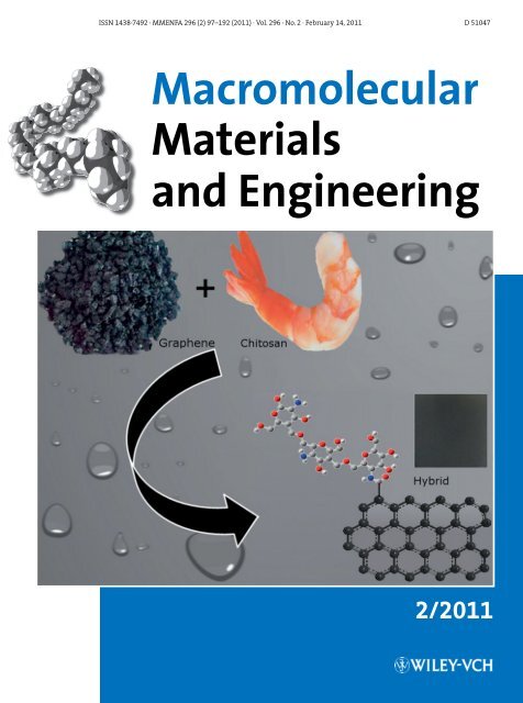
ISSN 1438–7492 · MMENFA 296 (2) 97–192 (2011) · Vol. 296 · No. 2 · February 14, 2011 D 51047<br />
Macromolecular<br />
Materials<br />
<strong>and</strong> Engineering<br />
2/2011
Full Paper<br />
<strong>Synthesis</strong> <strong>and</strong> <strong>Drug</strong>-<strong>Delivery</strong> <strong>Behavior</strong> <strong>of</strong><br />
<strong>Chitosan</strong>-<strong>Functionalized</strong> Graphene Oxide<br />
Hybrid Nanosheets a<br />
Vijay Kumar Rana, Myeon-Cheon Choi, Jin-Yeon Kong, Gwang Yeon Kim,<br />
Mi Ju Kim, Sun-Hee Kim, Satyendra Mishra, Raj Pal Singh, Chang-Sik Ha*<br />
<strong>Chitosan</strong>-functionalized graphene oxides (FGOCs) were successfully synthesized. FGOCs were<br />
found to significantly improve the solubility <strong>of</strong> the GO in aqueous acidic media. The presence<br />
<strong>of</strong> organic groups was confirmed by means <strong>of</strong> XPS <strong>and</strong> TGA. Restoration <strong>of</strong> the sp 2 carbon<br />
network <strong>and</strong> exfoliation <strong>of</strong> graphene sheets were confirmed<br />
by Raman spectroscopy, UV-visible spectroscopy<br />
<strong>and</strong> WAXD. The SEM <strong>and</strong> AFM investigations <strong>of</strong> the<br />
resultant FGOCs showed that most <strong>of</strong> the graphene<br />
sheets were individual <strong>and</strong> few were layered. Controlled<br />
release behavior <strong>of</strong> Ibupr<strong>of</strong>en <strong>and</strong> 5-fluorouracil was<br />
then investigated. We found that FGOCs are a promising<br />
new material for biological <strong>and</strong> medical applications.<br />
Introduction<br />
Graphene sheets, which are one atom thick <strong>and</strong> a 2D layer<br />
that is entirely made <strong>of</strong> carbon, have been an extensive<br />
V. K. Rana, M.-C. Choi, J.-Y. Kong, G. Y. Kim, C.-S. Ha<br />
Department <strong>of</strong> Polymer Science <strong>and</strong> Engineering, Pusan National<br />
University, Geumjeong-gu, Busan 609-735, Korea<br />
Fax: þ82 51 514 4331; E-mail: csha@pnu.edu<br />
M. J. Kim, S.-H. Kim<br />
Department <strong>of</strong> Biochemistry, School <strong>of</strong> Medicine, Pusan National<br />
University, Yangsan 626-870, South Korea<br />
S. Mishra<br />
Department <strong>of</strong> Chemical Technology, North Maharastra<br />
University, Jalgaon 425001, India<br />
R. P. Singh<br />
Division <strong>of</strong> Polymer Science <strong>and</strong> Engineering, National Chemical<br />
Laboratory, Dr. Homi Bhabha Road, Pune 411 008, India<br />
a : Supporting information for this article is available at the bottom<br />
<strong>of</strong> the article’s abstract page, which can be accessed from the<br />
journal’s homepage at http://www.mme-journal.de, or from the<br />
author.<br />
research material over the past decade. A single sheet <strong>of</strong> sp 2 -<br />
bonded carbon arranges in a honeycomb lattice, with<br />
interesting physical <strong>and</strong> electronic properties. [1,2] Its<br />
extraordinary properties, such as high carrier mobility,<br />
half-integer quantum Hall effect at room temperature, [3,4]<br />
spin transport, [5] high elasticity, [6] electromechanical modulation<br />
<strong>and</strong> ferromagnetism, [7] have made graphene a very<br />
promising c<strong>and</strong>idate as a robust atomic-scale scaffold in the<br />
design <strong>of</strong> new nanomaterials. [8] The fracture strength is<br />
comparable to fullerenes <strong>and</strong> carbon nanotubes (CNTs)<br />
with similar types <strong>of</strong> defects. [9] However, like C 60 <strong>and</strong> CNTs,<br />
graphene tends to aggregate in solution <strong>and</strong> in the solid<br />
state, giving rise to great technical difficulties during the<br />
fabrication <strong>of</strong> graphene-based devices in organic solvents.<br />
These aggregates are driven by the enhanced van der Waals<br />
attractive forces between graphene sheets. Graphene oxide,<br />
prepared by the chemical oxidization <strong>of</strong> graphite, possesses<br />
various reactive functional groups, including hydroxyl,<br />
epoxy <strong>and</strong> carboxylic acid groups. The reactive oxygen<br />
Macromol. Mater. Eng. 2011, 296, 131–140<br />
ß 2011 WILEY-VCH Verlag GmbH & Co. KGaA, Weinheim wileyonlinelibrary.com DOI: 10.1002/mame.201000307 131
V. K. Rana et al.<br />
www.mme-journal.de<br />
functional groups <strong>of</strong> graphene oxide (GO) can help GO<br />
exfoliated in various solvents to produce homogeneous<br />
colloidal suspensions, while influencing the properties <strong>of</strong><br />
graphene-based materials. One smart behavior <strong>of</strong> graphene<br />
is controlled drug delivery based on non/covalent dynamic<br />
bonding interactions, e.g., hydrogen bonding, hydrophobic,<br />
p-p stacking <strong>and</strong> electrostatic interactions, which respond<br />
to stimuli release by pH, temperature, ultraviolet or visible<br />
lights, chemical substances or electric fields. [10–12] Moreover,<br />
graphene sheets as a drug carrier are interesting<br />
because both sides <strong>of</strong> a single sheet could be accessible for<br />
drug binding. So far, rational functionalization chemistry<br />
has focused on imparting graphene with aqueous solubility<br />
<strong>and</strong> biocompatibility. [10] However, multifunctional graphene<br />
hybrid materials that take advantage <strong>of</strong> both the<br />
superior biological properties <strong>of</strong> graphene <strong>and</strong> various<br />
inherent properties <strong>of</strong> a functionalizing material have been<br />
largely unexplored. The polymer functionalization strategy<br />
is not only an effective way to solubilize graphene sheets in<br />
water, but this technique is also particularly important for<br />
the preparation <strong>of</strong> polymeric carbon packaging, metal-ion<br />
adsorption, novel drug delivery <strong>and</strong> gene composites.<br />
Furthermore, covalent techniques can be used to combine<br />
different polymers with graphene in order to create new<br />
hybrid materials with desirable properties. [11,12] Thus far,<br />
little has been done to explore graphene in biological<br />
systems. [13,14] <strong>Chitosan</strong> (CS) is a biocompatible, biodegradable<br />
<strong>and</strong> non-toxic natural polymer <strong>and</strong> has applications in<br />
wound healing, tissue repair, anti-microbial resistance, cell<br />
adhesion <strong>and</strong> food delivery. [15] Recently, a few reports were<br />
presented on CS/GO hybrid systems with enhanced<br />
mechanical properties. [16,17] No report has been published<br />
so far, however, on biological applications <strong>of</strong> the CS/GO<br />
hybrid systems.<br />
Herein, we demonstrate the biocompatibility <strong>and</strong> the<br />
drug release behavior <strong>of</strong> covalently CS-functionalized<br />
graphene (FGOCs; see Scheme 1). We evaluate the controlled<br />
release behavior <strong>of</strong> two drug molecules, i.e., ibupr<strong>of</strong>en (IBU)<br />
<strong>and</strong> 5-fluorouracil (5-FU), from the FGOCs. It is considered<br />
that the biological application <strong>of</strong> FGOCs is strongly<br />
dependent on the structural <strong>and</strong> physical characteristics<br />
<strong>of</strong> drugs. For example, the release behavior <strong>of</strong> drugs having a<br />
large molecular structure, e.g., doxorubicin, 7-ethyl-10-<br />
hydroxy-camptothecin (SN38) <strong>and</strong> camptothecin (CPT) etc.<br />
Scheme 1. <strong>Synthesis</strong> <strong>of</strong> the FGOCs <strong>and</strong> the dispersion <strong>of</strong> (a) GO <strong>and</strong> (b) the FGOCs in an aqueous acetic acid solution (CH 3 COOH/H 2 O 0.2/1).<br />
More details on the synthesis <strong>and</strong> characterization <strong>of</strong> FGOCs are given in the Supporting Information.<br />
132<br />
Macromol. Mater. Eng. 2011, 296, 131–140<br />
ß 2011 WILEY-VCH Verlag GmbH & Co. KGaA, Weinheim<br />
www.MaterialsViews.com
<strong>Synthesis</strong> <strong>and</strong> <strong>Drug</strong>-<strong>Delivery</strong> <strong>Behavior</strong> <strong>of</strong> <strong>Chitosan</strong>-<strong>Functionalized</strong> ...<br />
www.mme-journal.de<br />
from FGOCs nanosheets is different from the two small<br />
molecular drugs. We have chosen the two drugs used here<br />
based on their different affinity with FGOCs; IBU may have<br />
a higher attraction to FGOCs sheets because <strong>of</strong> its<br />
hydrophobic nature with a complete benzene ring (presumably<br />
higher p-stacking), while 5-FU has less attraction<br />
due to its hydrophilic nature <strong>and</strong> its diamide group that<br />
would contribute in the resonance <strong>of</strong> the benzenoid<br />
(presumably less p-stacking). We expect that the biocompatible<br />
FGOCs graphene sheets will be a novel promising<br />
material for biological applications. Infrared spectroscopy<br />
(FT-IR), thermogravimetric analysis (TGA), <strong>and</strong> X-ray<br />
photoelectron spectroscopy (XPS) confirmed the functionalization<br />
<strong>of</strong> graphene with chitosan <strong>and</strong> the exfoliation <strong>of</strong><br />
FGOCs. The restoration <strong>of</strong> the sp 2 carbon network on the<br />
basal planes <strong>of</strong> the graphene sheets was confirmed by<br />
Raman spectroscopy <strong>and</strong> wide-angle X-ray diffraction<br />
(WAXD). Moreover, the low cost, large scale production <strong>of</strong><br />
graphite <strong>and</strong> chitosan is unmatched by any hardcore<br />
carbon metals <strong>and</strong> polymers. In short, chitosan functionalized<br />
graphene sheets with a stable suspension in an<br />
aqueous acidic solution <strong>and</strong> their controlled releasing<br />
behavior with high biocompatibility are reported.<br />
Experimental Part<br />
Materials<br />
The materials that were used in this work included chitosan (degree<br />
<strong>of</strong> deacetylation 85%, molecular weight ¼ 190 kDa), lithium<br />
chloride (LiCl, 99%), pyridine (Py), dimethylformamide (DMF,<br />
99%), 5-FU (99%) <strong>and</strong> IBU (99%), supplied by Sigma-Aldrich<br />
Chemical Co. Natural graphite flakes (325 mesh) were purchased<br />
from Alfa Aesar. All chemicals were used as received without<br />
further purification. FGOCs were synthesized in this work with<br />
reference to the literature. [16–21] First, GO was prepared using<br />
Hummer’s method. [18] Then, the carboxylic acid groups <strong>of</strong> GO were<br />
converted into acyl chlorides (graphene-COCl) via st<strong>and</strong>ard<br />
chemistry, followed by a typical functionalization process for<br />
graphene with chitosan. More details on the synthesis are<br />
described in the Supporting Information.<br />
measurements, the base pressure was 1.33 10 7 –1.33 10 8 Pa.<br />
The binding energies were referenced to the C1s line at 284.6 eV<br />
from adventitious carbon. The survey spectra were obtained at a<br />
resolution <strong>of</strong> 1 eV from three scans. The atomic force microscopy<br />
(AFM) images <strong>of</strong> graphene were acquired in the tapping mode using<br />
a Multimode Nanoscope TM SPM, Digital Instruments (USA). The<br />
exfoliated surface morphology <strong>of</strong> the functionalized graphene<br />
(FGOCs) was measured using field emission scanning electron<br />
microscopy (FE-SEM, JSM-6700F, Korea Basic Science Institute)at an<br />
acceleration voltage <strong>of</strong> 20 kV with a Model 952888(8) microscope<br />
from Hitachi Ltd. UV absorption spectra were obtained using a UVvisible<br />
spectrophotometer (U-2010, HITACHI Co.). The fluorescence<br />
emission spectra were measured using a Hitachi F-4500 spectrometer<br />
at 25 8C. The excitation slit size was 10.0 nm <strong>and</strong> the emission<br />
slit size was also 10.0 nm. The scan speed was set at 240 nm min 1 .<br />
<strong>Drug</strong> Loading <strong>and</strong> Release<br />
100 mg <strong>of</strong> FGOCs graphene sheets were dispersed in 25 mL/60 mg<br />
hexane/IBU or water/5-FU solutions. The mixtures were incubated<br />
for 48 h to fabricate drug loaded FGOCs sheets. Solutions were<br />
covered with a polyethylene (PE) film to prevent the evaporation <strong>of</strong><br />
solvent. The drug loaded sample was separated from the solution<br />
by vacuum filtration, washed with solvent until complete removal<br />
<strong>of</strong> drugs was achieved <strong>and</strong> dried at room temperature. To check the<br />
removal <strong>of</strong> drugs from the FGOCs sheets, drug loaded FGOCs sheets<br />
were again washed with an appropriate solvent (hexane for IBU<br />
<strong>and</strong> water for 5-FU, respectively) <strong>and</strong> the filtrate was further<br />
measured using UV-vis spectrophotometry. The remaining drug<br />
loading amount was then also measured using UV-vis spectrophotometry.<br />
Then, 40 mg <strong>of</strong> drug loaded sample were dispersed in<br />
5 mL <strong>of</strong> phosphate-buffered saline (PBS, pH ¼ 7.4) <strong>and</strong> simulated<br />
stomach fluid (SSF, pH ¼ 1.4), placed into a dialysis membrane bag<br />
(molecular-weight cut<strong>of</strong>f 5 000 kDa), <strong>and</strong> then immersed into<br />
25 mL PBS <strong>and</strong> SSF, respectively, at 37 8C for 3 d.<br />
At periodic intervals, the release media was withdrawn <strong>and</strong><br />
another 1 mL <strong>of</strong> fresh buffer solution was added. The amounts <strong>of</strong><br />
IBU <strong>and</strong> 5-FU were determined by means <strong>of</strong> measuring the UV-vis<br />
spectrum at 224 <strong>and</strong> 265 nm, respectively. The reason behind the<br />
use <strong>of</strong> the two kinds <strong>of</strong> release media (PBS <strong>and</strong> SSF) is the different<br />
nature <strong>of</strong> the drug molecules at different pHs, i.e., IBU has a pK a <strong>of</strong><br />
4.43 <strong>and</strong> 5-FU has a pK a <strong>of</strong> 8.2. Moreover, the pH-dependent drug<br />
release from FGOCs nanosheets is important in the clinical setting,<br />
since the microenvironment in the extracellular tissues <strong>of</strong> tumors<br />
<strong>and</strong> intracellular lysosomes <strong>and</strong> endosomes are acidic. [10]<br />
Characterization<br />
FT-IR spectra were recorded using a Spectrum GX. The scan<br />
wavenumber was in the range 600–4 000 cm 1 with a resolution <strong>of</strong><br />
1cm 1 , <strong>and</strong> sixteen signals were averaged. Thermogravimetric<br />
analysis (TGA) was conducted under nitrogen on a TA instruments<br />
Q50 at a heating rate <strong>of</strong> 10 8C min 1 . The WAXD measurements<br />
were performed using a conventional X-ray diffractometer [Rigaku<br />
Miniflex, Cu K a , l ¼ 1.5418 Å]. The Raman spectra were recorded<br />
from 1 000 to 2 000 cm 1 using a FT Raman spectrometer (FRS-100S,<br />
Bruker) with an argon ion laser excitation <strong>of</strong> 514.5 nm. The XPS<br />
spectra were recorded using a VG-Scientific ESCALAB 250 spectrometer<br />
(UK) with a monochromatized Al K a X-ray source. During the<br />
Cell Proliferation Assays<br />
The 3-(4,5-dimethylthiazol-2-yl)-2,5-diphenyltetrazolium bromide<br />
(MTT) assay was used to determine the cytotoxicity <strong>of</strong> neat GO,<br />
FGOCs <strong>and</strong> drug loaded FGOCs graphene sheets. Herein, we have<br />
tried two types <strong>of</strong> in vitro cancer cell line viability, namely CEM<br />
human lymphoblastic leukemia <strong>and</strong> MCF7-human breast cancer<br />
for targeting the potent cancer cell killing effect with different<br />
concentrations <strong>of</strong> each sample. Cell proliferation was measured<br />
using the MTT (Sigma-Aldrich Co., St. Louis, MO) colorimetric dye<br />
reduction method. The cells were seeded in tissue culture flasks<br />
(5 10 3 cells) <strong>and</strong> incubated in a fully humidified atmosphere<br />
containing 5% CO 2 at 37 8C. For MTT assays, the cells were seeded in<br />
www.MaterialsViews.com<br />
Macromol. Mater. Eng. 2011, 296, 131–140<br />
ß 2011 WILEY-VCH Verlag GmbH & Co. KGaA, Weinheim<br />
133
V. K. Rana et al.<br />
www.mme-journal.de<br />
96 well plates at a density <strong>of</strong> 5 10 3 cells per well in 1 mL <strong>of</strong> culture<br />
medium, <strong>and</strong> then the cells were incubated with GO, FGOCs <strong>and</strong><br />
drug loaded FGOCs sheets. It is pointed out here that we present the<br />
cell line viability <strong>of</strong> all samples for 5 d for both cell lines. This is<br />
because, for CEM human lymphoblastic leukemia, it was hard to<br />
know thetoxic effect <strong>of</strong> FGOCs<strong>and</strong> drug loadedFGOCssheets up to 3<br />
d. In order to know the complete effect <strong>of</strong> the respective samples on<br />
both cell lines, therefore, we extended the cell proliferation assays<br />
to 5 d. Thesame trend wasfollowed in the case <strong>of</strong> MCF-7 cell lines for<br />
reliable assessment.<br />
After 5 d, the medium was aspirated after centrifugation <strong>and</strong><br />
MTT-formazan crystals were solubilized in 100 mL dimethyl<br />
sulfoxide (DMSO). The optical density <strong>of</strong> each sample was<br />
measured at 570 nm using an ELISA reader (Bio-Tec Instruments,<br />
VT, USA). The optical density <strong>of</strong> the media was proportional to the<br />
number <strong>of</strong> viable cells. Inhibition <strong>of</strong> proliferation was evaluated as<br />
a percentage <strong>of</strong> control growth (no drug in the sample). All<br />
experiments were repeated at least twice.<br />
Results <strong>and</strong> Discussion<br />
<strong>Chitosan</strong>-<strong>Functionalized</strong> Graphene<br />
Graphene oxide was prepared by oxidizing graphite via a<br />
modified Hummer’s method. [18] The resulting GO (single<br />
layered <strong>and</strong> few-layered) was dispersible in water but<br />
aggregated in solution. The FGOCs were obtained from the<br />
chitosan <strong>and</strong> graphene oxide molecules, which were<br />
covalently bonded together via an amide bond<br />
(Scheme 1) in DMF in the presence <strong>of</strong> pyridine, following<br />
st<strong>and</strong>ard chemistry. The covalent attachment <strong>of</strong> chitosan<br />
onto GO via the amide linkage was confirmed using IR<br />
spectroscopy, TGA <strong>and</strong> XPS. In Figure 1(a), the peaks at 1 726<br />
<strong>and</strong> 1 223 cm 1 in the FT-IR spectra were characteristic <strong>of</strong><br />
the C¼O <strong>and</strong> C O stretches <strong>of</strong> the carboxylic <strong>and</strong> epoxy<br />
groups, respectively, on graphene oxide. In the FGOCs<br />
spectrum, the peak at 1 726 cm 1 almost disappeared, <strong>and</strong> a<br />
new broad b<strong>and</strong> emerged at 1 653 cm 1 , corresponding to<br />
the C¼O characteristic stretching b<strong>and</strong> <strong>of</strong> the amide groups,<br />
which were further overlapped with C¼O in the ester<br />
groups. [19,20] Moreover, a broad peak at 1 554 cm 1 , as well<br />
as two weak peaks at 1 167 <strong>and</strong> 806 cm 1 , presumably<br />
confirmed the N–H bending <strong>and</strong> C–N stretching bonds <strong>of</strong><br />
the amide group. Additionally, the two characteristic b<strong>and</strong>s<br />
<strong>of</strong> the glucopyranose rings <strong>of</strong> the FGOCs appeared at 897<br />
<strong>and</strong> 1 115 cm 1 , respectively, implying that chitosan was<br />
attached. [21] The results from TGA in Figure 1(b) showed<br />
that three significant weight loss events were observed for<br />
GO powders, corresponding to the evaporation <strong>of</strong> water<br />
(below 100 8C) <strong>and</strong> the loss <strong>of</strong> what is likely to be carbon<br />
dioxide gas species (120–150 <strong>and</strong> 200–260 8C) from the<br />
decomposition <strong>of</strong> labile oxygen functional groups. [10] In<br />
contrast, the FGOCs have less than 2% weight loss below<br />
100 8C. The gradual weight loss about 15% below 350 8C is<br />
likely to be due to the loss <strong>of</strong> the glucopyranose ring <strong>of</strong><br />
Figure 1. a) FT-IR spectra <strong>of</strong> GO, FGOCs, <strong>and</strong> CS <strong>and</strong> b) TGA curve <strong>of</strong><br />
GO <strong>and</strong> FGOCs at a heating rate <strong>of</strong> 10 8C min 1 .<br />
chitosan <strong>and</strong> residual functional groups on the FGOCs<br />
sheets. These results are easy to underst<strong>and</strong>: the oxidation<br />
product <strong>of</strong> GO has a layered morphology with oxygencontaining<br />
functionality, thereby weakening the van der<br />
Waals forces between layers. This will disrupt the<br />
hexagonal carbon basal planes on the interior <strong>of</strong> multilayered<br />
stacks <strong>of</strong> GO, thus accelerating the process <strong>of</strong> weight<br />
loss, yielding CO, CO 2 <strong>and</strong> steam. [22] Remarkably, after<br />
functionalization <strong>of</strong> chitosan onto GO sheets, the total<br />
weight losses decreased to 42%, revealing that most <strong>of</strong> the<br />
epoxide <strong>and</strong> hydroxyl groups were successfully removed.<br />
Figure 2 shows the C1s XPS spectra <strong>of</strong> GO <strong>and</strong> the FGOCs<br />
(for the XPS survey spectra <strong>of</strong> GO <strong>and</strong> functionalized<br />
graphene hybrid sheets, i.e., FGOCs, see Figure S1, Supporting<br />
Information). In the C 1s XPS spectrum <strong>of</strong> GO in<br />
Figure 2(a), four types <strong>of</strong> carbon were clearly present with<br />
different chemical valences. The C 1s peaks <strong>of</strong> the graphite’s<br />
C O, C¼O <strong>and</strong> O C¼O were observed at 284.6, 286.7, 288.1<br />
134<br />
Macromol. Mater. Eng. 2011, 296, 131–140<br />
ß 2011 WILEY-VCH Verlag GmbH & Co. KGaA, Weinheim<br />
www.MaterialsViews.com
<strong>Synthesis</strong> <strong>and</strong> <strong>Drug</strong>-<strong>Delivery</strong> <strong>Behavior</strong> <strong>of</strong> <strong>Chitosan</strong>-<strong>Functionalized</strong> ...<br />
www.mme-journal.de<br />
Figure 2. a) C 1S XPS spectra <strong>of</strong> GO <strong>and</strong> b) <strong>of</strong> FGOCs. The C 1s XPS<br />
spectra <strong>of</strong> GO <strong>and</strong> the FGOCs showed that four types <strong>of</strong> carbon<br />
were clearly present with different chemical valences.<br />
<strong>and</strong> 289.0 eV. Although these four types <strong>of</strong> carbon were also<br />
present in the functionalized graphene, the C1s XPS<br />
spectrum <strong>of</strong> the FGOCs in Figure 2(b) clearly exhibited<br />
different intensity ratios. In contrast to the polymer<br />
reference (chitosan), an additional component at<br />
285.1 eV, corresponding to the carbonyl (C N) bond was<br />
observed in the C1s XPS spectrum <strong>of</strong> the FGOCs. Notably, the<br />
peak intensities <strong>of</strong> the oxidized carbon in the FGOCs were<br />
much lower than GO, <strong>and</strong> the C/O atomic ratio remarkably<br />
increased (from 1.72 to 4.6), indicating that most <strong>of</strong> the<br />
epoxide <strong>and</strong> hydroxyl groups disappeared after chemical<br />
modification by chitosan, suggesting that the GO was<br />
successfully modified. 23 Remarkably, a p-p transition took<br />
place in the basal plane after the reduction <strong>of</strong> the C1s peaks<br />
<strong>of</strong> O C¼O at 289.0 eV for the FGOCs. From these intensities,<br />
we can calculate the atomic percentages <strong>of</strong> the elements<br />
present after the functionalization procedure. This quantitative<br />
analysis is reported in Table S1 (Supporting<br />
Information) <strong>and</strong> shows a decrease <strong>of</strong> the oxygen levels<br />
<strong>and</strong> increase <strong>of</strong> the nitrogen levels.<br />
The chitosan functionalization <strong>of</strong> graphene facilitated its<br />
exfoliation in the aqueous acidic solution. Note that results<br />
were given here only for aqueous acetic acid solution, but it<br />
is also possible to disperse FGOCs graphene sheets in other<br />
organic or inorganic aqueous acidic solutions. The GO<br />
dispersion with aqueous acetic acid was light brown, as<br />
shown in Scheme 1. However, a black graphene dispersion<br />
(0.1 mg/1 mL) was observed for the FGOCs. This clear color<br />
change <strong>of</strong> the dispersion before <strong>and</strong> after the chemical<br />
modification was apparent evidence that GO was indeed<br />
reduced after the covalent functionalization with chitosan.<br />
Additionally, the resulting graphene dispersion was stable<br />
for several days to a few months. The negative charges <strong>of</strong><br />
the terminal carboxylic acid <strong>and</strong> protonated amine groups<br />
<strong>of</strong> chitosan were believed to supply an electrostatic<br />
repulsion, which provided stability for the graphene<br />
dispersions. Notably, after the addition <strong>of</strong> DMF into the<br />
solution (Figure S2, Supporting Information), the electrostatic<br />
repulsion was reduced, <strong>and</strong> graphene was recovered<br />
from the aqueous acetic solution.<br />
Significant structural changes occurred during the<br />
chemical processing from the pristine graphite (Gy) to<br />
GO <strong>and</strong> finally to the FGOCs. In the Raman spectrum <strong>of</strong> the<br />
Gy, the peak at 1 580 cm 1 (G b<strong>and</strong>) corresponded to the first<br />
order scattering <strong>of</strong> the E2g mode <strong>of</strong> the Gy in Figure 3(a) <strong>and</strong><br />
was related to the vibration <strong>of</strong> the sp 2 -bonded carbon atoms<br />
in a 2D hexagonal lattice. The (weak) disorder b<strong>and</strong> that was<br />
caused by the Gy edges (D b<strong>and</strong>) was observed at<br />
approximately 1 351 cm 1 . Comparing to the raw graphite,<br />
both the G <strong>and</strong> the D b<strong>and</strong>s underwent significant changes<br />
to values <strong>of</strong> 1 601 <strong>and</strong> 1 354 cm 1 respectively, upon the<br />
amorphization <strong>of</strong> graphite. The GO peaks for amorphous<br />
carbon containing a certain fraction <strong>of</strong> sp 3 carbons became<br />
weaker <strong>and</strong> broader, suggesting a higher level <strong>of</strong> disorder in<br />
the graphene layers. The Raman spectrum <strong>of</strong> the FGOCs also<br />
contained G <strong>and</strong> D b<strong>and</strong>s at 1 584 <strong>and</strong> 1 351 cm 1 ,<br />
respectively. Along the Gy-GO-FGOCs path, although the<br />
G b<strong>and</strong> peak was located at a higher frequency in GO than in<br />
graphite (1 601 vs. 1 580 cm 1 ), the G b<strong>and</strong> peaks were<br />
located at almost the same frequency in the FGOC <strong>and</strong> Gy.<br />
The blue shift, shifted to a higher frequency after the<br />
amorphization <strong>of</strong> graphite, can be explained by the<br />
transition from the graphite crystal to the single GO<br />
sheet. [24] In the FGOCs, the G b<strong>and</strong> shifted back to the<br />
position <strong>of</strong> the G b<strong>and</strong> in graphite, which was attributed to a<br />
graphitic ‘‘self-healing’’ that was similar as the decrease in<br />
the intensity <strong>of</strong> the D peak in heat-treated graphite. [25,26]<br />
The shoulder peak appearing at around 1 601 cm 1 also<br />
indicates that the FGOCs were partially reduced <strong>and</strong> a<br />
significant area in the FGOCs remained as its oxide form<br />
after the functionalization with chitosan. [27,28] The D/G<br />
intensity ratio <strong>of</strong> the FGOCs (1.44) increased compared to<br />
www.MaterialsViews.com<br />
Macromol. Mater. Eng. 2011, 296, 131–140<br />
ß 2011 WILEY-VCH Verlag GmbH & Co. KGaA, Weinheim<br />
135
V. K. Rana et al.<br />
www.mme-journal.de<br />
Figure 3. a) Raman spectra <strong>and</strong> b) X-ray diffraction patterns <strong>of</strong><br />
pristine graphite (Gy), GO, FGOCs <strong>and</strong> CS.<br />
that <strong>of</strong> GO (0.69), which means that the sp 3 carbon domain<br />
in the FGOCs increased with the functionalization <strong>of</strong> GO.<br />
Note that the covalent bonds between GO <strong>and</strong> chitosan<br />
might hinder the reduction <strong>of</strong> GO <strong>and</strong> the peak from<br />
chitosan chains can partially overlap with the D b<strong>and</strong> <strong>of</strong> the<br />
FGOCs. [29] Furthermore, the arrow peaks that were<br />
presumably assigned to the Ag2 mode <strong>of</strong> chitosan were<br />
shifted by several wavenumbers compared to the pristine<br />
chitosan. This relative shift sufficiently suggested a strong<br />
interaction between chitosan <strong>and</strong> the graphene sheets. [22]<br />
The self-healing effect after the functionalization <strong>of</strong> GO<br />
with chitosan was further confirmed by wide angle X-ray<br />
diffraction [Figure 3(b)]. The pristine graphite nanosheet<br />
(Gy) exhibited a (002) diffraction peak at 2u ¼ 26.458,<br />
corresponding to a d-spacing <strong>of</strong> 3.37 Å in Figure 1(b), <strong>and</strong><br />
the (001) peak <strong>of</strong> GO was located at 2u ¼ 10.848, corresponding<br />
to an interlayer distance <strong>of</strong> 8.4 Å. For the FGOCs, the<br />
highly broad diffraction peak [(002) plane], appearing<br />
at 2u ¼ 24.168, suggested that the interlayer distance <strong>of</strong> the<br />
FGOCs was slightly increased compared to that <strong>of</strong> pristine<br />
graphite <strong>and</strong> graphene layers <strong>of</strong> the FGOCs were partially<br />
exfoliated with a d-spacing <strong>of</strong> 3.68 Å. Additionally, the<br />
FGOCs exhibited an amorphous structure. The disruption in<br />
their structure <strong>and</strong> the significant reduction in the d-<br />
spacing suggested that attractive interactions existed<br />
between the layers, allowing this material to apply for<br />
drug carriers in addition to their excellent dispersibility in<br />
the solvents [30] (see Figure S2, Supporting Information).<br />
Activation <strong>and</strong> functionalization <strong>of</strong> GO led to increases in<br />
optical absorption in the visible <strong>and</strong> near-infrared range.<br />
The optical absorption peak at 235 nm, originating from the<br />
p-plasmon <strong>of</strong> carbon, [31] remained essentially unchanged.<br />
The FGOCs showed much higher absorbance in the vis-NIR<br />
range than GO [Figure 4(a)]. The significant increase in<br />
absorbance led to a solution color change (darkening) that is<br />
visible to eye (Scheme 1, inset). Similar darkening was<br />
observed in the hydration reduction <strong>of</strong> GO, which is<br />
attributed to restoration <strong>of</strong> electronic conjugation within<br />
graphene sheets. [32,33] According to the AFM image in<br />
Figure 5, FGOCs contains monolayer graphene with a<br />
thickness <strong>of</strong> 1.85 nm <strong>and</strong> is greater than unmodified<br />
monolayers <strong>of</strong> GO, i.e., 1 nm. In the scanning electron<br />
microscopy (SEM) image (Figure S3, Supporting Information),<br />
the FGOCs possessed lateral dimensions ranging from<br />
several hundred nanometers to several micrometers <strong>and</strong><br />
were arranged in an edge-to-edge configuration. Therefore,<br />
no significant folding or overlapping was observed. [10]<br />
Restoration <strong>of</strong> electronic conjugation will reboot the<br />
aromatic basal planes on FGOCs <strong>and</strong> therefore is capable <strong>of</strong><br />
absorbing aromatic compounds via inter/atomic interactions.<br />
This may be useful for drug carriers. Thus far, few<br />
reports have been published on the controlled loading <strong>and</strong><br />
targeted delivery <strong>of</strong> two or more different drugs using<br />
graphene-based nanocarriers. Dai et al. [13] <strong>and</strong> Zhang et<br />
al. [34] have reported poly(ethylene oxide) (PEO) functionalized<br />
graphene for biological applications. However, both<br />
authors used doxorubicin, SN38 <strong>and</strong> CPT as model anticancer<br />
drugs, having a high density <strong>of</strong> p-electron clouds <strong>and</strong><br />
more than two aromatic rings. These drugs are sometimes<br />
easy to attach onto the aromatic basal planes <strong>of</strong> functionalized<br />
GO on both sides by the hydrophobic <strong>and</strong> p-p<br />
stacking interaction. p-p interactions are caused by<br />
intermolecular overlap <strong>of</strong> p-orbitals in p-conjugated<br />
systems, so that they become stronger as the number <strong>of</strong><br />
p-electrons increases. Other non/covalent interactions,<br />
including hydrogen bonds, van der Waals forces, charge/<br />
transfer interactions <strong>and</strong> dipole/dipole interactions, also<br />
attract organic moieties.<br />
<strong>Drug</strong> <strong>Delivery</strong><br />
Until now, no reports have been published on drug<br />
molecules containing one aromatic moiety being loaded<br />
<strong>and</strong> their controlled release behavior through the graphene<br />
136<br />
Macromol. Mater. Eng. 2011, 296, 131–140<br />
ß 2011 WILEY-VCH Verlag GmbH & Co. KGaA, Weinheim<br />
www.MaterialsViews.com
<strong>Synthesis</strong> <strong>and</strong> <strong>Drug</strong>-<strong>Delivery</strong> <strong>Behavior</strong> <strong>of</strong> <strong>Chitosan</strong>-<strong>Functionalized</strong> ...<br />
www.mme-journal.de<br />
Figure 4. a) UV-vis absorbance spectra <strong>of</strong> GO, FGOCs <strong>and</strong> drug-loaded FGOCs. b) Fluorescence <strong>of</strong> GO <strong>and</strong> drug-loaded FGOCs in the visible<br />
range under excitation <strong>of</strong> 390 nm. c) IBU release <strong>and</strong> d) 5-FU release behavior <strong>of</strong> FGOCs/IBU <strong>and</strong> FGOCs/5-FU, respectively.<br />
sheets. Herein, we have used an anti-inflammatory drug,<br />
ibupr<strong>of</strong>en (pK a ¼ 4.43) <strong>and</strong> an anti-cancer drug, 5-fluorouracil<br />
(5-FU, pK a ¼ 8.2) as model <strong>and</strong> widely useful drugs in<br />
biomedical applications. [35] IBU is an aromatic drug that<br />
will create hydrophobic <strong>and</strong> p-p interactions, whereas 5-<br />
fluorouracil, a ‘‘benzenoid’’ resonance contributor, is<br />
aromatic. It has a continuous loop <strong>of</strong> p orbitals (the two<br />
nitrogen are sp 2 ) <strong>and</strong> its ‘‘diamide’’ contributor obeys all<br />
preference rules. Thus, it is believed to be the best resonance<br />
contributor (see Figure S4, Supporting Information). In both<br />
cases, inter/atomic interactions will remain. An aromatic<br />
hydrophobic drug <strong>and</strong> hydrophilic drug were loaded onto<br />
the FGOCs via simple physisorption (Figure S5, Supporting<br />
Information). We found that an aromatic drug (IBU) <strong>and</strong> a<br />
hydrophilic drug (5-FU) were complexed with FGOCs via<br />
simple physisorption <strong>of</strong> IBU <strong>and</strong> 5-FU dissolved in hexane<br />
<strong>and</strong> water with FGOCs solution, respectively (Figure S5,<br />
Supporting Information). The excess <strong>and</strong> uncoupled drugs<br />
were removed by centrifugation. Repeated washing <strong>and</strong><br />
filtration were used to remove residual free IBU <strong>and</strong> 5-FU<br />
through its respective solutions, i.e., hexane for IBU <strong>and</strong><br />
water for 5-FU, respectively. We found that the FGOCs were<br />
highly stable in physiological solution including buffer<br />
(Figure S6, Supporting Information). We also investigated<br />
the cytotoxicity <strong>and</strong> completed cell viability tests <strong>of</strong> the<br />
FGOCs sheets. The excess <strong>and</strong> uncoupled drugs were<br />
removed by centrifugation. Repeated washing <strong>and</strong> filtration<br />
were used to remove residual free IBU <strong>and</strong> 5-FU. UV-vis<br />
spectra <strong>of</strong> the resulting solutions revealed IBU <strong>and</strong> 5-FU<br />
peaks superimposed with the absorption curve <strong>of</strong> FGOCs in<br />
Figure 4(a), suggesting loading <strong>of</strong> IBU <strong>and</strong> 5-FU onto FGOCs<br />
sheets. On the basis <strong>of</strong> the extinction coefficients, we<br />
estimated that the drug loading ratio <strong>of</strong> IBU on FGOCs<br />
sheets was 0.097 mg mg 1 <strong>and</strong> higher than that <strong>of</strong> 5-FU<br />
(0.053 mg mg 1 ). The difference in the loading ability <strong>of</strong><br />
the drugs is ascribed to the difference in their chemical<br />
structures <strong>and</strong> interactions with FGOCs. IBU is a kind <strong>of</strong><br />
hydrophobic drug containing a complete aromatic ring <strong>and</strong><br />
would create complete p-p interaction with aromatic basal<br />
planes <strong>of</strong> FGOCs. Furthermore, IBU can create hydrogen<br />
www.MaterialsViews.com<br />
Macromol. Mater. Eng. 2011, 296, 131–140<br />
ß 2011 WILEY-VCH Verlag GmbH & Co. KGaA, Weinheim<br />
137
V. K. Rana et al.<br />
www.mme-journal.de<br />
graphene sheets under pH stimulation can be mainly<br />
attributed to the interaction <strong>of</strong> IBU with FGOCs <strong>and</strong><br />
ionization <strong>of</strong> IBU at different pH values.<br />
In both cases, the interaction <strong>of</strong> IBU with FGOCs would be<br />
same regardless <strong>of</strong> pH but at pH ¼ 7.5 IBU would be ionized<br />
(pK a ¼ 4.43), causing a higher release than that at pH ¼ 1.4,<br />
whereas at pH ¼ 1.4 IBU would be deionized <strong>and</strong> exhibit less<br />
release. At pH ¼ 7.5, the release rate <strong>of</strong> 5-FU becomes slower<br />
than that at pH ¼ 1.4, because 5-FU would be protonated at<br />
pH ¼ 1.4 (amine group) causing more easy release from<br />
FGOCs graphene sheets. Moreover, as mentioned above, IBU<br />
has a more hydrophobic <strong>and</strong> stronger p-p staking interaction<br />
with FGOCs than 5-FU <strong>and</strong> therefore IBU has more<br />
distinct controlled release behavior.<br />
We found that FGOCs/5-FU sheets afforded highly potent<br />
cell viability from 93.4 to 30.1% at 10 to 400 mg mL 1<br />
concentration with the CEM cell line after being incubated<br />
for 5 d, as shown in Figure 6(a). Consistency <strong>of</strong> cell line<br />
viability was almost the same in FGOCs/IBU samples.<br />
Importantly, no serious toxicity <strong>of</strong> the CEM cell line was<br />
measured for various concentrations <strong>of</strong> FGOCs graphene<br />
sheets without drug loading, suggesting that FGOCs sheets<br />
had very good biocompatibility.<br />
5-Fluorouracil is a widely used chemotherapy drug for<br />
targeting various cancers. 5-FU loaded graphene sheets<br />
(FGOCs/5-FU) again induced significant MCF-7 cancer cell<br />
Figure 5. a) Tapping mode AFM image <strong>of</strong> functionalized graphene<br />
FGOCs, deposited on freshly cleaved mica substrate <strong>and</strong> b) crosssection<br />
analysis <strong>of</strong> FGOCs AFM image, i.e., (a).<br />
bonding between the COOH <strong>of</strong> IBU <strong>and</strong> the glucopyranose<br />
ring <strong>of</strong> chitosan. Meanwhile, 5-FU has a relatively hydrophilic<br />
character <strong>and</strong> its diamide group would contribute in<br />
the resonance <strong>of</strong> benzenoid <strong>and</strong> therefore the p-p interaction<br />
<strong>of</strong> 5-FU with FGOCs would be less, causing less loading<br />
<strong>of</strong> 5-FU on FGOCs sheets. From the fluorescence spectra <strong>of</strong><br />
FGOCs/IBU, FGOCs/5-FU, FGOCs <strong>and</strong> GO at the same<br />
concentration (0.1 mg mL 1 ), a drastic increase <strong>of</strong> the<br />
fluorescence intensity was observed in FGOCs/IBU <strong>and</strong><br />
FGOCs/5-FU compared to in GO <strong>and</strong> FGOCs, as shown in<br />
Figure 4(b). This result suggests the close proximity <strong>of</strong> the<br />
drug molecule with FGOCs through non-covalent bonding<br />
<strong>of</strong> drug on FGOCs sheets. [36] The release <strong>of</strong> drugs from FGOCs<br />
sheets was carried out in PBS (pH ¼ 7.4) <strong>and</strong> SSF (pH ¼ 1.4)<br />
at 37 8C.<br />
We observed from Figure 4(c) that IBU was released only<br />
10% in SSF from FGOCs nanosheets, whereas the release<br />
rate was increased up to 70% into the respective release<br />
medium for 5-FU in 3 d, as shown in Figure 4(d). The same<br />
trend was observed in the release rate <strong>of</strong> both drugs for the<br />
PBS releasing medium. Here, IBU was released up to 19%<br />
only but, unlike IBU, 5-FU was released up to 50%. The<br />
significantly different IBU release behavior from FGOCs<br />
Figure 6. In vitro cell viability assay. a) Relative cell viability <strong>of</strong><br />
CEM cancer cell line <strong>and</strong> b) MCF-7 cancer cell line incubated with<br />
GO, FGOCs, <strong>and</strong> drug loaded FGOCs graphene sheets at different<br />
concentrations for 5 d (SD 3%).<br />
138<br />
Macromol. Mater. Eng. 2011, 296, 131–140<br />
ß 2011 WILEY-VCH Verlag GmbH & Co. KGaA, Weinheim<br />
www.MaterialsViews.com
<strong>Synthesis</strong> <strong>and</strong> <strong>Drug</strong>-<strong>Delivery</strong> <strong>Behavior</strong> <strong>of</strong> <strong>Chitosan</strong>-<strong>Functionalized</strong> ...<br />
www.mme-journal.de<br />
death, as shown in Figure 6(b). We suggest that 5-FU-loaded<br />
FGOCs sheets transported 5-FU inside the cells via<br />
endocytosis. One <strong>of</strong> the potential advantages <strong>of</strong> using<br />
FGOCs as a drug carrier compared to free drug is the ability<br />
to target delivery for selective destruction <strong>of</strong> certain types <strong>of</strong><br />
cells, reducing the toxicity to non-targeted cells. It was<br />
found that 5-FU loaded FGOCs exhibited higher concentration-dependent<br />
toxicity (10 to 100 mg mL 1 ) to MCF cells<br />
than FGOCs <strong>and</strong> GO sheets, suggesting that chitosan<br />
conjugation to graphene sheets afforded no enhancement<br />
in 5-FU delivery to MCF cells. These results suggest the<br />
potential for selectively enhancing the toxicity <strong>of</strong> drugs to<br />
certain types <strong>of</strong> cells by using FGOCs sheets with a targeting<br />
moiety as drug carriers. It is worth pointing out that we<br />
found targeting cell killing anti-cancer drug loaded<br />
graphene sheets having high <strong>and</strong> long term biocompatibility<br />
(5 d) with different kind <strong>of</strong> cancer cell lines (CEM &<br />
MCF-7). Moreover, FGOCs sheets exhibit higher (20%)<br />
biocompatibility than GO sheets for both cell lines.<br />
A drug delivery system is generally designed to improve<br />
the pharmacological <strong>and</strong> therapeutic pr<strong>of</strong>ile <strong>of</strong> a drug<br />
molecule. The ability <strong>of</strong> FGOCs to penetrate into the cells<br />
<strong>of</strong>fers the potential to use FGOCs as vehicles for the delivery<br />
<strong>of</strong> drug molecules. <strong>Delivery</strong> systems should be able to carry<br />
one or more therapeutic agents with recognition capacity<br />
<strong>and</strong> optical signals for imaging <strong>and</strong>/or specific targeting<br />
that is <strong>of</strong> fundamental advantage, for example, in the<br />
treatment <strong>of</strong> cancer <strong>and</strong> different types <strong>of</strong> infectious<br />
diseases. For this purpose, we have developed a new<br />
strategy for the functionalization <strong>of</strong> GO with chitosan. For<br />
future applications <strong>of</strong> FGOCs, it may be useful for clinical<br />
applications to incorporate a fluorescent probe into the<br />
FGOCs for tracking the cellular uptake <strong>of</strong> the material <strong>and</strong><br />
an antibiotic moiety as the active molecule which can<br />
covalently link to FGOCs.<br />
It is well known that carbon nanotubes (CNT) can be<br />
imaginatively produced by rolling up a single layer <strong>of</strong><br />
graphene sheet (single-walled CNT, SWCNT) [36,37] <strong>and</strong> by<br />
rolling up many layers to form concentric cylinders (multiwalled<br />
CNT, MWNT) [38] which show promise as materials<br />
for in vivo delivery <strong>and</strong> imaging applications. [39–43] Besides<br />
CNT having a specific emphasis in biological applications,<br />
graphene is currently an intensively investigated material<br />
for the same applications. In the present work, we have<br />
disclosed in vitro testing <strong>of</strong> both drug delivery <strong>and</strong> cell line<br />
studies <strong>of</strong> FGOCs. It may be useful as an in vivo<br />
administrated vehicle for drugs via intravenous therapy<br />
<strong>and</strong> oral administrations. Recently, Liu et al. have published<br />
a paper related to the in vivo behavior <strong>of</strong> nanographene<br />
sheets (NGS) with poly(ethylene glycol) (PEG) coating by a<br />
fluorescent labeling method using Cy7 dye for in vivo tumor<br />
uptake <strong>and</strong> efficient photothermal therapy. [44] They have<br />
mentioned that Cy7 dye was covalently conjugated to<br />
nanographene sheets via the formation <strong>of</strong> an amide bond<br />
instead <strong>of</strong> physical absorption by p-stacking. The same<br />
trend can be applied to our FGOCs as a future administrator.<br />
Moreover, using 64 Cu <strong>and</strong> 60 Co labeling, FGOCs may be<br />
useful as in vivo administrated vehicles for drugs via<br />
intravenous therapy, since chitosan is a well known<br />
biopolymer for in vivo treatment. [45–47]<br />
Conclusion<br />
In summary, GO was successfully functionalized with<br />
chitosan. The excellent exfoliation <strong>and</strong> dispersion <strong>of</strong> the<br />
functionalized graphene sheets in the aqueous acetic acid<br />
solution presented here suggests that this method could be<br />
used to produce exfoliated, reduced graphene sheets.<br />
Moreover, IBU <strong>and</strong> 5-FU were loaded successfully on FGOCs<br />
sheets, despite lower numbers <strong>of</strong> p electrons. Controlled<br />
release behavior <strong>and</strong> long term biocompatibility <strong>of</strong> FGOCs<br />
graphene sheets suggest that these graphene sheets are<br />
promising novel materials for biomedical applications. This<br />
facile <strong>and</strong> low cost process could improve the commercial<br />
applications <strong>of</strong> graphene materials.<br />
Acknowledgements: This work was supported by the National<br />
Research Foundation <strong>of</strong> Korea (NRF) through the Acceleration<br />
Research Program (No. 20100000790), the Pioneer Research Center<br />
Program (2010-0019308/2010-0019482), the WCU program, <strong>and</strong><br />
Brain Korea 21 Project funded by the Ministry <strong>of</strong> Education, Science<br />
<strong>and</strong> Technology, Korea.<br />
Received: August 20, 2010; Revised: October 20, 2010; Published<br />
online: December 22, 2010; DOI: 10.1002/mame.201000307<br />
Keywords: biocompatibility; chitosan; drug delivery systems;<br />
functionalization <strong>of</strong> polymers; solution properties<br />
[1] A. K. Geim, K. S. Novoselov, Nat. Mater. 2007, 6, 183.<br />
[2] X. L. Li, X. R. Wang, L. Zhang, S. W. Lee, H. J. Dai, Science 2008,<br />
319, 1229.<br />
[3] K. S. Novoselov, A. K. Geim, S. V. Morozov, D. Jiang, M. I.<br />
Katsnelson, I. V. Grigorieva, S. V. Dubonos, A. A. Firsov, Nature<br />
2005, 438, 197.<br />
[4] Y. Zhang, J. W. Tan, H. Stormer, L. P. Kim, Nature 2005, 438,<br />
201.<br />
[5] N. Tombros, C. Jozsa, M. Popinciuc, H. T. Jonkman, B. J. van<br />
Wees, Nature 2007, 448, 571.<br />
[6] K. S. Kim, Y. Zhao, H. Jang, S. Y. Lee, J. M. Kim, K. S. Kim, J.-H.<br />
Ahn, P. Kim, J. Choi, B. H. Hong, Nature 2009, 457, 706.<br />
[7] Y. Wang, Y. Huang, Y. Song, X. Y. Zhang, Y. F. Ma, J. J. Liang, Y. S.<br />
Chen, Nano Lett. 2009, 9, 220.<br />
[8] D. C. Elias, R. R. Nair, T. M. G. Mohiuddin, S. Morozov, V. P.<br />
Blake, M. P. Halsall, A. C. Ferrari, D. W. Boukvalov, M. I.<br />
Katsnelson, A. K. Geim, K. S. Novoselov, Science 2009, 323, 610.<br />
www.MaterialsViews.com<br />
Macromol. Mater. Eng. 2011, 296, 131–140<br />
ß 2011 WILEY-VCH Verlag GmbH & Co. KGaA, Weinheim<br />
139
V. K. Rana et al.<br />
www.mme-journal.de<br />
[9] M. F. Yu, O. Lourie, K. Moloni, T. F. Kelly, R. S. Ru<strong>of</strong>f, Science<br />
2000, 287, 637.<br />
[10] Y. Zhu, M. D. Stoller, W. Cai, A. Velamakanni, R. D. Piner,<br />
D. Chen, R. S. Ru<strong>of</strong>f, ACS Nano 2010, 4, 1227.<br />
[11] S. H. Lee, D. R. Dreyer, J. An, A. Velamakanni, R. D. Piner,<br />
S. Park, Y. Zhu, S. O. Kim, C. W. Bielawski, R. S. Ru<strong>of</strong>f, Macromol.<br />
Rapid Commun. 2010, 31, 281.<br />
[12] A. Chunder, J. Liu, L. Zhai, Macromol. Rapid Commun. 2010, 31,<br />
380.<br />
[13] X. Sun, Z. Liu, K. Welsher, J. T. Robinson, A. Goodwin, S. Zaric,<br />
H. Dai, Nano Res. 2008, 1, 203.<br />
[14] Z. Liu, J. T. Robinson, X. Sun, H. Dai, J. Am. Chem. Soc. 2008, 130,<br />
10876.<br />
[15] M. N. V. Ravi Kumar, R. A. A. Muzzarelli, C. Muzzarelli,<br />
H. Sashiwa, A. J. Domb, Chem. Rev. 2004, 104, 6017.<br />
[16] X. Yang, Y. Tu, L. Li, S. Shang, X.-M. Tao, ACS Appl. Mater. Int.<br />
2010, 2, 1707.<br />
[17] D. Han, L. Yan, W. Chen, W. Li, Carbohydrate Polym. 2011, 83,<br />
653.<br />
[18] S. Hummers, R. E. Offeman, J. Am. Chem. Soc. 1958, 80, 1339.<br />
[19] S. Niyogi, E. Bekyarova, M. E. Itkis, J. L. McWilliams, M. A.<br />
Hamon, R. C. Haddon, J. Am. Chem. Soc. 2006, 128, 7720.<br />
[20] X. Zhang, Y. Huang, Y. Wang, Y. Ma, Z. Liu, Y. Chen, Carbon<br />
2008, 47, 313.<br />
[21] G. Ke, W. Guan, C. Tang, W. Guan, D. Zeng, F. Deng, Biomacromolecules<br />
2007, 8, 322.<br />
[22] J. Shen, Y. Hu, M. Shi, N. Li, H. Ma, Ye. Mingxin, J. Phys. Chem. C<br />
2010, 114, 1498.<br />
[23] X. Fan, W. Peng, Y. Li, S. Wang, G. Zhang, F. Zhang, Adv. Mater.<br />
2008, 20, 4490.<br />
[24] R. Tuinstra, J. L. Koenig, J. Chem. Phys. 1970, 53, 1126.<br />
[25] C. C. Han, J. T. Lee, H. Chang, Chem. Mater. 2001, 13,<br />
4180.<br />
[26] K. Sato, R. Saito, Y. Oyama, J. Jiang, L. G. Cancado, M. A.<br />
Pimenta, A. Jorio, G. G. Samsonidze, G. Dresselhaus, M. S.<br />
Dresselhaus, Chem. Phys. Lett. 2006, 427, 117.<br />
[27] A. C. Ferrari, J. Robertson, Phys. Rev. B 2000, 61, 14095.<br />
[28] D. Graf, F. Molitor, K. Ensslin, C. Stampfer, A. Jungen,<br />
C. Hierold, L. Wirtz, Nano Lett. 2007, 7, 238.<br />
[29] K. N. Kudin, B. Ozbas, H. C. Schniepp, R. K. Prud’homme, I. A.<br />
Aksay, R. Car, Nano Lett. 2008, 8, 36.<br />
[30] M. J. McAllister, J. L. Li, D. H. Adamson, H. C. Schniepp, A. A.<br />
Abdala, J. L. Xo, M. Herrera-Alonso, D. L. Milius, R. Car, R. K.<br />
Prud’homme, I. A. Aksay, Chem. Mater. 2007, 19, 4396.<br />
[31] S. Attal, R. Thiruvengadathan, O. Regev, Anal. Chem. 2006, 78,<br />
8098.<br />
[32] S. Stankovich, D. A. Dikin, G. H. B. Dommett, K. M. Kohlhaas,<br />
E. J. Zimney, E. A. Stach, R. D. Piner, S. T. Nguyen, R. S. Ru<strong>of</strong>f,<br />
Nature 2006, 442, 282.<br />
[33] I. Jung, D. A. Dikin, R. D. Piner, R. S. Ru<strong>of</strong>f, Nano Lett. 2008, 8,<br />
4283.<br />
[34] L. Zhang, J. Xia, Q. Zhao, L. Liu, Z. Zhang, Small 2010, 6, 537.<br />
[35] S. J. Son, X. Bai, S. B. Lee, <strong>Drug</strong> Discovery Today 2007, 12, 650.<br />
[36] Z. Liu, X. Sun, N. Nakayama, H. Dai, ACS Nano 2007, 1, 50.<br />
[37] S. Iijima, T. Ichihashi, Nature 1993, 363, 603.<br />
[38] D. S. Bethune, C. H. Klang, M. S. de Vries, G. Gorman, R. Savoy,<br />
J. Vazquez, R. Beyers, Nature 1993, 363, 605.<br />
[39] V. L. Colvin, Nat. Biotechnol. 2003, 21, 1166.<br />
[40] Z. Li, T. Hulderman, R. Salmen, R. Chapman, S. S. Leonard, S. H.<br />
Young, A. Shvedova, M. I. Luster, P. P. Simeonova, Health<br />
Perspect. 2007, 115, 377.<br />
[41] A. Bianco, K. Kostarelos, M. Prato, Curr. Opin. Chem. Biol. 2005,<br />
9, 674.<br />
[42] L. Ma Hock, S. Treumann, V. Strauss, S. Brill, F. Luizi,<br />
M. Mertler, K. Wiench, A. O. Gamer, B. van Ravenzwaay,<br />
R. L<strong>and</strong>siedel, Toxicol. Sci. 2009, 112, 468.<br />
[43] Y. Sakamoto, D. Nakae, N. Fukumori, K. Tayama, A. Maekawa,<br />
K. Imai, A. Hirose, T. Nishimura, N. Ohashi, A. Ogata, J. Toxicol.<br />
Sci. 2009, 34, 65.<br />
[44] K. Yang, S. Zhang, G. Zhang, X. Sun, S.-T. Lee, Z. Liu, Nano Lett.<br />
2010, 10, 3318.<br />
[45] Y. Bai, Y. Zhang, J. Zhang, Q. Mu, W. Zhang, E. R. Butch, S. E.<br />
Snyder, B. Yan, Nature Nanotechnol. 2010, 5, 683.<br />
[46] G. Levitskaia, J. A. Creim, T. L. Curry, T. Luders, J. E. Morris, S. I.<br />
Sinkov, A. D. Woodstock, K. D. Thrall, Health Phys. 2009, 2,<br />
115.<br />
[47] X. L. Zhao, K. X. Li, X. F. Zhao, D. H. Pang, D. W. Chen, Chem.<br />
Pharm. Bull. (Tokyo) 2008, 7, 963.<br />
140<br />
Macromol. Mater. Eng. 2011, 296, 131–140<br />
ß 2011 WILEY-VCH Verlag GmbH & Co. KGaA, Weinheim<br />
www.MaterialsViews.com


