3D Visualization In Ophthalmology - TrueVision Systems
3D Visualization In Ophthalmology - TrueVision Systems
3D Visualization In Ophthalmology - TrueVision Systems

You also want an ePaper? Increase the reach of your titles
YUMPU automatically turns print PDFs into web optimized ePapers that Google loves.
COVER STORY<br />
<strong>3D</strong> <strong>Visualization</strong><br />
<strong>In</strong> <strong>Ophthalmology</strong><br />
New technology offers a “heads-up” way to perform surgery.<br />
BY ROBERT J. WEINSTOCK, MD, AND ERIC D. DONNENFELD, MD<br />
One of the most interesting and enjoyable<br />
aspects of cataract and refractive surgery is<br />
the opportunity to work with and evaluate<br />
new technology on a regular basis. Every<br />
year, the diagnostic and procedural equipment that we<br />
ophthalmologists use to improve our patients’ vision<br />
continues to evolve. Phaco machines, microkeratomes,<br />
IOLs, laser platforms, aberrometers, and topography<br />
machines are just some of the medical devices that continue<br />
to improve at a rapid pace.<br />
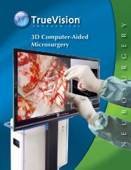
One of the newer developments in surgical technology<br />
is a <strong>3D</strong> high-definition surgical visualization device<br />
from <strong>TrueVision</strong> <strong>Systems</strong> (Santa Barbara, CA). <strong>In</strong> ophthalmic<br />
procedures, the <strong>3D</strong> high-definition system converts<br />
the optical image from the surgical microscope to<br />
a digital <strong>3D</strong> high-definition image projected onto a<br />
specialized “heads-up” viewing screen. <strong>In</strong> addition to<br />
the technology’s uses in the OR, the system has the<br />
potential for use in the education of fellow surgeons<br />
and telemedicine.<br />
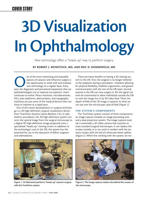
There are many benefits to having a <strong>3D</strong> viewing system<br />
in the OR. First, the surgeon is no longer tethered<br />
to the eyepieces during a procedure—freedom allowing<br />
for physical flexibility, healthier ergonomics, and greater<br />
communication with the rest of the OR team. Second,<br />
anyone in the OR can view surgery in <strong>3D</strong>; the signal can<br />
even be transmitted to allow individuals outside the OR<br />
to view the image live or by <strong>3D</strong> video feed. Third, the<br />
depth of field of the <strong>3D</strong> image is superior to what we<br />
can see over the microscopic optical field (Figure 1).<br />
THE SYSTEM’S COMPONENTS<br />
The <strong>TrueVision</strong> system consists of three components:<br />
an image capture module; an image processing unit;<br />
and a dual projection system. The image capture module<br />
is essentially a <strong>3D</strong> video camera that attaches to<br />
most standard surgical microscopes. It can replace the<br />
oculars entirely, or it can work in tandem with the primary<br />
oculars with the aid of a binocular beam splitter<br />
(Figure 2). When first working with the system, we rec-<br />
Figure 1. Dr. Weinstock performs “heads-up” cataract surgery<br />
with the <strong>TrueVision</strong> system.<br />
Figure 2. The image capture station sits behind the oculars of<br />
the microscope.<br />
62 I CATARACT & REFRACTIVE SURGERY TODAY I MAY 2008
COVER STORY<br />
ommend alternating<br />
between operating<br />
from the oculars<br />
and the screen (it is<br />
also a good way to<br />
compare the optical<br />
and digital images).<br />
The image capture<br />
module digitizes<br />
the stereo<br />
images obtained<br />
through the microscope<br />
and relays<br />
them to the image<br />
processing unit (a<br />
high-end computer)<br />
in real time. The<br />
computer enhances<br />
Figure 3. The image processing<br />
unit, dual projectors, and screen.<br />
and processes the images and then sends one from<br />
each ocular view to a dual projection system. Finally,<br />
the dual projection system shows the images on a specialized<br />
rear projection screen (Figure 3.)<br />
The rear projection screen may be positioned in the<br />
OR so that, by slightly tilting his head, the surgeon can<br />
adequately view the screen from the side while operating.<br />
Three-dimensional polarizing glasses are required to<br />
view the screen in <strong>3D</strong> high definition. As with any projection<br />
system, the room’s lights may either be on or off,<br />
but the resolution and contrast are better with a slightly<br />
darkened OR (Figure 4).<br />
INDICATIONS AND CONSIDERATIONS<br />
FOR USE<br />
We have found that the depth of field and magnification<br />
with the system are superior to the ocular view with<br />
a microscope. It is important to note that the resolution<br />
of the <strong>TrueVision</strong> system is between 80% to 90% of that<br />
normally seen through the oculars, so surgeons may prefer<br />
to look through the oculars when performing maneuvers<br />
for which they need the highest resolution.<br />
For routine cataract surgeries, surface ablation procedures,<br />
corneal transplants, phakic IOL procedures,<br />
and LASIK, the <strong>TrueVision</strong> system may be preferable to<br />
a traditional ocular view. The technology allows the<br />
surgeon to sit up or lean back in a chair and to assume<br />
a comfortable position rather than hunch over the eyepieces<br />
with his shoulders drooped—the norm when<br />
operating though oculars. Ergonomic positioning is<br />
especially helpful for longer procedures such as corneal<br />
transplants, endothelial transplants, and other surface<br />
reconstructions that may require the surgeon to look<br />
through the oculars for an extended period of time.<br />
The <strong>TrueVision</strong> system may be similarly helpful for<br />
lengthy retinal procedures such as vitrectomies and<br />
internal limiting membrane peels.<br />
COMMUNICATION AND EDUCATION<br />
As a communication medium, the <strong>TrueVision</strong> system<br />
enables the OR staff, nurses, residents, fellows, and<br />
even members of patients’ families to view intraocular<br />
surgery on the <strong>3D</strong> screen just as the surgeon does. As a<br />
result, the scrub technicians and other operating personnel<br />
can fully appreciate what is happening during a<br />
case and better anticipate the surgeon’s needs for additional<br />
instrumentation (Figure 5).<br />
The ability to send the <strong>3D</strong> high-definition signal to a<br />
remote location may be particularly advantageous for<br />
teaching institutions. A group of surgeons or students<br />
can view the procedure from an auditorium while the<br />
surgeon operates in a nearby suite. Real-time viewing<br />
allows an exchange of comments, questions, and feed-<br />
(Continued on page 65)<br />
Figure 4. The surgeon must slightly turn his head to see the<br />
screen.<br />
Figure 5. The scrub technician and other OR personnel also<br />
have a stereoscopic view of the surgical field.<br />
MAY 2008 I CATARACT & REFRACTIVE SURGERY TODAY I 63
COVER STORY<br />
Figure 6. Surgeons watch recorded <strong>3D</strong> cases at a Kansas City<br />
<strong>Ophthalmology</strong> Society meeting.<br />
(Continued from page 63)<br />
back between the surgeon and his audience (Figure 6).<br />
<strong>In</strong> addition, the video recording and playback features<br />
facilitate teaching and presentation of surgical procedures<br />
in <strong>3D</strong>.<br />
The <strong>TrueVision</strong> system also has utility for marketing<br />
efforts and lay education. Potential patients can watch<br />
surgical procedures from the waiting room or during<br />
LASIK or cataract surgery seminars, adding dramatic<br />
educational value.<br />
FUTURE APPLICATIONS<br />
<strong>TrueVision</strong> <strong>Systems</strong> is developing applications that<br />
will enhance the image that is projected on the screen<br />
and plans to create software to help ophthalmologists<br />
during surgery. <strong>In</strong> the works is a <strong>3D</strong> cursor with which<br />
surgeons or their assistants can draw on the screen to<br />
guide limbal relaxing incisions, the sizing and formation<br />
of the capsulorhexis, and more. Digital applications will<br />
make it possible to color code the anterior chamber<br />
depth, which will assist surgeons in determining the<br />
required depth of instrumentation inside the eye. ■<br />
Eric D. Donnenfeld, MD, is a partner with<br />
Ophthalmic Consultants of Long Island in<br />
Rockville Centre, New York, and a trustee of<br />
Dartmouth Medical School in Hanover, New<br />
Hampshire. He is a consultant to <strong>TrueVision</strong><br />
<strong>Systems</strong>. Dr. Donnenfeld may be reached at (516) 766-<br />
2519; eddoph@aol.com.<br />
Robert J. Weinstock, MD, practices ophthalmology<br />
at The Eye <strong>In</strong>stitute of West Florida in<br />
Largo. He is a consultant to <strong>TrueVision</strong><br />
<strong>Systems</strong>. Dr. Weinstock may be reached at<br />
(727) 244-1958; rjweinstock@yahoo.com.<br />
MAY 2008 I CATARACT & REFRACTIVE SURGERY TODAY I 65