Preparation of iron oxide nanoparticles supported ... - ScienceDirect
Preparation of iron oxide nanoparticles supported ... - ScienceDirect
Preparation of iron oxide nanoparticles supported ... - ScienceDirect

Create successful ePaper yourself
Turn your PDF publications into a flip-book with our unique Google optimized e-Paper software.
Wei-Wen Liu et al. / New Carbon Materials, 2011, 26(4): 255–261<br />
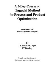
Fig.6 (a) Raman spectrum <strong>of</strong> SWCNTs. (b) The low-frequency region <strong>of</strong> Raman spectrum for the SWCNTs<br />
3.4 Growth mechanism <strong>of</strong> SWCNTs<br />
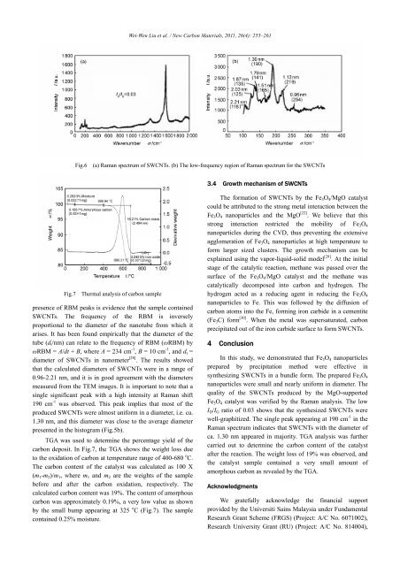
Fig.7 Thermal analysis <strong>of</strong> carbon sample<br />
presence <strong>of</strong> RBM peaks is evidence that the sample contained<br />
SWCNTs. The frequency <strong>of</strong> the RBM is inversely<br />
proportional to the diameter <strong>of</strong> the nanotube from which it<br />
arises. It has been found empirically that the diameter <strong>of</strong> the<br />
tube (d t /nm) can relate to the frequency <strong>of</strong> RBM (ωRBM) by<br />
ωRBM = A/dt + B, where A = 234 cm -1 , B = 10 cm -1 , and d t =<br />
diameter <strong>of</strong> SWCNTs in nanometer [28] . The results showed<br />
that the calculated diameters <strong>of</strong> SWCNTs were in a range <strong>of</strong><br />
0.96-2.21 nm, and it is in good agreement with the diameters<br />
measured from the TEM images. It is important to note that a<br />
single significant peak with a high intensity at Raman shift<br />
190 cm -1 was observed. This peak implies that most <strong>of</strong> the<br />
produced SWCNTs were almost uniform in a diameter, i.e. ca.<br />
1.30 nm, and this diameter was close to the average diameter<br />
presented in the histogram (Fig.5b).<br />
TGA was used to determine the percentage yield <strong>of</strong> the<br />
carbon deposit. In Fig.7, the TGA shows the weight loss due<br />
to the oxidation <strong>of</strong> carbon at temperature range <strong>of</strong> 400-680 o C.<br />
The carbon content <strong>of</strong> the catalyst was calculated as 100 X<br />
(m 1 -m 2 )/m 1 , where m 1 and m 2 are the weights <strong>of</strong> the sample<br />
before and after the carbon oxidation, respectively. The<br />
calculated carbon content was 19%. The content <strong>of</strong> amorphous<br />
carbon was approximately 0.19%, a very low value as shown<br />
by the small bump appearing at 325 o C (Fig.7). The sample<br />
contained 0.25% moisture.<br />
The formation <strong>of</strong> SWCNTs by the Fe 3 O 4 /MgO catalyst<br />
could be attributed to the strong metal interaction between the<br />
Fe 3 O 4 <strong>nanoparticles</strong> and the MgO [22] . We believe that this<br />
strong interaction restricted the mobility <strong>of</strong> Fe 3 O 4<br />
<strong>nanoparticles</strong> during the CVD, thus preventing the extensive<br />
agglomeration <strong>of</strong> Fe 3 O 4 <strong>nanoparticles</strong> at high temperature to<br />
form larger sized clusters. The growth mechanism can be<br />
explained using the vapor-liquid-solid model [29] . At the initial<br />
stage <strong>of</strong> the catalytic reaction, methane was passed over the<br />
surface <strong>of</strong> the Fe 3 O 4 /MgO catalyst and the methane was<br />
catalytically decomposed into carbon and hydrogen. The<br />
hydrogen acted as a reducing agent in reducing the Fe 3 O 4<br />
<strong>nanoparticles</strong> to Fe. This was followed by the diffusion <strong>of</strong><br />
carbon atoms into the Fe, forming <strong>iron</strong> carbide in a cementite<br />
(Fe 3 C) form [30] . When the metal was supersaturated, carbon<br />
precipitated out <strong>of</strong> the <strong>iron</strong> carbide surface to form SWCNTs.<br />
4 Conclusion<br />
In this study, we demonstrated that Fe 3 O 4 <strong>nanoparticles</strong><br />
prepared by precipitation method were effective in<br />
synthesizing SWCNTs in a bundle form. The prepared Fe 3 O 4<br />
<strong>nanoparticles</strong> were small and nearly uniform in diameter. The<br />
quality <strong>of</strong> the SWCNTs produced by the MgO-<strong>supported</strong><br />
Fe 3 O 4 catalyst was verified by the Raman analysis. The low<br />
I D /I G ratio <strong>of</strong> 0.03 shows that the synthesized SWCNTs were<br />
well-graphitized. The single peak appearing at 190 cm -1 in the<br />
Raman spectrum indicates that SWCNTs with the diameter <strong>of</strong><br />
ca. 1.30 nm appeared in majority. TGA analysis was further<br />
carried out to determine the carbon content <strong>of</strong> the catalyst<br />
after the reaction. The weight loss <strong>of</strong> 19% was observed, and<br />
the catalyst sample contained a very small amount <strong>of</strong><br />
amorphous carbon as revealed by the TGA.<br />
Acknowledgments<br />
We gratefully acknowledge the financial support<br />
provided by the Universiti Sains Malaysia under Fundamental<br />
Research Grant Scheme (FRGS) (Project: A/C No. 6071002),<br />
Research University Grant (RU) (Project: A/C No. 814004),