3D Fourier analysis methods for digital processing and 3D ...
3D Fourier analysis methods for digital processing and 3D ...
3D Fourier analysis methods for digital processing and 3D ...
You also want an ePaper? Increase the reach of your titles
YUMPU automatically turns print PDFs into web optimized ePapers that Google loves.
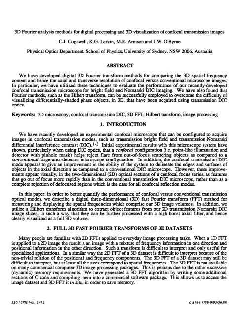
<strong>3D</strong> <strong>Fourier</strong> <strong>analysis</strong> <strong>methods</strong> <strong>for</strong> <strong>digital</strong> <strong>processing</strong> <strong>and</strong> <strong>3D</strong> visualization of confocal transmission images<br />
.<br />
c.J. Cogswell, K.G. Larkin, M.R. Arnison <strong>and</strong> J.W. OByrne<br />
Physical Optics Department, School of Physics, University of Sydney, NSW 2006, Australia<br />
ABSTRACT<br />
We have developed <strong>digital</strong> <strong>3D</strong> <strong>Fourier</strong> trans<strong>for</strong>m <strong>methods</strong> <strong>for</strong> comparing the <strong>3D</strong> spatial frequency<br />
content <strong>and</strong> hence the axial <strong>and</strong> transverse resolution of confocal versus conventional microscope images.<br />
In particular, we have utilized these techniques to evaluate the per<strong>for</strong>mance of our recently-developed<br />
confocal transmission microscope <strong>for</strong> bright field <strong>and</strong> Nomarski DIC imaging. We have also found that<br />
<strong>Fourier</strong> <strong>methods</strong>, such as the Hibert trans<strong>for</strong>m, can be successfully employed to overcome the difficulty of<br />
visualizing differentially-shaded phase objects, in <strong>3D</strong>, that have been acquired using transmission DIC<br />
optics.<br />
Keywords: <strong>3D</strong> microscopy, confocal transmission DIC, <strong>3D</strong> FF1', Hubert trans<strong>for</strong>m, image <strong>processing</strong><br />
1. INTRODUCTION<br />
We have recently developed an experimental confocal microscope that can be configured to acquire<br />
images in confocal transmission modes, such as transmission bright field <strong>and</strong> transmission Nomarski<br />
differential interference contrast (DIC).13 Initial experimental results with this microscope system have<br />
shown, particularly when using DIC optics, that a confocal configuration (i.e. point-like illumination <strong>and</strong><br />
detector with pinhole mask) helps reject flare from out-of-focus scattering objects as compared to a<br />
conventional large-area-detector microscope configuration. In addition, the confocal transmission DIC<br />
mode appears to give an improvement in the ability of the system to delineate the edges <strong>and</strong> surfaces of<br />
objects in the axial direction as compared to a conventional DIC microscope. However, these improvements<br />
appear visually, in the two-dimensional (2D) optical sections of a confocal focus series, as features<br />
that go out of focus more rapidly than in the conventional transmission DIC microscope, rather than as a<br />
complete rejection of defocused regions which is the case <strong>for</strong> all confocal reflection modes.<br />
In this paper, in order to better quantify the per<strong>for</strong>mance of confocal versus conventional transmission<br />
optical modes, we describe a <strong>digital</strong> three-dimensional (<strong>3D</strong>) fast <strong>Fourier</strong> trans<strong>for</strong>m (FVF) method <strong>for</strong><br />
measuring <strong>and</strong> displaying the spatial frequencies which comprise our <strong>3D</strong> image volumes. In addition, we<br />
utilize a Hilbert trans<strong>for</strong>m algorithm to extract object features from our 2D transmission Nomarski DIC<br />
image slices, in such a way that they can be further processed with a high boost axial filter, <strong>and</strong> hence<br />
clearly visualized as a full <strong>3D</strong> volume.<br />
2. FULL <strong>3D</strong> FAST FOURIER TRANSFORMS OF <strong>3D</strong> DATASETS<br />
Many people are familiar with 2D FFTs applied to everyday image <strong>processing</strong> tasks. When a 1D FFT<br />
is applied to a 2D image the result is an image with a mixture of frequency in<strong>for</strong>mation in one direction <strong>and</strong><br />
positional in<strong>for</strong>mation in the other direction. Such a trans<strong>for</strong>m is difficult to interpret <strong>and</strong> only useful <strong>for</strong><br />
specialized applications. In a similar way the 2D FF1' of a <strong>3D</strong> dataset is difficult to interpret because of the<br />
non-trivial relation of the positional <strong>and</strong> frequency components. The <strong>3D</strong> FF1' of a <strong>3D</strong> dataset may still be<br />
difficult to interpret, but at least all the axes correspond to spatial frequencies. The <strong>3D</strong> FFT is not available<br />
on many commercial computer <strong>3D</strong> image <strong>processing</strong> packages. This is perhaps due to the rather excessive<br />
(dynamic) memory requirements. We have generated a <strong>3D</strong> FF1' algorithm by writing some additional<br />
sections of C code <strong>and</strong> compiling them into a commercial software package. This allows us to access the<br />
image dataset <strong>and</strong> <strong>3D</strong> FF1' it in situ, in order to save memory.<br />
230 / SPIE Vol. 2412 0-8194-1 759-9/95/$6.00
If the imaging process satisfies certain constraints (such as being linear <strong>and</strong> space invariant) then the <strong>3D</strong><br />
FFT can be expressed as the product of the object spectrum multiplied by the optical transferfunction of<br />
the imaging process. Thus, in a comparison of imaging processes using the same object, relative responses<br />
can be assessed. For example, Figure 1 (left) shows the <strong>3D</strong> FVF of a transmission Nomarski DIC image of<br />
a thin, lightly stained, biological specimen. Figure 1 (right) shows the <strong>3D</strong> FFT of the same object viewed<br />
using a transmission bright field mode (i.e. using identical optics except with Wollaston prisms <strong>and</strong><br />
analyzer removed). The horizontal axes correspond to spatial frequencies in the lateral (x <strong>and</strong> y) dimensions<br />
of the microscope, while the vertical axis corresponds to spatial frequencies in the axial or focus<br />
direction (z dimension). The DIC <strong>3D</strong> FF1' (left) shows a broader (asymmetrical) base than the bright field<br />
case. This demonstrates the higher lateral spatial frequency content plus the direcüonal asymmetry characteristic<br />
of Nomarski DIC imaging modes. Of particular interest in the renditions of <strong>3D</strong> FETs is the<br />
response along the z spatial frequency axis. Components along this axis are indicative of the degree of<br />
optical sectioning in the imaging process. Some care has to be taken in interpretation because of the possibility<br />
of artefacts arising from image defects such as intensity noise <strong>and</strong> misregistration (displayed here as<br />
strong central peaks).<br />
Figure 1. <strong>3D</strong> FFTs of transmission Nomarski DIC (left) <strong>and</strong> transmission bright field (right) image<br />
volumes of the same biological specimen (lightly stained orchid root tip chromosomes). Horizontal<br />
axes correspond to x <strong>and</strong> y image spatial frequencies while the vertical axes correspond to axial (z)<br />
frequencies. The z axis (vertical scale) has been stretched by a factor of 7 times the x <strong>and</strong> y scales.<br />
3. <strong>3D</strong> VISUAUZATION OF NOMARSKI DIC IMAGES USING THE HILBERT TRANSFORM<br />
<strong>3D</strong> rendering of image sections obtained using Nomarski DIC produces the following dilemma:<br />
i) Because Nomarski DIC makes phase structure visible <strong>and</strong> enhances higher spatial frequency features<br />
compared to bright field, it is the preferred transmission optical mode <strong>for</strong> many biological applications.<br />
However, in 2D Nomarski DIC images, the object features appear in bas relief, that is to say phase<br />
gradients (refractive index boundaries) in the specimen can appear as shadow or highlight (depending<br />
on their orientation). This bas relief effect produces the visual appearance of <strong>3D</strong>, even though the<br />
image is obtained from a single plane of focus within the specimen volume.<br />
ii) Typical <strong>digital</strong> <strong>3D</strong> rendering techniques are based upon controlling either the opacity or the reflectivity<br />
of the final displayed image using the total projection of the individual voxel "values" along the<br />
SPIE Vol. 2412/231
line of sight. Connected segments of a rendered feature are there<strong>for</strong>e composed of voxels with comparable<br />
values. In the case of a transmission DIC focus series, when attempting to render the image data<br />
volume in <strong>3D</strong>, the shadow <strong>and</strong> highlight regions of a particular feature will have dramatically different<br />
values, hence will not, in general, appear connected.<br />
There are a number of ways around the problem of visualizing <strong>3D</strong> Nomarski DIC images. The most<br />
obvious is to convert individual DIC image slices back to something resembling bright field. For this<br />
method to be of any utility the features gained by Nomarski DIC (such as enhanced high spatial frequencies)<br />
must not be lost. The obvious way to convert from Nomarski to bright field is to reverse the<br />
(differential) DIC process, i.e., to integrate along the direction of Nomarski shear. This solution has a<br />
number of problems which are shown in Figure 2. The image at left is a single 2D slice from a confocal<br />
transmission DIC dataset of lightly-stained orchid root tip chromosomes. Figure 2 (middle) shows the<br />
result of per<strong>for</strong>ming a simple integration operation on the original DIC image. Streaks are due to difficulties<br />
initializing the integration. Worse still, the integration process is equivalent to a type of low pass filtering<br />
or blurring so that the enhanced high spatial frequencies are lost.<br />
Another way of modelling the Nomarski process is to consider it in the spatial frequency domain. In<br />
this system the (real space) differentiation corresponds to multiplication by the spatial frequency parallel to<br />
the shear. Integration is then just the inverse, namely division by the spatial frequency. Both these<br />
processes are antisymmetric (or odd) but the division selectively attenuates higher frequency components.<br />
What is needed is an antisymmetric trans<strong>for</strong>m or process which does not change the relative balance of the<br />
various frequency components. Such a trans<strong>for</strong>m exists <strong>and</strong> is known as the Hubert trans<strong>for</strong>m4. In essence<br />
the trans<strong>for</strong>m just keeps all the positive frequency components the same but reverses the sign of all the<br />
negative frequency components. The effect of the Hubert trans<strong>for</strong>m on a Nomarski image is shown in<br />
Figure 2 (right). The chromosomes which had gray/darklgray <strong>and</strong> gray/light/gray shading, on the left <strong>and</strong><br />
right edges respectively, convert to a lighter central region with dark/light transitions on one side <strong>and</strong><br />
light/dark transitions on the other side. The main effect is to "symmetrize" the image <strong>and</strong> render connected<br />
regions with similar pixel values.<br />
Figure 2. Comparison of <strong>digital</strong> <strong>processing</strong> techniques <strong>for</strong> confocal transmission Nomarski DIC images.<br />
Left: an original confocal transmission Nomarski DIC image of metaphase chromosomes from an<br />
orchid root tip preparation. Middle: the resulting image after a computer integration was per<strong>for</strong>med.<br />
Right: the resulting image after a Hilbert trans<strong>for</strong>m was per<strong>for</strong>med. Chromosome width equals<br />
approximately 1 j.Lm.<br />
The implementation of the 2D Hubert trans<strong>for</strong>m (HT) is shown schematically in Figure 3. It is possible<br />
to implement an approximate HT in real space4'5 using a special convolution kernel, but we chose the<br />
232 1SPIE Vol. 2412
Figure 3<br />
Algorithm <strong>for</strong> generating Hilbert-trans<strong>for</strong>med Nomarski DIC images<br />
SPIEVo!. 2412/233
exact method <strong>for</strong> the images shown in this paper. Briefly the process shown in Figure 3 is as follows:<br />
. Fast <strong>Fourier</strong> trans<strong>for</strong>m (FFT) the image<br />
. Multiply both the real <strong>and</strong> imaginary parts of the FF1.' by the Hubert frequency response image<br />
(which merely consists of +1 or -1 depending on lateral position).<br />
. Swap the resultant real <strong>and</strong> imaginary parts<br />
. InverseFVF <strong>and</strong> take the real part as the new image (the imaginary part is essentially zero).<br />
Once the DIC image slices were Hubert trans<strong>for</strong>med, we assembled them into a <strong>3D</strong> dataset using a<br />
volume rendering software package. Next we per<strong>for</strong>med a high boost filtering operation which enhanced<br />
the high spatial frequencies in the axial direction. Finally, lighting <strong>and</strong> colour were added <strong>and</strong> opacity<br />
adjusted to improve visibility of the individual chromosomes. Figure 4 shows the results of these image<br />
<strong>processing</strong> steps. At left is a confocal transmission Nomarski DIC isometric projection <strong>and</strong> at right is a<br />
conventional transmission DIC dataset taken at the same time using the same optics, except that a second,<br />
large-area photodetector was utilized <strong>and</strong> its amplifier gain was increased to match its contrast to that of the<br />
confocal detector. Identical image <strong>processing</strong> <strong>and</strong> <strong>3D</strong> rendering (as described above) were per<strong>for</strong>med on<br />
both datasets. The results show that our confocal transmission DIC microscope (left) has more clearly<br />
delineated the chromosomes as compared to the conventional case (right) which shows regions where the<br />
chromosomes appear smudged.<br />
Figure 4. Comparison of confocal (left) versus conventional (right) transmission Nomarski DIC microscopy<br />
techniques. These two <strong>3D</strong> datasets were acquired simultaneously from the same biological<br />
preparation (metaphase chromosomes from an orchid root tip) <strong>and</strong> were processed <strong>and</strong> rendered using<br />
identical <strong>digital</strong> imaging <strong>methods</strong>. The confocal transmission DIC image (left) shows better delineation<br />
of chromosomes than the conventional DIC case.<br />
234 ISPIE Vol. 2412
4. CONCLUSION<br />
Utilizing <strong>3D</strong> FF1' techniques, such as Hilbert trans<strong>for</strong>ms <strong>and</strong> high boost filtering, has allowed us to<br />
analyze <strong>and</strong> evaluate the per<strong>for</strong>mance of our confocal transmission microscope as compared to a conventional<br />
transmission system. In particular, image <strong>processing</strong> in the <strong>Fourier</strong> domain using a Hilbert trans<strong>for</strong>m<br />
has provided the means by which differentially-shaded Nomarski DIC images can be converted into a <strong>for</strong>m<br />
that can be rendered <strong>and</strong> visualized as a full <strong>3D</strong> volume, without losing the enhanced high spatial frequencies<br />
characteristic of our confocal transmission DIC microscope.<br />
5. ACKNOWLEDGMENTS<br />
The authors would like to thank the staff of VISLAB (the visualisation laboratory at Sydney University)<br />
<strong>for</strong> help in preparing some of the <strong>3D</strong> renditions of the images. The Physical Optics Department is<br />
supported by funds from the University of Sydney, the Science Foundation <strong>for</strong> Physics within the<br />
University of Sydney, <strong>and</strong> the Australian Research Council.<br />
6. REFERENCES<br />
1 Cogswell C.J., Larkin, K.G., O'Byme, J.W. <strong>and</strong> Arnison, M.R., High-resolution, multiple optical mode<br />
confocal microscope: I. System design, image acquisition <strong>and</strong> <strong>3D</strong> visualization. SPIE 2184,48-54,<br />
1994.<br />
2 Cogswell C.J. <strong>and</strong> O'Byrne J.W., A high resolution confocal transmission microscope: I. System<br />
design. SPIE, 1660,503-511, 1992.<br />
3 Dixon, A. E. <strong>and</strong> Cogswell, C. J., Confocal microscopy with transmitted light, in H<strong>and</strong>book of<br />
Biological Confocal Microscopy, 2nd edition, J. B. Pawley, ecL, Plenum, NY, in press, 1995.<br />
4 Oppenheim, A., <strong>and</strong> R, S., Discrete-Time Signal Processing, Prentice-Hall, Engelwood Cliffs, N.J.,<br />
1989.<br />
5 Chim, S. S. C., <strong>and</strong> Kino, G. S., Three-dimensional image realization in interference microscopy,<br />
Applied Optics 31, (14), 2550-2553, 1992.<br />
SPIE Vol. 24121235


