3D Fourier analysis methods for digital processing and 3D ...
3D Fourier analysis methods for digital processing and 3D ...
3D Fourier analysis methods for digital processing and 3D ...

You also want an ePaper? Increase the reach of your titles
YUMPU automatically turns print PDFs into web optimized ePapers that Google loves.
exact method <strong>for</strong> the images shown in this paper. Briefly the process shown in Figure 3 is as follows:<br />
. Fast <strong>Fourier</strong> trans<strong>for</strong>m (FFT) the image<br />
. Multiply both the real <strong>and</strong> imaginary parts of the FF1.' by the Hubert frequency response image<br />
(which merely consists of +1 or -1 depending on lateral position).<br />
. Swap the resultant real <strong>and</strong> imaginary parts<br />
. InverseFVF <strong>and</strong> take the real part as the new image (the imaginary part is essentially zero).<br />
Once the DIC image slices were Hubert trans<strong>for</strong>med, we assembled them into a <strong>3D</strong> dataset using a<br />
volume rendering software package. Next we per<strong>for</strong>med a high boost filtering operation which enhanced<br />
the high spatial frequencies in the axial direction. Finally, lighting <strong>and</strong> colour were added <strong>and</strong> opacity<br />
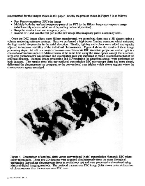
adjusted to improve visibility of the individual chromosomes. Figure 4 shows the results of these image<br />
<strong>processing</strong> steps. At left is a confocal transmission Nomarski DIC isometric projection <strong>and</strong> at right is a<br />
conventional transmission DIC dataset taken at the same time using the same optics, except that a second,<br />
large-area photodetector was utilized <strong>and</strong> its amplifier gain was increased to match its contrast to that of the<br />
confocal detector. Identical image <strong>processing</strong> <strong>and</strong> <strong>3D</strong> rendering (as described above) were per<strong>for</strong>med on<br />
both datasets. The results show that our confocal transmission DIC microscope (left) has more clearly<br />
delineated the chromosomes as compared to the conventional case (right) which shows regions where the<br />
chromosomes appear smudged.<br />
Figure 4. Comparison of confocal (left) versus conventional (right) transmission Nomarski DIC microscopy<br />
techniques. These two <strong>3D</strong> datasets were acquired simultaneously from the same biological<br />
preparation (metaphase chromosomes from an orchid root tip) <strong>and</strong> were processed <strong>and</strong> rendered using<br />
identical <strong>digital</strong> imaging <strong>methods</strong>. The confocal transmission DIC image (left) shows better delineation<br />
of chromosomes than the conventional DIC case.<br />
234 ISPIE Vol. 2412