Oral Melanotic Macule - A Case Report - sygdoms
Oral Melanotic Macule - A Case Report - sygdoms
Oral Melanotic Macule - A Case Report - sygdoms
You also want an ePaper? Increase the reach of your titles
YUMPU automatically turns print PDFs into web optimized ePapers that Google loves.
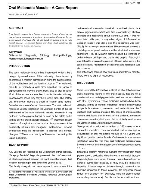
<strong>Oral</strong> <strong>Melanotic</strong> <strong>Macule</strong> - A <strong>Case</strong> <strong>Report</strong><br />
ISSN 0970-4388<br />
PAIS S a , HEGDE S K b , BHAT S S C<br />
ABSTRACT<br />
A melanotic macule is a benign pigmented lesion of oral cavity<br />
characterised by increase in melanin pigmentation. Presented here is<br />
a case report of 12 year old girl with black pigmented area on right<br />
buccal mucosa. An exciscon biopsy was done which confirmed the<br />
diognosis by as melanotic macule.<br />
Key Words<br />
Differential diagnosis, Etiology, Histopathology,<br />
Management, <strong>Melanotic</strong> mecule.<br />
INTRODUCTION<br />
The term melanotic macule has been used to describe a<br />
benign pigmented lesion of the oral cavity, characterized by<br />
an increase in melanin pigmentation along the basal cell layer<br />
of the epithelium and the lamina propria. The melanotic<br />
macule is typically a well circumscribed flat area of<br />
pigmentation that may be brown, black, blue or grey in colour.<br />
Most of the lesions are less than 1 cm in diameter, although<br />
in occasional cases they may be larger in size. The solitary<br />
oral melanotic macule is seen in middle aged adults.<br />
Females are more affected than males. The oral melanotic<br />
macule is usually located on the vermilion border of the lips<br />
and termed as the labial melanotic macule. Intraorally, it may<br />
be found on the gingiva, buccal mucosa or the palate and is<br />
termed as the oral melanotic macule. 1,2,3 Treatment usually<br />
consists of surgical excision, and a biopsy to rule out the<br />
possibility of an early malignant melanoma. Periodic<br />
evaluation may be necessary to assess any clinical<br />
changes. 4 There is a paucity of literature concerning this<br />
lesion in children.<br />
CASE REPORT<br />
A12 year old girl reported to the Department of Pedodontics,<br />
Yenepoya Dental College Mangalore with the chief complaint<br />
of black pigmented area on the right buccal mucosa, that<br />
kept on increasing in size since one year (Fig. 1).<br />
The family history did not reveal any such occurrences. Intra<br />
a. Assistant Professor, b. Associate Professor, c. Professor and<br />
Head Department of Pediatric Dentistry, Yenepoya Dental College,<br />
Mangalore.<br />
oral examination revealed a well circumscribed bluish black<br />
area of pigmentation which was firm in consistency, elliptical<br />
in shape and measuring about 1.0x0.5x0.1 cms. It was not<br />
associated with pain or any other signs of inflammation.<br />
Excision biopsy was done and the specimen was sent<br />
(Fig.2) for histologic examination. Biopsy report showed a<br />
mild degree of parakeratosis in the stratified squamous<br />
epithelium (Fig. 3). Melanin pigment could be identified in<br />
both the basal cell layer and the lamina propria. Although it<br />
was difficult to evaluate the amount of found to be more in the<br />
basal cell layer. Proliferation of capillaries and fibroblasts was<br />
also observed.<br />
The patient was recalled after one week and after six months.<br />
There were no signs of recurrence.<br />
DISCUSSION<br />
There is very little information in literature about the brown or<br />
black melanotic lesions of the oral mucosa, that are not a<br />
manifestation of racial pigmentation and are not associated<br />
with other syndromes. These melanotic macules have been<br />
variously termed as ephelis, melanosis, lentigo, solitary labial<br />
lentigo, labial melanotic macule and oral melanotic macules. 3<br />
Buchner and Hansen analyzed 105 cases of oral melanotic<br />
macule and found that in most of the patients, melanotic<br />
macule was a solitary lesion and the most likely location was<br />
the vermilion border, followed by the gingiva.<br />
George et al, conducted a review of 353 cases of oral<br />
melanotic macule 4 . They concluded that mean age of<br />
occurrence of oral melanotic macule is 43.1 years with<br />
significant predilection for female. <strong>Oral</strong> melanotic macule was<br />
located on lower lip. The most oral melanotic macules were<br />
Brown in colour and the mean size of the lesion was about<br />
6.8 mm.<br />
Regarding etiology, melanotic macules may result from racial<br />
pigmentation, endocrine disturbance, antimalarial therapy,<br />
Peutz-Jeghers syndrome, trauma, hemochromatosis, or<br />
chronic pulmonary disease, or they may be idiopathic.<br />
Majority of these require clinicopathologic correlation for<br />
definitive diagnosis. In these instances, the diagnosis should<br />
reflect the etiology (for example, melanin pigmentation<br />
secondary to trauma). For those lesions without an<br />
J Indian Soc Pedo Prev Dent June (2004) 22 (2) 73 - 75
<strong>Oral</strong> <strong>Melanotic</strong> <strong>Macule</strong><br />
identifiable etiologic factor, the term oral melanotic macule<br />
has been suggested 2 .<br />
In the literature, the oral melanotic macule has been given<br />
various inappropriate and erroneous names, such as<br />
ephelis 5,6 and lentigo 7,8 . Ephelis (freckle) is a circumscribed<br />
brown macule over skin that has been exposed to sunlight.<br />
Histologically, ephelis shows increased melanin<br />
pigmentation in the basal-cell layer without an increase in the<br />
number of melanocytes 3 .<br />
Ephelides are not found on mucous membranes. Although<br />
the histologic appearance of the melanotic macule of the oral<br />
mucosa is similar to that of ephelis of the skin, it is not at all<br />
related to exposure to sun and thus the term ephelis for the<br />
intraoral lesion is a misnomer.<br />
The term lentigo has also been suggested for oral melanotic<br />
macules. But since melanotic macules of the oral mucosa do<br />
not exhibit a significant increase in the number of<br />
melanocytes, the term lentigo is not considered appropriate<br />
for this type of lesion.<br />
Weather et al 1 and Page et al 2 , recently introduced the terms<br />
labial melanotic macule for lesions on the vermilion border<br />
and oral melanotic macule for lesions within the oral cavity.<br />
The term melanotic macule should be reserved for lesions in<br />
which there is a clinicopathologic correlation between the<br />
clinical feature of a discrete pigmented macule and the<br />
histologic feature of hyperpigmentation of the basal-cell layer<br />
and / or the lamina propria 3 .<br />
The term focal melanosis should be used as a histologic<br />
designation when hyperpigmentation of the basal-cell layer<br />
and/or the lamina propria is associated with clinically<br />
nonpigmented pathologic conditions 3 .<br />
Regarding histopathology, the dark colour of the lesion is<br />
due to increase in melanin pigment of the basal cell layer, not<br />
from an increased number of melanocytes. Melanin may also<br />
be found in the lamina propria. Further histologic criteria are<br />
absence of elongated rete ridges and lack of prominent<br />
melanocytic activity. If there is an elogation of rete ridges, a<br />
heavily pigmented basal cell layer, and an increase in the<br />
Fig: 1 Intra oral photograph showing <strong>Melanotic</strong> <strong>Macule</strong> in<br />
the Right Buccal Mucosa<br />
Fig: 2 Post- operative Intra oral photograph after the<br />
excision of <strong>Oral</strong> <strong>Melanotic</strong> <strong>Macule</strong>.<br />
Fig: 3 Histo-pathologic features suggestive of <strong>Oral</strong><br />
<strong>Melanotic</strong> <strong>Macule</strong>.<br />
sygdom.info J Indian Soc Pedo Prev Dent June (2004) 22 (2) 74
<strong>Oral</strong> <strong>Melanotic</strong> <strong>Macule</strong><br />
number of normal-appearing basal layer melanocytes, a<br />
junctional nevus has to be considered. If the melanocytes<br />
show proliferation, atypia, and some irregularity in their<br />
arrangement, the histopathologic diagnosis is atypical<br />
melanocytic hyperplasia, which may correspond clinically to<br />
early malignant melanoma (melanoma in situ) 9 .<br />
In our case, we found melanin in both the basal cell layer and<br />
the lamina propira. This agrees with the study conducted by<br />
Buchner and Hansen 3 . Page et al 2 found that 30% of their<br />
cases had melanin only in the basal cell layer and 3.8% only<br />
in the lamina propria 2 .<br />
The small size, slow growth rate, and flat clinical appearance<br />
favour a benign diagnosis.<br />
<strong>Oral</strong> melanotic macule has to be differentiated from certain<br />
other similar conditions exhibiting hyperpigmentation. Racial<br />
pigmentation is generally diffuse, is genetically acquired and<br />
seen at birth. It is more common in Caucasians and has been<br />
termed as oral melanosis.<br />
The two most important causes of post inflammatory hyper<br />
pigmentation are lichen planus and lupus erythematosis.<br />
There are certain endocrinal hyper pigmentations. Addison's<br />
disease causes a darkening of the oral mucosa which is<br />
irregular, patchy and found on the gums.<br />
Metal deposition can cause discolouration either from copper<br />
as in Wilson's disease or from amalgam as in an amalgam<br />
tattoo.<br />
It could be associated with syndromes as in Peutz Jeghers<br />
syndrome where freckles are seen not only in the oral cavity,<br />
but also at the distal extremities, Leopard syndrome where<br />
pigmentation is seen all over the body.<br />
Antimalarial drugs like chloroquine can also cause muscosal<br />
hyperpigmentation which also occurs on other body parts like<br />
the shinns.<br />
The treatment of oral melanotic macule is debatable 5 .<br />
Although the lesion is completely benign 2 and shows no<br />
tendency to recur or to become malignant, sometimes it is<br />
difficult if to distinguish it cliniclly from other pigmented<br />
lesions, such as nevus, malignant melanoma in situ, and<br />
incipient malignant melanoma. Thus, complete excision of<br />
oral melanotic macule is indicated and histologic<br />
examination or at least be checked at frequent intervals for<br />
any change in size, shape, or colour. This is especially<br />
necessary for lesions of the palate - a location for which<br />
oral malignant . melanoma has a strong predilection 10 .<br />
<strong>Melanotic</strong> lesions having a duration of fewer than 5 years,<br />
which have exhibited changes in size or colour or which<br />
exhibit tumefaction, ulceration, or bleeding, should be<br />
excised. Lesions with reliable history of more than 5 years<br />
without change in character in which a known cause seems<br />
evident (trauma, etc.), may be followed or excised, although<br />
we prefer the latter.<br />
REFERENCES<br />
1. Weathers D. R., Corio. R. L, Crawford B. E., Giansant J. S.<br />
and Page. L. R. : The labial melanotic macule. <strong>Oral</strong> Surg <strong>Oral</strong><br />
Med <strong>Oral</strong> Pathol 1976 ; 42 : 196 - 205<br />
2. Page L. R., Corio R. L., Crawford B. E., Giansant J. S. and<br />
Weathers D. R. : The oral melanotic macule. <strong>Oral</strong> Surg <strong>Oral</strong><br />
Med <strong>Oral</strong> Pathol 1977 ; 44: 219 - 226<br />
3. Buchner A. and Hansen L. S. : <strong>Melanotic</strong> <strong>Macule</strong> of the oral<br />
mucosa. <strong>Oral</strong> Surg <strong>Oral</strong> Med <strong>Oral</strong> Pathol 1979 ; 48 : 244 - 249.<br />
4. George E. K., Andrew P. H., William T. R., Louis M. A. and John<br />
A. S. : <strong>Oral</strong> <strong>Melanotic</strong> <strong>Macule</strong> - A Review of 353 cases. <strong>Oral</strong><br />
Surg <strong>Oral</strong> Med <strong>Oral</strong> Pathol 1993 ; 76 : 59 - 62<br />
5. Trodohl J. N. and Spragul W. G. : Benign and Malignant<br />
Mucosa, an Analysis of 135 cases. Cancer 1970 ; 25 : 812 -<br />
823<br />
6. Bhaskar S. N.: <strong>Oral</strong> Lesions in the Aged population - a survey<br />
of 785 cases. Geriatrics 1968 ; 23 : 137 - 149<br />
7. Beeker S. W. Jr. : Pigmentary Lesions oral melanotic macule<br />
on to the skin and oral cavity. <strong>Oral</strong> surg 1969 ; 28 : 526 -533.<br />
8. Shapiro. L. and Zegarelli D. J. : The solitary Labial Lentigo - a<br />
clinicopathologic study of 20 cases. <strong>Oral</strong> surg 1971 ; 31 : 87 -<br />
92<br />
9. Bork. K., Hoede. N., Korting. G. W., Burgdorf. W. H. C. and<br />
Young S. K. : Diseases of the oral Mucosa and the Lips. 2nd<br />
Edition, W. B. Saunders Company, 1996 ; 310 - 312<br />
10.Chaudhary A. P., Hampel A. and Gorlin R. L. : Primary<br />
Malignant Melanoma of the <strong>Oral</strong> Cavity-a Review of 105<br />
cases. Cancer 1958 ; 11 : 923 - 928<br />
11.Bengel W., Veltman G., Loevy H. T. and Pierangelo T. :<br />
Differential Diagnosis of Diseases of the <strong>Oral</strong> Mucosa.<br />
Quintessence Publishing Co., Inc., Chicago / Illinosi / 1989 :<br />
153 - 201<br />
Reprint Requests to :<br />
Dr. Sham Bhat<br />
Head of the Department of paediatric Dentistry,<br />
Yenepoya Dental College,<br />
Kodialbail, Mangalore,<br />
Karnataka,<br />
INDIA.<br />
J Indian Soc Pedo Prev Dent June (2004) 22 (2) 75<br />
sygdom.info


