SYBR Green Master Mix
SYBR Green Master Mix
SYBR Green Master Mix

Create successful ePaper yourself
Turn your PDF publications into a flip-book with our unique Google optimized e-Paper software.
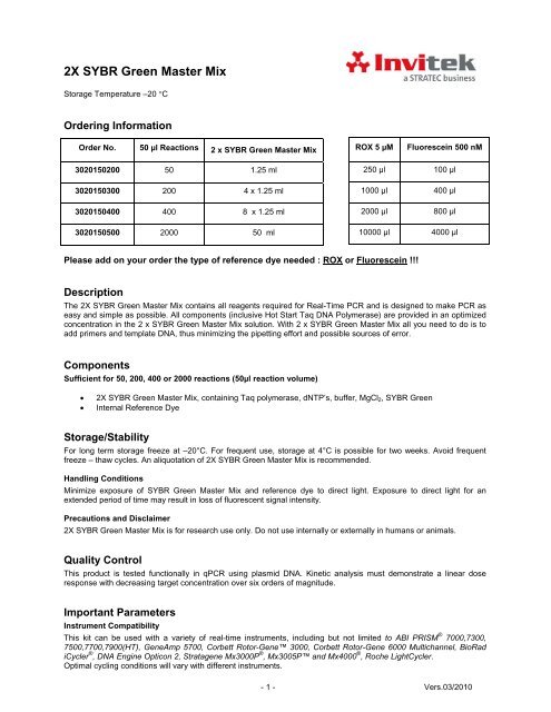
2X <strong>SYBR</strong> <strong>Green</strong> <strong>Master</strong> <strong>Mix</strong><br />
Storage Temperature –20 °C<br />
Ordering Information<br />
Order No. 50 µl Reactions 2 x <strong>SYBR</strong> <strong>Green</strong> <strong>Master</strong> <strong>Mix</strong><br />
ROX 5 µM<br />
Fluorescein 500 nM<br />
3020150200 50 1.25 ml<br />
3020150300 200 4 x 1.25 ml<br />
3020150400 400 8 x 1.25 ml<br />
3020150500 2000 50 ml<br />
250 µl 100 µl<br />
1000 µl 400 µl<br />
2000 µl 800 µl<br />
10000 µl 4000 µl<br />
Please add on your order the type of reference dye needed : ROX or Fluorescein !!!<br />
Description<br />
The 2X <strong>SYBR</strong> <strong>Green</strong> <strong>Master</strong> <strong>Mix</strong> contains all reagents required for Real-Time PCR and is designed to make PCR as<br />
easy and simple as possible. All components (inclusive Hot Start Taq DNA Polymerase) are provided in an optimized<br />
concentration in the 2 x <strong>SYBR</strong> <strong>Green</strong> <strong>Master</strong> <strong>Mix</strong> solution. With 2 x <strong>SYBR</strong> <strong>Green</strong> <strong>Master</strong> <strong>Mix</strong> all you need to do is to<br />
add primers and template DNA, thus minimizing the pipetting effort and possible sources of error.<br />
Components<br />
Sufficient for 50, 200, 400 or 2000 reactions (50µl reaction volume)<br />
• 2X <strong>SYBR</strong> <strong>Green</strong> <strong>Master</strong> <strong>Mix</strong>, containing Taq polymerase, dNTP’s, buffer, MgCl 2 , <strong>SYBR</strong> <strong>Green</strong><br />
• Internal Reference Dye<br />
Storage/Stability<br />
For long term storage freeze at –20°C. For frequent use, storage at 4°C is possible for two weeks. Avoid frequent<br />
freeze – thaw cycles. An aliquotation of 2X <strong>SYBR</strong> <strong>Green</strong> <strong>Master</strong> <strong>Mix</strong> is recommended.<br />
Handling Conditions<br />
Minimize exposure of <strong>SYBR</strong> <strong>Green</strong> <strong>Master</strong> <strong>Mix</strong> and reference dye to direct light. Exposure to direct light for an<br />
extended period of time may result in loss of fluorescent signal intensity.<br />
Precautions and Disclaimer<br />
2X <strong>SYBR</strong> <strong>Green</strong> <strong>Master</strong> <strong>Mix</strong> is for research use only. Do not use internally or externally in humans or animals.<br />
Quality Control<br />
This product is tested functionally in qPCR using plasmid DNA. Kinetic analysis must demonstrate a linear dose<br />
response with decreasing target concentration over six orders of magnitude.<br />
Important Parameters<br />
Instrument Compatibility<br />
This kit can be used with a variety of real-time instruments, including but not limited to ABI PRISM ® 7000,7300,<br />
7500,7700,7900(HT), GeneAmp 5700, Corbett Rotor-Gene 3000, Corbett Rotor-Gene 6000 Multichannel, BioRad<br />
iCycler ® , DNA Engine Opticon 2, Stratagene Mx3000P ® , Mx3005P and Mx4000 ® , Roche LightCycler.<br />
Optimal cycling conditions will vary with different instruments.<br />
- 1 - Vers.03/2010
Template<br />
The template should be of high molecular weight and its purity and integrity checked on an agarose gel prior to use.<br />
Contamination of the DNA template with RNA can result in reduced yield in PCR due to chelating of Mg 2+ and hence<br />
should be pre-purified. Template concentration will have a profound effect on PCR performance.<br />
Recommended amounts for standard PCR and reaction volume 25 µl are:<br />
• Human DNA up to 500 ng<br />
• Plasmid DNA 0.1–10 ng<br />
• Bacterial DNA 1–10 ng<br />
Difficult Templates<br />
For better amplification results use additional substances for example glycerol (5 – 10 %), DMSO (2 – 20 %),<br />
formamide (2 – 20 %), tetramethylammonium chloride (0.01 – 10 mM) or combinations of these.<br />
The role of DMSO, betaine and glycerol can significantly reduce the presence of non-specific binding products, which<br />
results in band smearing and presence of non-target sequence products.<br />
DNA Polymerase Activation Time<br />
The Hot Start DNA Polymerase is activated in the 10-minute incubation at 95°C before PCR cycling.<br />
ROX Reference Dye<br />
The passive ROX reference dye has an excitation maximum at 584 nm and an emission maximum at 612 nm. It can<br />
be included in the reaction to normalize the fluorescent reporter signal for instruments that are compatible with that<br />
option. ROX is supplied at a 5 µM concentration. Use the following table to determine the amount of ROX to use with<br />
a particular instrument:<br />
Instrument Amount of ROX per 50 µl reaction Final ROX Concentration<br />
ABI PRISM<br />
7000,7300,7700,7900(HT) /<br />
GeneAmp 5700***<br />
3 - 5 µl 300 nM - 500 nM<br />
Stratagene Mx3000P, Mx3005P and<br />
Mx4000, ABI 7500***<br />
DNA Engine Opticon 2, Chromo 4***<br />
Dilute <strong>Mix</strong> 1:10 500 nM<br />
1.5 –2.5 µl<br />
Dilute <strong>Mix</strong> 1:10 500 nM<br />
1.5 –2.0 µl<br />
30 nM - 50 nM<br />
30 nM - 50 nM<br />
Fluorescein<br />
The Fluorescein reference dye has an excitation maximum at 494 nm and an emission maximum at 525 nm. The Bio-<br />
Rad iCycler ® allows the use of fluorescein as a reference dye to normalize the fluorescent reporter signal in reactions<br />
with 2X <strong>SYBR</strong> <strong>Green</strong> <strong>Master</strong> <strong>Mix</strong>. The starting point of optimization is 50 nM Fluorescein. Optimal results are obtained<br />
between 10 - 100 nM Fluorescein.<br />
Instrument Amount of Fluorescein per 50 µl<br />
reaction<br />
Final Fluorescein Concentration<br />
BioRad iCycler, MyiQ, iQ5 ***<br />
Dilute <strong>Mix</strong> 1:5 100 nM<br />
5 µl<br />
10 nM<br />
*** Names of the instruments are registered trade marks of the respective firms<br />
- 2 - Vers.03/2010
Basic Protocol<br />
Combine the following components in a PCR-reaction tube:<br />
Component Concentration Volume Final concentration<br />
PCR grade H 2 O<br />
variable (added to the final<br />
volume of 25 µl)<br />
2X <strong>SYBR</strong> <strong>Green</strong> <strong>Master</strong> <strong>Mix</strong> 2x 12.5 µl 1 x<br />
Primer A 1µM variable 0.2 – 1 µM<br />
Primer B 1µM variable 0.2 – 1 µM<br />
Template DNA 10 ng / µl variable 1 – 150 ng* 1<br />
Total volume 25 µl<br />
* 1 : 10 ng/ 25 µl reaction for linearized plasmid DNA<br />
50 ng/ 25 µl – 125 ng/ 25 µl for mammalian genomic DNA<br />
Total volume of 50 µl is also possible.<br />
<strong>Mix</strong> well, centrifuge and place in cycler. Optimal conditions for concentration of primer, template and MgCl 2 and<br />
temperature profile need to be determined for each reaction.<br />
Cycling conditions<br />
Step Temperature Time Number of cycles<br />
initial denaturation 95°C 10 min 1<br />
denaturation 95°C 20 sec – 1 min<br />
annealing* 1 53°C - 68°C 20 sec – 1 min 35 – 45* 2<br />
elongation 72°C 0.6 – 0.7 min / kb<br />
Melting Curve 50°C - 95°C depends on machine 1<br />
* 1 : depends on primer<br />
* 2 : 35 cycles for plasmid DNA; 40 – 45 cycles for genomic DNA<br />
Troubleshooting<br />
Observation<br />
Check<br />
No Amplification<br />
Pipetting error / reagents missing<br />
Enzyme not activated<br />
Annealing step<br />
Extension step<br />
Primers<br />
- Repeat experiment checking concentration of all reagents.<br />
- Check that the full 5 minute activation step is performed before cycling.<br />
- Check that you have the optimal annealing temperature by performing a<br />
temperature gradient (2°C increments). Annealing time should be<br />
carried out as specified in the protocol.<br />
- Extension time can be increased for longer amplicons.<br />
- Amplification of products over 300 bp is not recommended.<br />
- Poor primer design - Check for primer dimers on gel.<br />
- Wrong primer concentration - 0.4µM recommended.<br />
- Primers degraded - Check on polyacrylamide gel.<br />
Re-order new primers if necessary.<br />
Detection step - Detection reading taken at the end of the elongation phase .<br />
- 3 - Vers.03/2010
Product too long<br />
To few cycles<br />
Template<br />
Wrong dye layer<br />
- The ideal amplicon size is between 80-200bp.<br />
Amplification of products over 300 bp is not recommended.<br />
- 40 cycles is recommended.<br />
- Impure template - Purify template before use. For templates isolated<br />
from difficult sources (such as plant) use a commercial mix containing<br />
enhancers/detergents.<br />
- Wrong concentration - A concentration of up to 500 ng can usually be<br />
used.<br />
- Degraded - Make fresh dilution from stock. Check storage conditions.<br />
- Check that machine settings correspond with experiment.<br />
Wavy/ Erratic Lines<br />
No ROX<br />
Too many cycles performed<br />
Wrong detection step<br />
Machine needs calibrating<br />
Baseline for ∆ Rn set at wrong cycle<br />
- Check machine settings. ABI Prism requires ROX for normalization.<br />
ROX is available as a separate vial.<br />
- Reduce number of cycles.<br />
- Check detection step is set in the correct stage of the cycle.<br />
- Wavy lines can be caused by mirror misalignment or lamp problems.<br />
Consult machine manufacturer.<br />
- ∆ Rn should be set between 3-15 cycles and at least 2 cycles before 1st<br />
dilution amplifies.<br />
Reaction volume too low.<br />
- Some QPCR instruments (e.g. ABI Prism 7700) are set to read<br />
accurately only at volumes of at least 15 µl.<br />
Amplification in No Template Control<br />
Primer dimers<br />
Contamination<br />
- Primer dimers can also be identified by using a serial dilution of your<br />
template and running products on a gel. As template concentration<br />
increases, the primer dimer bands should decrease in intensity. If the<br />
presence of primer dimers is observed, then it may help to do one of the<br />
following:<br />
1) re-design the primers,<br />
2) try increasing the annealing temperature, or<br />
3) decrease the primer concentration.<br />
- If using a standard curve, a sub-optimal gradient can indicate inhibition<br />
from primer dimers.<br />
- Template contaminated - Purify template before use.<br />
- If doing QRT-PCR, treat RNA template with recombinant DNase I or<br />
design exon-spanning primers.<br />
- DNA polymerase contaminated - All recombinant DNA polymerases will<br />
contain small amounts of E. coli DNA. However, if contamination<br />
remains a problem, a "BLAST"search can be performed to check for<br />
homology to the E. coli genome.<br />
- Reagents contaminated - Repeat with fresh reagents and always use<br />
filter tips.<br />
High Well-to-Well Variance<br />
Poor plate choice<br />
Low quality sealing material<br />
Machine needs re-calibrating<br />
Evaporation<br />
Concentration gradient formed in vial<br />
- Do not use frosted or black plates.<br />
- Use only high quality optically clear seals that have been specifically<br />
designed for fluorescence applications.<br />
- Follow manufacturer's guidelines.<br />
- Do not use corner wells or use a more robust seal.<br />
- Invert the mixture a couple of times before use.<br />
Low Sensitivity (High Cycle Threshold (Ct))<br />
Evaporation<br />
- Do not use corner wells or use a more robust seal.<br />
- 4 - Vers.03/2010
Low quality sealing material<br />
Primer dimers<br />
Annealing step<br />
Extension step<br />
Primers<br />
Efficiency is poor<br />
Pipetting errors<br />
- Use only high quality optically clear seals that have been specifically<br />
designed for fluorescence applications.<br />
- Primer dimers can also be identified by using a serial dilution of your<br />
template and running products on a gel. As template concentration<br />
increases, the primer dimer bands should decrease in intensity. If the<br />
presence of primer dimers is observed, then it may help to do one of the<br />
following:<br />
1) re-design the primers,<br />
2) try increasing the annealing temperature, or<br />
3) decrease the primer concentration.<br />
- If using a standard curve, a sub-optimal gradient can indicate inhibition<br />
from primer dimers.<br />
- Check that you have the optimal annealing temperature by performing a<br />
temperature gradient (2°C increments).<br />
- Annealing time should be carried out as specified in the protocol. -Start<br />
with the recommended annealing/extension time (60 seconds) and<br />
increase with 10 seconds steps.<br />
- Extension time can be increased for longer amplicons. Increase<br />
annealing/extension temperature in steps of 2°C.<br />
- Amplification of products over 300 bp is not recommended.<br />
- Wrong primer concentration - 0.4 µM recommended.<br />
- To optimize use different ratios of reverse/forward primer mixes e.g.<br />
50/50nM 300/50nM 900/50nM 300/50nM 300/300nM ....<br />
- Check for degradation of primers via denaturing PAGE.<br />
- This is usually caused by the length of the amplicon. The length of<br />
amplicons should be between 80 and 150 bp. With adjusted reaction<br />
times it is possible to amplify up to 500 bp. Primer design is not optimal<br />
or primers have been designed containing secondary structures. If<br />
possible, we recommend redoing primer design according to usual<br />
primer design guidelines. If not, try a 3 - step protocol, this could<br />
enhance the PCR efficiency.<br />
- As Real Time PCR is a highly sensitive tool, error will also be amplified<br />
easily. The use of <strong>Master</strong> <strong>Mix</strong>es can reduce this, as variability is kept to<br />
a minimum. A standard curve should always be used to check for<br />
irregularities. Check for PCR efficiency and pipetting errors.<br />
No Ct value<br />
Number of cycles is insufficient<br />
Detection during wrong PCR step<br />
Primers degraded<br />
Insufficient amount of starting DNA<br />
template<br />
Template DNA degraded<br />
Primer design sub-optimal<br />
- Start with 35 cycles, and then increase up to 50 cycles.<br />
- Make sure that detection occurs at the end of the elongation step<br />
(at 72°C).<br />
- Check for degradation of primers via denaturing PAGE.<br />
- Start with a high concentration and make dilution series if the concentration<br />
is unknown. The recommended maximum amount of template is 500 ng<br />
genomic DNA.<br />
- Check DNA on agarose gel for degradation. Check storage conditions if<br />
DNA is degraded and prepare a new DNA.<br />
- Check via gel electrophoresis for presence of any PCR product. If no<br />
specific products can be detected redo the design. To do so, use primer<br />
design software that checks for primer Tm, complementarity’s,<br />
secondary structures, amplicon size, number of identical nucleotides,<br />
etc....<br />
When studying expression profiles in eucaryotic targets: choose PCR<br />
primers that span at least one exon-exon junction in the target mRNA to<br />
prevent amplification of the target from contaminating genomic DNA.<br />
- 5 - Vers.03/2010
∆Tm between forward and reverse<br />
primer is too high<br />
No linearity in the Ct values of a dilution series<br />
Low ∆ Rn<br />
- If no amplification occurs and if everything else has been excluded as<br />
cause of this result check ∆Tm between forward and reverse primers.<br />
If ∆Tm is more than 4°C the PCR may possibly not perform well or not at<br />
all.<br />
- Annealing/extension time too short<br />
- Annealing/extension temperature too high or to low<br />
- <strong>SYBR</strong> <strong>Green</strong> hydrolysis<br />
- Primer concentration or ratio sub-optimal<br />
- <strong>SYBR</strong> <strong>Green</strong> may have bleached<br />
- Pipetting errors<br />
Less step growth curve<br />
- Annealing/extension time too short<br />
- Primer concentration or ratio sub-optimal<br />
- PCR product too long<br />
High fluorescence in the negative control<br />
Contamination from previous PCR<br />
Detection of primer-dimers<br />
- Clean work practices should be used to avoid DNA template<br />
contamination<br />
- Primer dimers can also be identified by using a serial dilution of your<br />
template and running products on a gel. As template concentration<br />
increases, the primer dimer bands should decrease in intensity. If the<br />
presence of primer dimers is observed, then it may help to do one of the<br />
following:<br />
1) re-design the primers,<br />
2) try increasing the annealing temperature, or<br />
3) decrease the primer concentration.<br />
- If using a standard curve, a sub-optimal gradient can indicate inhibition<br />
from primer dimers.<br />
More than one peak visible in meltcurve<br />
- Primer concentration or ratio sub-optimal<br />
Standard curve: R2-3.32 (