OptiPrep™ The ideal density gradient medium for isolation of cells
OptiPrep™ The ideal density gradient medium for isolation of cells
OptiPrep™ The ideal density gradient medium for isolation of cells

You also want an ePaper? Increase the reach of your titles
YUMPU automatically turns print PDFs into web optimized ePapers that Google loves.
Axis-Shield<br />
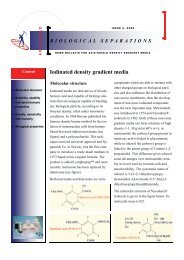
ISSUE 8, 2009<br />
BIOLOGICAL SEPARATIONS<br />
NEWS BULLETIN FOR AXIS-SHIELD DENSITY GRADIENT MEDIA<br />
Applications<br />
• Alveolar <strong>cells</strong><br />
• Neuronal <strong>cells</strong><br />
• Dendritic <strong>cells</strong><br />
• Islets <strong>of</strong> Langerhans<br />
• Stellate <strong>cells</strong><br />
• Gastric mucosa <strong>cells</strong><br />
• Sperm <strong>cells</strong><br />
• Removal <strong>of</strong> nonviable<br />
<strong>cells</strong><br />
• Islets <strong>of</strong> Langerhans<br />
• Plant protoplasts<br />
• Progenitor <strong>cells</strong><br />
• Non-mammalian <strong>cells</strong><br />
• Fractionation <strong>of</strong> a<br />
mixed population <strong>of</strong><br />
<strong>cells</strong><br />
• Analysis <strong>of</strong> apoptosis<br />
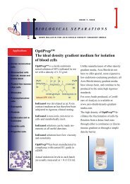
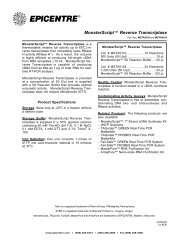
OptiPrep<br />
<strong>The</strong> <strong>ideal</strong> <strong>density</strong> <strong>gradient</strong> <strong>medium</strong> <strong>for</strong><br />
<strong>isolation</strong> <strong>of</strong> <strong>cells</strong><br />
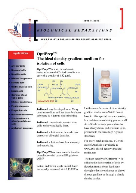
OptiPrep is a sterile endotoxin<br />
tested solution <strong>of</strong> 60% iodixanol in water<br />
with a <strong>density</strong> <strong>of</strong> 1.32 g/ml.<br />
Iodixanol was developed as an X-ray<br />
contrast <strong>medium</strong> and has there<strong>for</strong>e been<br />
subjected to rigorous clinical testing.<br />
Iodixanol is non-ionic, non-toxic to<br />
<strong>cells</strong> and metabolically inert.<br />
Iodixanol solutions can be made isoosmotic<br />
at all useful densities.<br />
Iodixanol solutions have low viscosity<br />
and osmolarity.<br />
OptiPrep has been manufactured in<br />
compliance with current EU guide to<br />
cGMP<br />
Actual endotoxin levels in each batch<br />
are usually measured at < 0.13 EU/ml.<br />
Unlike manufacturers <strong>of</strong> other <strong>density</strong><br />
<strong>gradient</strong> media, Axis-Shield do not<br />
have to <strong>of</strong>fer special, more expensive,<br />
low endotoxin-containing products; all<br />
Axis-Shield <strong>density</strong> <strong>gradient</strong> media<br />
have always been, and continue to be,<br />
produced to the same high rigorous<br />
standards.<br />
For every batch produced, a Certificate<br />
<strong>of</strong> Analysis is available at:<br />
www.axis-shield-<strong>density</strong>-<strong>gradient</strong>media.com<br />
<strong>The</strong> high <strong>density</strong> <strong>of</strong> OptiPrep facilitates<br />
the fractionation <strong>of</strong> <strong>cells</strong> by<br />
flotation from a dense load zone<br />
through either a continuous or discontinuous<br />
<strong>gradient</strong> or through a simple<br />
<strong>density</strong> barrier.
Biological Separations<br />
Page 2<br />
Isolation <strong>of</strong> alveolar <strong>cells</strong> from lung tissue<br />
Alveolar (pneumocyte) type II <strong>cells</strong> are widely studied<br />
because they synthesize and secrete the phospholipidrich<br />
lung surfactant, which lines the air-alveolar interface<br />
and prevents alveolar collapse by lowering surface<br />
tension at low lung volumes. Isolation <strong>of</strong> these <strong>cells</strong><br />
from both adult and foetal lung is an important prerequisite<br />
<strong>for</strong> their culture and study. Viscardi et al, who successfully<br />
developed a Nycodenz® <strong>gradient</strong> technique,<br />
emphasised the importance <strong>of</strong> the non-toxic, noninvasive<br />
properties <strong>of</strong> this <strong>gradient</strong> <strong>medium</strong> and pointed<br />
out that although Percoll® <strong>gradient</strong>s had been used previously<br />
<strong>for</strong> purifying these <strong>cells</strong>, the potentially toxic<br />
nature <strong>of</strong> a polyvinyl-pyrollidone-coated silica colloid<br />
was <strong>of</strong> considerable concern in studies <strong>of</strong> their function.<br />
<strong>The</strong> technique <strong>for</strong> <strong>isolation</strong> <strong>of</strong> pneumocytes is a multistep<br />
one; after tracheostomy <strong>of</strong> the anaesthetized animal,<br />
the alveolar vasculature is perfused to remove blood<br />
<strong>cells</strong> and the lungs lavaged to remove alveolar macrophages.<br />
Lung tissue <strong>cells</strong> are then dispersed by enzymic<br />
disaggregation and mincing. Following filtration, the<br />
mixed cell suspension is loaded on to a continuous Opti-<br />
Prep <strong>gradient</strong> (1.03-1.15 g/ml) and centrifuged at<br />
1500g <strong>for</strong> 20 min at 15°C. <strong>The</strong> pneumocytes band at<br />
approx. 1.056 g/ml just below the sample/<strong>gradient</strong> interface,<br />
while the broad band at approx. 1.086 g/ml contains<br />
fibroblasts, endothelial <strong>cells</strong> and macrophages and<br />
any erythrocytes band close to the bottom <strong>of</strong> the <strong>gradient</strong>.<br />
For a detailed protocol and references see C44 on<br />
the OptiPrep Application CD or online at:<br />
www.axis-shield-<strong>density</strong>-<strong>gradient</strong>-media.com<br />
Isolation <strong>of</strong> rat and human hippocampal neuron fractions<br />
As with any tissue, the first step in fractionating the <strong>cells</strong><br />
from neural tissue is to release the <strong>cells</strong> by enzymic disaggregation<br />
and (in the case <strong>of</strong> the hippocampus) triturition.<br />
Both OptiPrep and Nycodenz® <strong>gradient</strong>s have<br />
been used, primarily to isolate a motoneuron-rich fraction<br />
from both spinal cord and from hippocampus, using<br />
either a simple <strong>density</strong> barrier or a discontinuous <strong>gradient</strong>.<br />
<strong>The</strong> discontinuous <strong>gradient</strong>s may additionally provide<br />
a partial purification <strong>of</strong> some other neural <strong>cells</strong>.<br />
Generally the <strong>gradient</strong>s <strong>for</strong> rat hippocampal tissue are<br />
three or four-step discontinuous ones and not simple<br />
<strong>density</strong> barriers. In the method devised by Brewer et al<br />
the cell suspension is layered over a discontinuous <strong>gradient</strong><br />
consisting <strong>of</strong> 1.029, 1.036, 1.043 and 1.057 g/ml<br />
Nycodenz® in Hibernate A/B27 <strong>medium</strong>.<br />
Brewer et al was one <strong>of</strong> the first groups to shift from<br />
Nycodenz® to OptiPrep <strong>for</strong> hippocampal neuron puri-<br />
fication using a four-layer <strong>gradient</strong> covering a similar<br />
<strong>density</strong> range. Marks et al and Kretz et al reported the<br />
use <strong>of</strong> an almost identical <strong>gradient</strong>. Neurons from cortical<br />
tissue, hypothalamus and brain stem have been isolated<br />
in similar <strong>gradient</strong>s. In all cases the motor neurons<br />
band towards the bottom <strong>of</strong> the <strong>gradient</strong> (fraction D).<br />
For a detailed protocol and references see C29 on the<br />
OptiPrep Applications CD or online at: www.axisshield-<strong>density</strong>-<strong>gradient</strong>-media.com
Issue 8, 2009<br />
Page 3<br />
Isolation <strong>of</strong> dendritic <strong>cells</strong> from blood and tissue<br />
More recently OptiPrep has been used <strong>for</strong> the <strong>isolation</strong><br />
<strong>of</strong> DCs from a variety <strong>of</strong> tissue types and blood. As<br />
with many low <strong>density</strong> <strong>cells</strong>, flotation through a “low<br />
<strong>density</strong> barrier” has been found to give improved results.<br />
Once the tissue has been disaggregated enzymically,<br />
the <strong>cells</strong> are suspended in a relatively dense <strong>medium</strong><br />
and layered beneath the low <strong>density</strong> <strong>medium</strong> as<br />
described originally by Ruedl et al. <strong>The</strong> low <strong>density</strong><br />
dendritic <strong>cells</strong> float to the top <strong>of</strong> the low <strong>density</strong> solution<br />
to band at the interface between this solution and a<br />
small volume <strong>of</strong> culture <strong>medium</strong> (or balanced salt solution),<br />
which is always layered on top (see figure). This<br />
methodology is widely used and has been applied to the<br />
following sources: blood, bone marrow, inflammatory<br />
<strong>cells</strong>, Langerhans <strong>cells</strong>, liver, lung, lymph, lymph<br />
nodes, Peyer’s patch tissue, spleen and thymus. In<br />
early publications the <strong>density</strong> <strong>of</strong> the barrier was 1.068<br />
Isolation <strong>of</strong> stellate <strong>cells</strong> from liver and pancreas<br />
g/ml but more recently this has been reduced to 1.065 g/<br />
ml. <strong>The</strong>re are also a few examples <strong>of</strong> sedimentation on<br />
to a <strong>density</strong> barrier.<br />
For a detailed protocol and references see C20 on the<br />
OptiPrep Applications CD or online at www.axisshield-<strong>density</strong>-<strong>gradient</strong>-media.com<br />
Stellate <strong>cells</strong> are present in both the liver and pancreas<br />
and represent a major and clinically important cell<br />
population in both tissues. In liver the <strong>cells</strong> are important<br />
in the development <strong>of</strong> hepatic fibrosis and in the<br />
pancreas they mediate the fibrosis associated with<br />
chronic pancreatitis.<br />
In the case <strong>of</strong> liver, the purification <strong>of</strong> other cell types is<br />
also <strong>of</strong> major consideration. Stellate <strong>cells</strong> are just one<br />
type <strong>of</strong> hepatic non-parenchymal <strong>cells</strong> (the others include<br />
sinusoidal <strong>cells</strong>, Kupffer <strong>cells</strong>, endothelial <strong>cells</strong><br />
etc) and the separation is carried out in at least two<br />
stages. <strong>The</strong> first stage is the removal <strong>of</strong> the parenchymal<br />
<strong>cells</strong>, followed by the fractionation <strong>of</strong> the nonparenchymal<br />
<strong>cells</strong>.<br />
Stellate <strong>cells</strong> are the least dense <strong>of</strong> the different cell<br />
types in both tissues and they can be purified either by<br />
sedimentation on to, or flotation through, a suitable<br />
<strong>density</strong> barrier. Both strategies have been used effectively<br />
but flotation is <strong>of</strong>ten regarded as superior to sedimentation<br />
as stellate <strong>cells</strong> tend to adhere to other <strong>cells</strong><br />
when sedimenting across a sample/<strong>gradient</strong> interface.<br />
<strong>The</strong> method has been per<strong>for</strong>med with both Nycodenz®<br />
and OptiPrep.<br />
Barriers prepared from OptiPrep have densities ranging<br />
from 1.053 to 1.067 g/ml and it is generally accepted<br />
that the lower the <strong>density</strong>, the purer the stellate cell<br />
preparation. As with Nycodenz® <strong>gradient</strong>s the crude<br />
cell suspension may be layered on top <strong>of</strong> the <strong>density</strong><br />
barrier or adjusted to a higher <strong>density</strong> and layered beneath<br />
the barrier.<br />
Iodixanol barriers have been used <strong>for</strong> the preparation <strong>of</strong><br />
stellate <strong>cells</strong> from the following sources: human liver,<br />
human pancreas, mouse liver, rat liver and rat pancreas.<br />
For a detailed protocol and references see C33 on the<br />
OptiPrep Applications CD or online at www.axisshield-<strong>density</strong>-<strong>gradient</strong>-media.com
Biological Separations<br />
Page 4<br />
Isolation <strong>of</strong> gastric mucosal <strong>cells</strong> using continuous or discontinuous <strong>gradient</strong>s<br />
Probably the most intensively studied cell present in the<br />
gastric mucosa is the acid-secreting parietal cell as this<br />
provides a very useful model <strong>for</strong> studying both the<br />
regulation <strong>of</strong> ion-transport and intracellular signalling<br />
pathways. ECL <strong>cells</strong> are the main endocrine/paracrine<br />
cell type and they play an important role in controlling<br />
acid-secretion. <strong>The</strong> other major cell type is the chief<br />
cell, which secretes pepsin.<br />
Parietal <strong>cells</strong><br />
Since the late eighties the most widely used strategy is<br />
to purify parietal <strong>cells</strong> from Pronase/ collagenasedisaggregated<br />
gastric mucosa (from both rats and rabbits)<br />
on either continuous or discontinuous Nycodenz®<br />
<strong>gradient</strong>s, followed by centrifugal elutriation. More recently<br />
Nycodenz® has been replaced by OptiPrep as<br />
the latter gave improved purities (80-90%) and <strong>for</strong><br />
some studies this may be a sufficient degree <strong>of</strong> purification<br />
without the need to carry out a subsequent elutriation<br />
step.<br />
ECL <strong>cells</strong><br />
Rat ECL <strong>cells</strong> are routinely purified by centrifugal elutriation,<br />
but are contaminated by largely denser particles<br />
which can be removed in a discontinuous Nycodenz®<br />
<strong>gradient</strong> <strong>of</strong> 1.046 and 1.058 g/ml. <strong>The</strong> corresponding<br />
iodixanol <strong>gradient</strong>s are slightly denser, ρ = 1.061 and<br />
1.084 g/ml, the <strong>cells</strong> banding above the lower <strong>density</strong><br />
layer. A simple iodixanol <strong>density</strong> barrier ρ = 1.061 g/ml<br />
can be used as an alternative (see figure below).<br />
In a continuous iodixanol <strong>gradient</strong> covering the range<br />
1.049-1.139 g/ml; the parietal <strong>cells</strong> band at approx<br />
1.052 g/ml (A), while the chief <strong>cells</strong> have a much<br />
higher <strong>density</strong> (D) (see top figure).<br />
For a detailed protocol and references see C28 on<br />
the OptiPrep Applications CD or online at:<br />
www.axis-shield-<strong>density</strong>-<strong>gradient</strong>-media.com<br />
For a detailed protocol see C28 on the OptiPrep<br />
Applications CD or online at: www.axis-shield<strong>density</strong>-<strong>gradient</strong>-media.com<br />
Isolation <strong>of</strong> spermatozoa from experimental animals<br />
Ejaculates contain variable proportions <strong>of</strong> viable spermatozoa<br />
<strong>of</strong> normal morphology, sometimes a very low<br />
percentage <strong>of</strong> the total cell population. OptiPrep <strong>gradient</strong>s<br />
can be used <strong>for</strong> the recovery <strong>of</strong> highly viable<br />
fractions from bovine ejaculates. Previously, a method<br />
<strong>for</strong> the separation <strong>of</strong> the viable fraction from bovine<br />
ejaculates using Nycodenz® <strong>gradient</strong>s was developed.<br />
In field tests, using a split ejaculate, one part separated<br />
on a Nycodenz® <strong>gradient</strong> and the other processed nor-<br />
mally; the Nycodenz® purified <strong>cells</strong> fertilized as efficiently<br />
as those treated by routine methods.<br />
OptiPrep has certain significant advantages over its<br />
predecessor, Nycodenz®, <strong>for</strong> the separation <strong>of</strong> sperm<br />
<strong>cells</strong> in particular, because <strong>of</strong> their relatively high <strong>density</strong>.<br />
Adjustment <strong>of</strong> the raw ejaculate to a sufficiently<br />
high <strong>density</strong> (1.17 g/ml) and maintenance <strong>of</strong> isoosmotic<br />
conditions can only be achieved with OptiPrep.
Issue 8, 2009<br />
Page 5<br />
OptiPrep thus <strong>of</strong>fers convenience in sample handling.<br />
Bovine semen in a sorbitol-containing diluent (<strong>density</strong><br />
1.018 g/ml) is mixed with an equal volume <strong>of</strong> OptiPrep<br />
so that the <strong>density</strong> <strong>of</strong> the suspension is approx 1.17 g/ml.<br />
This is then overlayered with two <strong>density</strong> barriers <strong>of</strong><br />
<strong>density</strong> 1.154 and 1.119 g/ml (see figure). After centrifugation<br />
at 1500g <strong>for</strong> 20 min at 20°C the de<strong>for</strong>med<br />
<strong>cells</strong> and cytoplasmic droplets will have floated to the<br />
meniscus, non-viable <strong>cells</strong> will remain in the load zone<br />
and the viable motile sperm <strong>cells</strong> <strong>of</strong> normal morphology<br />
will band at the 1.119/1.154 g/ml interface.<br />
<strong>The</strong> <strong>medium</strong> used <strong>for</strong> washing and storing sperm <strong>cells</strong><br />
varies from laboratory to laboratory and if, <strong>for</strong> example,<br />
it is based on a simple culture <strong>medium</strong> containing 10%<br />
serum rather than a sorbitol-containing <strong>medium</strong>, then it<br />
will have a <strong>density</strong> <strong>of</strong> approx 1.009 g/ml. <strong>The</strong> amount <strong>of</strong><br />
OptiPrep required to raise the <strong>density</strong> <strong>of</strong> the semen to<br />
1.17 g/ml and <strong>for</strong> ratios <strong>of</strong> OptiPrep and <strong>medium</strong><br />
used to make the low <strong>density</strong> layers will vary.<br />
<strong>The</strong> quality <strong>of</strong> the semen has been assessed by using<br />
membrane integrity as an indicator <strong>of</strong> general cell function<br />
and viability (<strong>The</strong> Osmotic Resistance test). <strong>The</strong><br />
motile band from the ρ = 1.12/1.14 g/ml interface<br />
shows over 95% viability by this test, while the pelleted<br />
material and particulate material remaining in the loading<br />
layer are found to be 99% non-viable <strong>cells</strong> by this<br />
method.<br />
OptiPrep has also been used as a dense cushion to<br />
concentrate spermatozoa and remove contaminating<br />
debris with equine, boar, gazelle and mouse semen.<br />
A huge advantage <strong>of</strong> using OptiPrep <strong>for</strong> human semen<br />
fractionation is that iodixanol has been clinically<br />
tested and screened as an X-ray imaging agent and it is<br />
the only <strong>gradient</strong> <strong>medium</strong> that is routinely tested <strong>for</strong><br />
endotoxin, levels <strong>of</strong> which are < 1 EU/ml (well within<br />
the limits <strong>for</strong> human applications).<br />
For a detailed protocol see C16, C17 and C46 on the<br />
OptiPrep Applications CD or online at: www.axisshield-<strong>density</strong>-<strong>gradient</strong>-media.com<br />
Removal <strong>of</strong> non-viable <strong>cells</strong> from tissue and cultured <strong>cells</strong><br />
Cell suspensions prepared from a lung or peritoneal lavage,<br />
or from enzymically dissociated tissue, will comprise<br />
live and dead <strong>cells</strong> together with cell debris. <strong>The</strong><br />
<strong>cells</strong> are <strong>of</strong>ten contained in a comparatively large volume<br />
at low concentration and need to be concentrated<br />
prior to further processing in smaller volumes. Pelleting<br />
and resuspending the <strong>cells</strong> causes further damage and<br />
attempts to separate the various cell-types in the suspension<br />
by <strong>density</strong> <strong>gradient</strong> centrifugation are usually unsuccessful<br />
because the DNA, which is released by broken<br />
<strong>cells</strong>, causes aggregation <strong>of</strong> <strong>cells</strong> throughout the<br />
<strong>gradient</strong>.<br />
<strong>The</strong>re is also a need to remove large numbers <strong>of</strong> non-<br />
viable <strong>cells</strong> from cultured cell lines after some operational<br />
failure has caused a major loss <strong>of</strong> cell viability.<br />
Simple <strong>density</strong> barrier techniques can provide a means<br />
<strong>of</strong> eliminating non-viable <strong>cells</strong> and removing contamination<br />
by released intracellular contents, thereby facilitating<br />
the success <strong>of</strong> subsequent separation techniques<br />
or cell culture.<br />
Non-viable <strong>cells</strong> tend to lose their osmotic integrity,<br />
thus their aqueous cytosolic compartment becomes accessible<br />
to solute molecules in the surrounding <strong>medium</strong>.<br />
When this occurs the <strong>density</strong> <strong>of</strong> the cell increases,<br />
usually to a value above 1.15 g/ml; such non-viable <strong>cells</strong>
Biological Separations<br />
Page 6<br />
can there<strong>for</strong>e be easily separated from the viable ones.<br />
A simple approach to separate viable and non-viable<br />
<strong>cells</strong> is to centrifuge the cell suspension over a barrier<br />
solution <strong>of</strong> <strong>density</strong> approx 1.15 g/ml (see figure). <strong>The</strong><br />
barrier can be prepared from OptiPrep or Nycodenz®.<br />
OptiPrep will provide a slightly higher <strong>density</strong> and<br />
there is some evidence that the smaller molecular<br />
weight Nycodenz® may be more accessible to the cytosolic<br />
compartment <strong>of</strong> nonviable <strong>cells</strong>. <strong>The</strong> viable <strong>cells</strong><br />
will <strong>for</strong>m a band at the interface, while the non-viable<br />
<strong>cells</strong> will <strong>for</strong>m a pellet.<br />
<strong>cells</strong> band at the top <strong>of</strong> the upper layers while non-viable<br />
<strong>cells</strong> and contaminants remain in the load zone (see figure<br />
below). This has been used very successfully <strong>for</strong><br />
retrieving a "damaged" cultured cell line.<br />
Both methods are extremely gentle as the viable <strong>cells</strong><br />
are concentrated without pelleting.<br />
Iodixanol barriers have been used <strong>for</strong> the removal <strong>of</strong><br />
nonviable <strong>cells</strong> from: amphibian <strong>cells</strong>, cultured <strong>cells</strong>,<br />
hepatocytes, muscle <strong>cells</strong> and progenitor <strong>cells</strong>.<br />
An alternative approach, which may be more efficient<br />
with OptiPrep is to adjust the <strong>density</strong> <strong>of</strong> the cell suspension<br />
to 1.15-1.16 g/ml and to overlay this with a<br />
solution <strong>of</strong> 1.12 g/ml. After centrifugation the viable<br />
For detailed protocols and references see C13 on the<br />
OptiPrep Applications CD or online at: www.axisshield-<strong>density</strong>-<strong>gradient</strong>-media.com<br />
Isolation <strong>of</strong> Islets <strong>of</strong> Langerhans from pancreatic tissue<br />
<strong>The</strong> development <strong>of</strong> a routine method <strong>for</strong> the <strong>isolation</strong><br />
<strong>of</strong> Islets <strong>of</strong> Langerhans from pancreas will have important<br />
clinical and research applications. Islet transplants<br />
are used in the treatment <strong>of</strong> diabetes and a simple procedure<br />
<strong>for</strong> the preparation <strong>of</strong> functional islets in bulk<br />
would be a significant improvement on present methods.<br />
Islets are first prepared by disaggregation <strong>of</strong> the pancreas<br />
by enzymic digestion in order to release the islets<br />
from the acinar <strong>cells</strong>. <strong>The</strong> optimal methods <strong>of</strong> disaggregation<br />
<strong>of</strong> tissue and choice <strong>of</strong> <strong>medium</strong> will vary with<br />
the source <strong>of</strong> tissue and from laboratory to laboratory.<br />
In OptiPrep <strong>gradient</strong>s it has been demonstrated that<br />
islets have a lower buoyant <strong>density</strong> than acinar <strong>cells</strong>.<br />
This allows them to be separated by flotation through a<br />
low <strong>density</strong> barrier. Dr van der Burg, University Hospital,<br />
Leiden, Netherlands has developed a method using<br />
OptiPrep which produces islets from porcine pancreas<br />
with a high viability. Note that the method uses the University<br />
<strong>of</strong> Wisconsin Solution (UWS) as the suspending<br />
<strong>medium</strong>, other customized media which are used to retain<br />
high islet viability may have different densities and<br />
the method may consequently require adaptation. Tissues<br />
from other species may also require some protocol<br />
optimization.<br />
Cells from the pancreatic digest are sedimented and suspended<br />
in a solution <strong>of</strong> <strong>density</strong> 1.10 g/ml and then overlayered<br />
with a low-<strong>density</strong> barrier (ρ = 1.09 g/ml) and a<br />
layer <strong>of</strong> UWS (see figure). After centrifugation the islets<br />
band at the interface below the UWS. <strong>The</strong> load zone not<br />
only retains the acinar <strong>cells</strong> but also any residual
Issue 8, 2009<br />
Page 7<br />
digestive enzymes used <strong>for</strong> the disaggregation <strong>of</strong> the<br />
tissue. <strong>The</strong> Working Solution is simply produced from<br />
equal volumes <strong>of</strong> OptiPrep and double-strength UWS<br />
- the slightly raised osmolality <strong>of</strong> the Working Solution<br />
is beneficial to the separation.<br />
<strong>The</strong> use <strong>of</strong> OptiPrep has been reported to contribute<br />
significantly to improved recovery <strong>of</strong> islets from pancreas<br />
digests and the retention <strong>of</strong> islet integrity and viability.<br />
Diabetic single-donor pig allograft recipients<br />
became normoglycemic within 24 h following transplantation;<br />
similar results were obtained with transplantation<br />
into primates. More recently Shibata et al have<br />
proposed reducing the RCF used to band the islets in<br />
the iodixanol <strong>gradient</strong> to 100g. At the lower RCF, recovery,<br />
purity, resistance to fragmentation and insulin<br />
response to glucose, were all improved compared to<br />
that at the higher RCF.<br />
Although the method as described originally and illustrated<br />
in the figure was designed <strong>for</strong> use with tubes in a<br />
low-speed centrifuge, it has been scaled up to use with<br />
the Cobe 2991 centrifuge.<br />
This iodixanol separation technology has been used in<br />
the following studies on pancreatic islets: Human: gene<br />
delivery, in vitro studies, transplantation, yield, viability<br />
and function; mouse; porcine: immune reactions, insulin<br />
release, transplantation, viruses, yield, viability and<br />
function; Primate- non-human; rat. <strong>The</strong>re are also several<br />
review articles.<br />
For a detailed protocol and references see C15 on<br />
the OptiPrep Applications CD or online at:<br />
www.axis-shield-<strong>density</strong>-<strong>gradient</strong>-media.com<br />
Isolation <strong>of</strong> plant protoplasts from wheat or barley leaves<br />
Once the cellulose walls are removed from plant <strong>cells</strong>,<br />
they become sensitive to osmotic changes in the environment,<br />
shrinking and swelling in response to the environment,<br />
with consequent changes to their buoyant<br />
densities. In the preparation <strong>of</strong> plant protoplasts the osmolality<br />
<strong>of</strong> the <strong>medium</strong> used to digest the plant wall is<br />
important: use <strong>of</strong> a digest mixture whose osmolality is<br />
1.8x that <strong>of</strong> the living plant tissue is a widely used<br />
guideline. A raised osmolality promotes shrinkage <strong>of</strong><br />
the protoplast away from the cell wall and facilitates its<br />
<strong>isolation</strong>.<br />
Using wheat or barley leaves as starting material, excellent<br />
populations <strong>of</strong> intact protoplasts can be prepared<br />
using a simplified modification <strong>of</strong> the Bethke et al<br />
method which omits the densest layer and also modulates<br />
the <strong>density</strong> <strong>of</strong> the lighter layers. OptiPrep is<br />
added directly to the plant digest to make a suspension<br />
with a final <strong>density</strong> <strong>of</strong> 1.08 g/ml. <strong>The</strong> separation procedure<br />
is outlined in the figure.<br />
Centrifugation is actually not necessary to band the pro-<br />
toplasts: their size means that if the tube is allowed to<br />
stand on the bench (1g), they will float to the upper interface<br />
in about 20-30 min.<br />
<strong>The</strong> top band contains over 95% intact protoplasts. <strong>The</strong><br />
number <strong>of</strong> intact protoplasts remaining in the ρ = 1.08 g/<br />
ml layer is insignificant.<br />
For detailed a protocol and references see C18 on the<br />
OptiPrep Applications CD or online at: www.axisshield-<strong>density</strong>-<strong>gradient</strong>-media.com
Web:<br />
www.axis-shield-<strong>density</strong>-<strong>gradient</strong>media.com<br />
Axis-Shield<br />
PO Box 6863 Rodelokka<br />
N-0504 Oslo<br />
Norway<br />
In this leaflet we have presented some <strong>of</strong> the applications<br />
available <strong>for</strong> the <strong>isolation</strong> <strong>of</strong> <strong>cells</strong> using OptiPrep. More<br />
in<strong>for</strong>mation can be found on: www.axis-shield-<strong>density</strong><strong>gradient</strong>-media.com.<br />
On the front page click on<br />
“Mammalian and non-mammalian <strong>cells</strong>”.<br />
Altogether there are now 55 applications available <strong>for</strong> the<br />
<strong>isolation</strong> <strong>of</strong> mammalian and non-mammalian <strong>cells</strong> using<br />
OptiPrep<br />
Percoll is a trademark <strong>of</strong> GE Healthcare companies<br />
Phone: +47 24 05 60 00<br />
Fax: +47 24 05 60 10<br />
Email: bjh@no.axis-shield.com or<br />
john@jgrescon.fsbusiness.co.uk<br />
Isolation <strong>of</strong> a progenitor enriched fraction from bone marrow<br />
Although the resolving power <strong>of</strong> <strong>density</strong> <strong>gradient</strong>s is<br />
insufficient to allow megakaryocytic progenitor <strong>cells</strong> to<br />
be isolated in a sufficiently pure <strong>for</strong>m <strong>for</strong> further analysis<br />
and culture; they do provide a useful preliminary<br />
purification step, which can usually achieve a twothreefold<br />
enrichment <strong>of</strong> these <strong>cells</strong> from crude bone<br />
marrow samples. This allows a more economical and<br />
effective use <strong>of</strong> expensive immuno-magnetic beads to<br />
remove lineage <strong>cells</strong> with a cocktail <strong>of</strong> lineage specific<br />
monoclonal antibodies.<br />
OptiPrep has been used as a simple <strong>density</strong> barrier <strong>of</strong><br />
<strong>density</strong> 1.080 g/ml or 1.077 g/ml <strong>for</strong> enrichment <strong>of</strong><br />
progenitor <strong>cells</strong> from bone marrow; the method is summarized<br />
in the figure. Interfacial <strong>cells</strong> are harvested <strong>for</strong><br />
subsequent depletion <strong>of</strong> lineage-committed <strong>cells</strong> using<br />
antibody-beads. A discontinuous <strong>gradient</strong> <strong>of</strong> 1.050,<br />
1.080 and 1.090 g/ml is another option.<br />
Iodixanol <strong>gradient</strong>s have also been used to enrich <strong>for</strong><br />
progenitor <strong>cells</strong> from blood and thymus. In the case <strong>of</strong><br />
hippocampal progenitor <strong>cells</strong>, the denser fractions from<br />
a 1.029, 1.036, 1.043 and 1.057 g/ml discontinuous <strong>gradient</strong><br />
were harvested (Porritt, H.E. et al, Tabrizifard, S.<br />
et al).<br />
For a detailed protocol and references see C23 on the<br />
OptiPrep Application CD or online at: www.axisshield-<strong>density</strong>-<strong>gradient</strong>-media.com