Investigation of the Emission Mechanism in Milled SrAl2O4:Eu, Dy ...
Investigation of the Emission Mechanism in Milled SrAl2O4:Eu, Dy ...
Investigation of the Emission Mechanism in Milled SrAl2O4:Eu, Dy ...

You also want an ePaper? Increase the reach of your titles
YUMPU automatically turns print PDFs into web optimized ePapers that Google loves.
RESEARCH ARTICLE<br />
www.acsami.org<br />
<strong>Investigation</strong> <strong>of</strong> <strong>the</strong> <strong>Emission</strong> <strong>Mechanism</strong> <strong>in</strong> <strong>Milled</strong> SrAl 2 O 4 :<strong>Eu</strong>, <strong>Dy</strong><br />
Us<strong>in</strong>g Optical and Synchrotron X-ray Spectroscopy<br />
Sanjeev Kumar Kandpal, † Ben Goundie, ‡ Joshua Wright, ‡ Rachel A. Pollock, ‡ Michael D. Mason, † and<br />
Robert W. Meulenberg* ,‡,§<br />
† Department <strong>of</strong> Chemical and Biological Eng<strong>in</strong>eer<strong>in</strong>g, University <strong>of</strong> Ma<strong>in</strong>e, Orono, Ma<strong>in</strong>e 04469, United States<br />
‡ Department <strong>of</strong> Physics and Astronomy, University <strong>of</strong> Ma<strong>in</strong>e, Orono, Ma<strong>in</strong>e 04469, United States<br />
§ Laboratory for Surface Science and Technology, University <strong>of</strong> Ma<strong>in</strong>e, Orono, Ma<strong>in</strong>e 04469, United States<br />
ABSTRACT: There currently exists much debate as to <strong>the</strong> active state related to <strong>the</strong> “long<br />
afterglow” effect <strong>in</strong> europium doped oxide materials. Redox couples that consist <strong>of</strong> <strong>Eu</strong> +/2+<br />
and <strong>Eu</strong> 2+/3+ are discussed, but no common answer is currently accepted. Here, we present<br />
a comparison <strong>of</strong> <strong>the</strong> optical properties <strong>of</strong> a commercially available SrAl 2 O 4 :<strong>Eu</strong>, <strong>Dy</strong><br />
phosphor, as a function <strong>of</strong> nanoparticle size reduction via dry mechanical mill<strong>in</strong>g. X-ray<br />
and optical spectroscopic data <strong>in</strong>dicate a significant decrease <strong>in</strong> phosphorescence efficiency<br />
and an <strong>in</strong>crease <strong>in</strong> laser stimulated emission efficiency as near surface <strong>Eu</strong> 2+ ions are oxidized<br />
to <strong>Eu</strong> 3+ as a consequence <strong>of</strong> <strong>in</strong>creased exposure dur<strong>in</strong>g <strong>the</strong> mill<strong>in</strong>g process. These results<br />
show evidence only for <strong>Eu</strong> 2+/3+ oxidation states, suggest<strong>in</strong>g <strong>the</strong> mechanism related to long<br />
afterglow effect does not arise from <strong>Eu</strong> + species. We also suggest that size reduction, as a rule, cannot be universally applied to<br />
improve optical properties <strong>of</strong> nanostructures.<br />
KEYWORDS: rare earth dop<strong>in</strong>g, EXAFS, electronic structure, <strong>the</strong>rmolum<strong>in</strong>escence, long afterglow phosphorescence<br />
’ INTRODUCTION<br />
In recent years, rare earth doped particles have found widespread<br />
use as phosphors <strong>in</strong> commercial products rang<strong>in</strong>g from<br />
temperature detection, 1 structural damage sens<strong>in</strong>g, 2 emergency<br />
light<strong>in</strong>g, and even children’s toys. 3 The attractiveness <strong>of</strong> <strong>the</strong>se<br />
materials can generally be attributed to <strong>the</strong>ir bright long-lived<br />
visible phosphorescence, wavelength tunability, ease <strong>of</strong> syn<strong>the</strong>sis,<br />
good chemical stability, and relative low cost. Increas<strong>in</strong>gly, <strong>the</strong>se<br />
materials are be<strong>in</strong>g <strong>in</strong>vestigated for applications requir<strong>in</strong>g reduced<br />
particle dimensions, such as <strong>in</strong> th<strong>in</strong> films for optical or<br />
electronic devices, 4 or medical diagnostics. 5 Recent work suggests<br />
that some doped phosphors exhibit little degradation <strong>in</strong><br />
optical efficiency at reduced dimensions; however, <strong>the</strong> emission<br />
mechanism <strong>in</strong> <strong>the</strong>se systems is still controversial 6 and may be<br />
affected by particle size. 7 Typically, nanoscale phosphors are<br />
prepared by solid state reaction, vacuum deposition, or combustion<br />
methods, 8 which can be prohibitively expensive for most<br />
commercial applications. An alternative more cost-effective<br />
approach <strong>in</strong>volves size reduction via mechanical mill<strong>in</strong>g. While<br />
this approach is not universally applicable to all nanomaterials, it<br />
may have value for nanoparticles where <strong>the</strong> property <strong>of</strong> <strong>in</strong>terest is<br />
determ<strong>in</strong>ed by <strong>the</strong> presence <strong>of</strong> dopant ions with<strong>in</strong> <strong>the</strong> particle<br />
and not <strong>the</strong> f<strong>in</strong>al particle dimensions.<br />
In this manuscript, we present a comparison <strong>of</strong> <strong>the</strong> optical<br />
properties <strong>of</strong> a commercially available SrAl 2 O 4 :<strong>Eu</strong>, <strong>Dy</strong> phosphor<br />
before and after mill<strong>in</strong>g. To improve dispersity, a low cost surfactant<br />
is also considered. Both X-ray and visible spectroscopic data<br />
is presented and used to describe <strong>the</strong> observed changes <strong>in</strong> optical<br />
efficiency. Our work suggests that size reduction leads to degradation<br />
<strong>of</strong> <strong>the</strong> phosphorescence and <strong>the</strong>rmolum<strong>in</strong>escence <strong>of</strong> <strong>the</strong> phosphor<br />
but an <strong>in</strong>crease <strong>in</strong> laser <strong>in</strong>duced emission, provid<strong>in</strong>g fur<strong>the</strong>r<br />
<strong>in</strong>sight <strong>in</strong>to <strong>the</strong> emission mechanism <strong>in</strong> this system.<br />
’ EXPERIMENTAL METHODS<br />
Sample Preparation. A 3 lb capacity benchtop ball mill was<br />
used to mechanically reduce <strong>the</strong> size <strong>of</strong> a commercially available<br />
SrAl 2 O4:<strong>Eu</strong>, <strong>Dy</strong> phosphor (United Nuclear, LumiNova G-300).<br />
The mill was charged with ∼0.31 cm diameter Alum<strong>in</strong>a-silica<br />
beads (50% total capacity) and <strong>the</strong> dry granular phosphor (25%<br />
total capacity). Cont<strong>in</strong>uous mill<strong>in</strong>g was carried for 48 h where<br />
∼100 mg <strong>of</strong> test samples was extracted every 12 h. The samples<br />
were sized by transmission electron microscopy (TEM) and<br />
<strong>Dy</strong>namic Light Scatter<strong>in</strong>g (DLS, Zetasizer Nano) <strong>in</strong> water (data<br />
not shown). The milled samples exhibited a dramatic <strong>in</strong>crease <strong>in</strong><br />
solution stability with only m<strong>in</strong>imal settl<strong>in</strong>g <strong>in</strong> 6 h. This procedure<br />
resulted <strong>in</strong> an apparent reduction <strong>in</strong> diameter from >1000 nm<br />
(volume weighted) to 150 nm after 12 h. For longer mill<strong>in</strong>g<br />
times, DLS data <strong>in</strong>dicated a small <strong>in</strong>crease <strong>in</strong> size, which, by<br />
analysis <strong>of</strong> TEM data, was attributed to aggregation <strong>of</strong> smaller<br />
particles. In a second trial, polyethylene glycol (MW 20 000) was<br />
added (5 wt % <strong>of</strong> phosphor) to improve particle dispersity dur<strong>in</strong>g<br />
both <strong>the</strong> mill<strong>in</strong>g and siz<strong>in</strong>g procedures. The result<strong>in</strong>g particles<br />
showed only a small additional decrease <strong>in</strong> size after 12 h,<br />
Received: June 1, 2011<br />
Accepted: August 4, 2011<br />
Published: August 19, 2011<br />
r 2011 American Chemical Society 3482 dx.doi.org/10.1021/am200710j | ACS Appl. Mater. Interfaces 2011, 3, 3482–3486
ACS Applied Materials & Interfaces<br />
RESEARCH ARTICLE<br />
Figure 1. Powder X-ray diffraction measurements for <strong>the</strong> (a) orig<strong>in</strong>al,<br />
(b) dry milled, and (c) PEG milled samples alongside <strong>the</strong> calculated<br />
peak <strong>in</strong>tensities based on PDF card 04-010-5403 (bottom trace). The<br />
data <strong>in</strong> both panels are equivalent, with (B) plott<strong>in</strong>g an expanded region<br />
to make peak shifts visible. Spectra are <strong>of</strong>fset for clarity.<br />
reach<strong>in</strong>g a limit<strong>in</strong>g diameter <strong>of</strong> around 150 nm. Solution stability<br />
was fur<strong>the</strong>r <strong>in</strong>creased <strong>in</strong> <strong>the</strong>se samples.<br />
Characterization. X-ray Diffraction. Nanoparticle structure<br />
was studied us<strong>in</strong>g X-ray diffraction (XRD) by θ 2θ scans with<br />
Cu KR radiation us<strong>in</strong>g l<strong>in</strong>e focus on a PANalytical X 0 PertPro<br />
Diffractometer (Figure 1). In all <strong>the</strong> samples studied, <strong>the</strong> XRD<br />
peaks were well <strong>in</strong>dexed to s<strong>in</strong>gle phase monocl<strong>in</strong>ic SrAl 2 O 4<br />
(PDF# 04-010-5403).<br />
Synchrotron Methods. S<strong>of</strong>t X-ray absorption near edge spectroscopy<br />
(SXANES) was performed at beaml<strong>in</strong>e 8-2 at <strong>the</strong><br />
Stanford Synchrotron Radiation Lightsource (SSRL), Stanford<br />
L<strong>in</strong>ear Accelerator Center. Powders were affixed to carbon tape<br />
and <strong>in</strong>serted <strong>in</strong>to an ultrahigh vacuum chamber. SXANES experimental<br />
measurements were taken at base pressures <strong>of</strong> less than<br />
5 10 9 Torr. SXANES experiments were conducted us<strong>in</strong>g <strong>the</strong><br />
total electron yield (TEY) detection method where <strong>the</strong> total photocurrent<br />
is measured as <strong>the</strong> photon energy is scanned through <strong>the</strong><br />
absorption edges. The experimental energy resolution was ∼0.10<br />
0.20 eV for <strong>the</strong> various edges (O, K; <strong>Eu</strong>, M 4,5 ; <strong>Dy</strong>, M 4,5 ) studied<br />
<strong>in</strong> this letter. For <strong>the</strong> hard X-ray measurements, both X-ray<br />
absorption near edge spectroscopy and extended X-ray absorption<br />
f<strong>in</strong>e structure were analyzed. X-ray absorption f<strong>in</strong>e structure<br />
(XAFS) spectroscopy was performed at beaml<strong>in</strong>e 10-2 at <strong>the</strong><br />
SSRL us<strong>in</strong>g Si(220) crystals detuned about 30%; a slit height <strong>of</strong><br />
2 mm was used. Powder samples were mounted on 5 10 mm<br />
pieces <strong>of</strong> Kapton tape. <strong>Eu</strong> L 3 -edge XAFS spectra were collected<br />
at room temperature <strong>in</strong> partial fluorescence mode us<strong>in</strong>g a 13-<br />
element Ge detector to collect <strong>Eu</strong> Lβ fluorescence photons.<br />
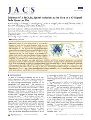
Figure 2. (A) O K-edge, (B) <strong>Dy</strong> M 5 -edge, and (C)/(D) <strong>Eu</strong> M 5 -edge<br />
XAFS <strong>of</strong> <strong>the</strong> as purchased (black) and 48 h dry milled (gray) samples.<br />
The bottom two traces <strong>in</strong> (D) are multiplet simulations <strong>of</strong> <strong>the</strong> <strong>Eu</strong> 2+ and<br />
<strong>Eu</strong> 3+ , respectively. Some spectra are <strong>of</strong>fset for clarity.<br />
I 0 was monitored us<strong>in</strong>g a nitrogen filled ionization chamber. An<br />
<strong>Eu</strong> foil was placed after <strong>the</strong> sample for energy calibration.<br />
Optical Methods. For optical characterization, an equivalent<br />
mass (0.15 g) <strong>of</strong> <strong>the</strong> as purchased and 48 h milled samples was<br />
placed <strong>in</strong> a welled slide and capped with a standard coverslip.<br />
Samples were stored overnight, <strong>in</strong> <strong>the</strong> dark, 9 <strong>in</strong> a 80 °C freezer<br />
prior to characterization to ensure complete <strong>the</strong>rmal deactivation<br />
consistent with previously reported methods. 10 Lum<strong>in</strong>escence<br />
was generated under laser (473, 632 nm) or <strong>the</strong>rmal excitation. A<br />
home-built confocal microscope equipped with a 0.85 NA air<br />
objective was used for optical excitation and signal collection.<br />
The result<strong>in</strong>g emission was directed to a 1/4 m spectrometer<br />
(Acton Instruments) and liquid nitrogen cooled CCD camera<br />
(Pr<strong>in</strong>ceton Instruments, SPEC-10). Excitation power (6 mW)<br />
was held fixed for all samples at each wavelength, and spectra<br />
were similarly collected over constant <strong>in</strong>tegration periods. Phosphorescence<br />
spectra (10 s <strong>in</strong>tegration) were collected every 10 s<br />
for 1 h follow<strong>in</strong>g a 10 s exposure to 473 nm radiation (6 mW).<br />
For photo<strong>the</strong>rmal emission, a s<strong>of</strong>tware controlled hot plate was<br />
used to provide a well determ<strong>in</strong>ed temperature ramp (∼1 °C<br />
per second). Photo<strong>the</strong>rmal emission was collected via an optical<br />
fiber (Thorlabs, AFS105/125Y) placed <strong>in</strong> direct contact with <strong>the</strong><br />
sample coverslip and directed to an avalanche photodiode<br />
module (EGG, SPCM). Total temperature dependent emission<br />
was determ<strong>in</strong>ed by <strong>in</strong>tegrat<strong>in</strong>g each time sampled spectrum.<br />
’ RESULTS AND DISCUSSION<br />
As observed <strong>in</strong> Figure 1A, <strong>the</strong> structure <strong>of</strong> <strong>the</strong> doped SrAl 2 O 4<br />
is not strongly affected with mill<strong>in</strong>g; <strong>in</strong>deed, <strong>the</strong> monocl<strong>in</strong>ic<br />
phase that occurs is <strong>the</strong> undoped material, present <strong>in</strong> all stages <strong>of</strong><br />
3483 dx.doi.org/10.1021/am200710j |ACS Appl. Mater. Interfaces 2011, 3, 3482–3486
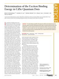
ACS Applied Materials & Interfaces<br />
Figure 3. <strong>Eu</strong> L 3 -edge (A) hard XANES, (B) k 3 -weighted χ(k) EXAFS,<br />
and (C) Fourier transforms (not phase shift corrected) <strong>of</strong> <strong>the</strong> EXAFS for<br />
<strong>the</strong> (i) as prepared, (ii) dry milled, and (iii) PEG milled samples. The<br />
black dashed l<strong>in</strong>es <strong>in</strong> (A) are described <strong>in</strong> <strong>the</strong> text. The bottom trace <strong>in</strong><br />
(C) is <strong>the</strong> <strong>the</strong>oretical FT EXAFS <strong>of</strong> <strong>Eu</strong>:SrAl 2 O 4 . All spectra are <strong>of</strong>fset for<br />
clarity.<br />
<strong>the</strong> nanosized doped material. The large scale <strong>of</strong> Figure 1A,<br />
however, obscures any small changes that may occur <strong>in</strong> peak<br />
positions. Plotted <strong>in</strong> Figure 1B is an expanded region <strong>of</strong> <strong>the</strong> data<br />
<strong>in</strong> Figure 1A; notably, this expanded region encompasses peak<br />
<strong>in</strong>dices <strong>of</strong> Æ004æ, Æ222æ, and Æ 203æ. The relationship between<br />
lattice plane spac<strong>in</strong>g and lattice parameters for a monocl<strong>in</strong>ic<br />
lattice is<br />
1<br />
d 2 ¼ 1<br />
s<strong>in</strong> 2 β<br />
h 2<br />
a 2 þ k2 s<strong>in</strong> 2 β<br />
b 2 þ l2<br />
c 2<br />
!<br />
2hlcos β<br />
ac<br />
For <strong>the</strong> Æ004æ peak, only <strong>the</strong> c parameter contributes to <strong>the</strong><br />
lattice spac<strong>in</strong>g while <strong>the</strong> o<strong>the</strong>r peaks have contributions from <strong>the</strong><br />
o<strong>the</strong>r lattice parameters. Most notably, <strong>the</strong> Æ 203æ peak exhibits<br />
a decrease <strong>in</strong> d-spac<strong>in</strong>g with mill<strong>in</strong>g; this implies that <strong>the</strong> a lattice<br />
parameter changes as <strong>the</strong> particle size is reduced dur<strong>in</strong>g <strong>the</strong><br />
mill<strong>in</strong>g process. This suggests a reduction <strong>in</strong> bond length occurs<br />
<strong>in</strong> <strong>the</strong>se materials (see EXAFS results, Figure 3).<br />
Oxygen K-edge, <strong>Dy</strong> M 5 -edge, and <strong>Eu</strong> M 5 -edge SXANES are<br />
presented <strong>in</strong> Figure 2A,B,C/D, respectively. The O K-edge X-ray<br />
spectra for <strong>the</strong> as purchased and dry milled samples, <strong>in</strong> general,<br />
look similar, suggest<strong>in</strong>g <strong>the</strong> local oxygen environment is not<br />
strongly affected with mill<strong>in</strong>g. The spectral features resemble<br />
those found <strong>in</strong> strontium oxide based materials 11 and do not<br />
agree with published O K-edge SXANES data for alum<strong>in</strong>um<br />
oxide, 12 europium oxide, 13 or lanthanum oxide. 14 Interest<strong>in</strong>gly,<br />
however, we observe a shift <strong>in</strong> <strong>the</strong> pre-edge peak <strong>of</strong> ∼0.4 eV with<br />
mill<strong>in</strong>g. The pre-edge peak represents transitions from O 1s<br />
states to O 2p antibond<strong>in</strong>g states hybridized with <strong>Eu</strong> 4f states. 15<br />
RESEARCH ARTICLE<br />
A shift <strong>of</strong> ∼0.4 eV has been observed <strong>in</strong> <strong>the</strong> O K-edge XAS for<br />
<strong>Eu</strong>FeO 3 when compared to SmFeO 3 . This energy shift occurs<br />
because <strong>the</strong> electronic configuration <strong>of</strong> Sm is such that it conta<strong>in</strong>s<br />
one less f electron than <strong>Eu</strong>. Similar behavior is more than likely<br />
occurr<strong>in</strong>g <strong>in</strong> our materials. Dur<strong>in</strong>g mill<strong>in</strong>g, we observe an <strong>in</strong>crease<br />
<strong>in</strong> <strong>Eu</strong> 3+ species, especially on <strong>the</strong> surface (vida <strong>in</strong>fra). As our<br />
spectroscopy is sensitive to <strong>the</strong> surface, a shift <strong>in</strong> ∼0.4 eV is<br />
consistent with “oxidation” <strong>of</strong> <strong>the</strong> <strong>Eu</strong>. The change <strong>in</strong> <strong>in</strong>tensity<br />
may be simply due to a size effect due to changes <strong>in</strong> <strong>the</strong> multiple<br />
scatter<strong>in</strong>g processes.<br />
The <strong>Dy</strong> and <strong>Eu</strong> M 5 -edge data suggest that <strong>the</strong> codopant <strong>Dy</strong><br />
exists <strong>in</strong> <strong>the</strong> 3+ oxidation state 4 while <strong>the</strong> <strong>Eu</strong> atoms exist <strong>in</strong> a<br />
mixed 2+/3+ oxidation state, agree<strong>in</strong>g with o<strong>the</strong>r reports on<br />
similar materials. 4,16 The f<strong>in</strong>e structure <strong>in</strong> <strong>the</strong> <strong>Eu</strong> M 5 -edge has<br />
been shown to provide a spectroscopic signature <strong>of</strong> <strong>Eu</strong> 2+ and<br />
<strong>Eu</strong> 3+ . 17 As <strong>the</strong> particles undergo a dry mill<strong>in</strong>g process [gray trace,<br />
Figure 2C], a decrease <strong>in</strong> <strong>the</strong> amount <strong>of</strong> <strong>Eu</strong> 2+ is observed. As this<br />
data is taken with <strong>the</strong> total electron yield detection method, we<br />
expect some level <strong>of</strong> surface sensitivity to exist <strong>in</strong> our data. Therefore,<br />
<strong>the</strong> data suggests a surface layer (on <strong>the</strong> order <strong>of</strong> 10 nm) that<br />
is rich <strong>in</strong> <strong>Eu</strong> 3+ . For quantification <strong>of</strong> <strong>the</strong> ratio between <strong>the</strong><br />
oxidation states <strong>of</strong> <strong>Eu</strong> (<strong>Eu</strong> 3+ /<strong>Eu</strong> 2+ ), multiplet simulations us<strong>in</strong>g<br />
<strong>the</strong> CTM4XAS program 18 were performed (bottom traces,<br />
Figure 2D). A “best fit” to <strong>the</strong> <strong>Eu</strong> M 5 -edge data is achieved assum<strong>in</strong>g<br />
a l<strong>in</strong>ear comb<strong>in</strong>ation <strong>of</strong> <strong>the</strong> <strong>the</strong>oretical <strong>Eu</strong> 3+/2+ components.<br />
In our analysis, we assume <strong>the</strong> photoionization cross sections<br />
do not change between <strong>Eu</strong> 2+ and <strong>Eu</strong> 3+ which allows us to<br />
<strong>in</strong>terpret <strong>the</strong> peak <strong>in</strong>tegrated area ratios as <strong>the</strong> valence ratios.<br />
Prior literature 19 suggests that XAS derived peak ratios agree<br />
quantitatively with <strong>Eu</strong> oxidation state ratios obta<strong>in</strong>ed via o<strong>the</strong>r<br />
analytical methods. As seen by eye, <strong>the</strong> <strong>Eu</strong> 2+ component decreases<br />
with mill<strong>in</strong>g. This data <strong>in</strong>dicates a <strong>Eu</strong> 3+ /<strong>Eu</strong> 2+ <strong>of</strong> ∼75%/<br />
25% and ∼85%/15% ((5%), for <strong>the</strong> as purchased and dry milled<br />
samples, respectively. We note, however, that any changes <strong>in</strong> <strong>the</strong><br />
photoionization cross sections would affect <strong>the</strong> derived values<br />
equally, as <strong>the</strong> <strong>Eu</strong> 3+ cross section could be a bit larger, 20<br />
suggest<strong>in</strong>g <strong>the</strong> <strong>Eu</strong> 3+ component could be slightly overestimated<br />
<strong>in</strong> our analysis. It should be noted, however, that any small<br />
differences <strong>in</strong> photoionization cross section could not account<br />
for any changes <strong>in</strong> our conclusions, as <strong>the</strong> surface will still be<br />
populated primarily with <strong>Eu</strong> 3+ .<br />
As <strong>the</strong> SXANES experiments are sensitive to <strong>the</strong> surface, we<br />
used <strong>Eu</strong> L 3 -edge HXANES measurements to <strong>in</strong>vestigate <strong>the</strong> bulk<br />
electronic structure <strong>of</strong> <strong>the</strong>se materials. The <strong>Eu</strong> L 3 -edge HXANES<br />
is plotted <strong>in</strong> Figure 3A and, like <strong>the</strong> s<strong>of</strong>t X-ray experiments,<br />
suggests a mixed <strong>Eu</strong> 2+/3+ oxidation state. Unlike <strong>the</strong> SXANES,<br />
however, <strong>the</strong> HXANES, which are recorded <strong>in</strong> <strong>the</strong> partial fluorescence<br />
yield (PFY) mode, <strong>in</strong>dicates a predom<strong>in</strong>ance <strong>of</strong> <strong>Eu</strong> 2+<br />
over <strong>the</strong> <strong>Eu</strong> 3+ species. Us<strong>in</strong>g a data reduction method <strong>of</strong> an arctangent<br />
function to represent free election contribution to <strong>the</strong><br />
l<strong>in</strong>e shape, 21 two Gaussian peaks were used to deconvolute <strong>the</strong><br />
fraction <strong>of</strong> <strong>Eu</strong> 2+ and <strong>Eu</strong> 3+ present <strong>in</strong> <strong>the</strong> system (Figure 3A).<br />
Us<strong>in</strong>g this method, we obta<strong>in</strong> a valence state population <strong>of</strong><br />
∼80% for <strong>Eu</strong> 2+ and 20% for <strong>Eu</strong> 3+ with this ratio not chang<strong>in</strong>g<br />
with mill<strong>in</strong>g (with<strong>in</strong> experimental error). As <strong>the</strong> PFY technique is<br />
sensitive to <strong>the</strong> bulk, our comb<strong>in</strong>ed XAFS measurements suggest<br />
that <strong>the</strong> particles are predom<strong>in</strong>ately <strong>Eu</strong> 2+ , with a surface rich <strong>Eu</strong> 3+<br />
layer. The extended XAFS (EXAFS) region (Figure 3B) is<br />
extracted from Figure 3A us<strong>in</strong>g <strong>the</strong> ATHENA s<strong>of</strong>tware package.<br />
22 We note that, although <strong>the</strong> data is noisy, we can observe<br />
scatter<strong>in</strong>g contributions out to k ∼ 10 Å 1 for <strong>the</strong> as purchased<br />
sample. Follow<strong>in</strong>g mill<strong>in</strong>g, a reduction <strong>in</strong> EXAFS oscillations is<br />
3484 dx.doi.org/10.1021/am200710j |ACS Appl. Mater. Interfaces 2011, 3, 3482–3486
ACS Applied Materials & Interfaces<br />
RESEARCH ARTICLE<br />
Figure 4. Comparison <strong>of</strong> (A) phosphorescence spectra and (B) <strong>in</strong>tegrated <strong>the</strong>rmolum<strong>in</strong>escence versus sample temperature for <strong>the</strong> as purchased (black)<br />
and 48 h milled (gray) samples. Laser stimulated emission spectra, <strong>of</strong> <strong>the</strong> same samples, under cont<strong>in</strong>uous (C) 632 nm and (D) 473 nm excitation.<br />
observed, likely to changes <strong>in</strong> <strong>the</strong> Deybe-Waller factors as <strong>the</strong><br />
particle size is reduced. 23<br />
The Fourier transform (FT) <strong>of</strong> <strong>the</strong> EXAFS signal (2 < k <<br />
10 Å 1 ) is plotted <strong>in</strong> Figure 3C and represents a pseudoradial<br />
function that allows determ<strong>in</strong>ation <strong>of</strong> <strong>the</strong> backscatter<strong>in</strong>g atoms<br />
(i.e., nearest neighbors). The first scatter<strong>in</strong>g contribution at ∼2Å<br />
is from O atoms, consistent with EXAFS results on <strong>Eu</strong> 2 O 3 . 24 The<br />
second scatter<strong>in</strong>g contribution at ∼2.7 Å is due to scatter<strong>in</strong>g from<br />
Al atoms. This assignment is consistent with reported EXAFS<br />
derived R values for rare earth/alum<strong>in</strong>um alloys 25 and most likely<br />
excludes <strong>Eu</strong> <strong>Eu</strong> scatter<strong>in</strong>g events as it has been shown that <strong>the</strong>se<br />
contributions occur at R > 3.5 Å. 24,26 To fully understand <strong>the</strong><br />
EXAFS results, we performed XAFS calculations us<strong>in</strong>g <strong>the</strong> ab<br />
<strong>in</strong>itio code FEFF9. 27 For <strong>the</strong> calculations, a FEFF <strong>in</strong>put file was<br />
created us<strong>in</strong>g <strong>the</strong> SrAl 2 O 4 crystal structure with Sr as <strong>the</strong><br />
absorb<strong>in</strong>g atom. We <strong>the</strong>n modified <strong>the</strong> FEFF <strong>in</strong>put file with <strong>Eu</strong><br />
as <strong>the</strong> absorb<strong>in</strong>g atom, leav<strong>in</strong>g all <strong>the</strong> o<strong>the</strong>r structural parameters<br />
<strong>the</strong> same. After <strong>the</strong> calculation, <strong>the</strong> <strong>the</strong>oretical spectrum was imported<br />
<strong>in</strong>to <strong>the</strong> ATHENA program and followed <strong>the</strong> same<br />
data reduction parameters as performed on <strong>the</strong> experimental<br />
samples. The FT EXAFS <strong>of</strong> <strong>the</strong> <strong>the</strong>oretical spectrum is plotted<br />
<strong>in</strong> Figure 3C (bottom trace). The overlap between experimental<br />
spectrum for <strong>the</strong> as purchased sample and <strong>the</strong>ory is very good,<br />
suggest<strong>in</strong>g <strong>the</strong> local environment is not strongly affected by<br />
mill<strong>in</strong>g. Upon mill<strong>in</strong>g, we see a shift to lower R <strong>in</strong> <strong>the</strong> FT EXAFS<br />
for <strong>the</strong> first shell contributor <strong>of</strong> oxygen, a result consistent with<br />
<strong>the</strong> XRD data which suggests a mill<strong>in</strong>g <strong>in</strong>duced reduction <strong>in</strong><br />
lattice parameters (Figure 1B). Our comb<strong>in</strong>ed XAFS and XRD<br />
results provide a good chemical and structural model for our<br />
system: <strong>Eu</strong> ions substitute for Sr sites <strong>in</strong> <strong>the</strong> monocl<strong>in</strong>ic SrAl 2 O 4<br />
lattice, do not cluster, and exist <strong>in</strong> a dual oxidation state, with <strong>the</strong><br />
<strong>Eu</strong> 3+ located near <strong>the</strong> surface <strong>of</strong> <strong>the</strong> nanoparticle. Upon mill<strong>in</strong>g,<br />
we see a reduction <strong>in</strong> average nanoparticle size, an <strong>in</strong>crease <strong>in</strong><br />
<strong>Eu</strong> 3+ species, and a reduction <strong>in</strong> lattice parameters.<br />
For optical characterization, equivalent masses <strong>of</strong> <strong>the</strong> as<br />
purchased and 48 h milled samples were compared. Consistent<br />
with previously reported results, <strong>the</strong> phosphorescence spectra for<br />
both <strong>the</strong> milled and as purchased samples <strong>in</strong>dicates broad green<br />
emission as shown <strong>in</strong> Figure 4A where <strong>the</strong> phosphorescence<br />
spectra collected after 1 m<strong>in</strong> follow<strong>in</strong>g optical excitation are<br />
compared. The decay <strong>in</strong>dicates a persistent reduced phosphorescence<br />
emission for <strong>the</strong> milled samples (decay curve not shown).<br />
This decrease <strong>in</strong> <strong>the</strong> total emission <strong>of</strong> <strong>the</strong> milled samples can be<br />
attributed to an <strong>in</strong>creased <strong>Eu</strong> 3+ /<strong>Eu</strong> 2+ ratio, caused by oxidation <strong>of</strong><br />
<strong>Eu</strong> 2+ dur<strong>in</strong>g <strong>the</strong> mill<strong>in</strong>g process, as described by <strong>the</strong> XAFS results<br />
above. Interest<strong>in</strong>gly, no apparent blue shift <strong>in</strong> <strong>the</strong> phosphorescence<br />
spectrum is observed for <strong>the</strong> ∼150 nm milled particles,<br />
<strong>in</strong>dicat<strong>in</strong>g a m<strong>in</strong>imal effect on <strong>the</strong> oxidation state or <strong>the</strong> electronic<br />
energy levels <strong>of</strong> <strong>the</strong> codoped <strong>Dy</strong> 3+ ions, which are thought to<br />
act as long-lived hole traps result<strong>in</strong>g <strong>in</strong> long afterglow <strong>in</strong> this<br />
system, 28 a result consistent with <strong>the</strong> SXANES analysis (Figure 2B).<br />
This is fur<strong>the</strong>r evidenced by <strong>the</strong> conserved <strong>the</strong>rmolum<strong>in</strong>escence<br />
behavior (Figure 4B), which exhibits a proportional decrease <strong>in</strong><br />
signal, but no change <strong>in</strong> l<strong>in</strong>e shape. We note that, although <strong>Dy</strong> 3+<br />
is generally thought to act as an electronic trap, <strong>in</strong> certa<strong>in</strong> conditions<br />
<strong>Dy</strong> 3+ has been shown to act as a hole trap. 29 As our syn<strong>the</strong>sized<br />
materials resemble those found <strong>in</strong> ref 29, we <strong>in</strong>voke a<br />
hole trapp<strong>in</strong>g mechanism, although we do note that our data does<br />
not exclusively po<strong>in</strong>t to such a mechanism.<br />
While <strong>the</strong> phosphorescence and <strong>the</strong>rmolum<strong>in</strong>escence data<br />
<strong>in</strong>dicates an approximately 50% decrease <strong>in</strong> emission efficiency,<br />
<strong>the</strong> emission due to cont<strong>in</strong>uous laser stimulation at both 473 nm<br />
(cont<strong>in</strong>uous wave diode pumped solid laser) and 632 nm<br />
(cont<strong>in</strong>uous wave He Ne), shown <strong>in</strong> Figure 4B,C, respectively,<br />
exhibits an <strong>in</strong>crease <strong>in</strong> efficiency for <strong>the</strong> milled samples. While <strong>the</strong><br />
specific mechanism for <strong>the</strong> strong short-lived visible emission<br />
(fluorescence) is still be<strong>in</strong>g <strong>in</strong>vestigated, it also appears to be<br />
commensurate with <strong>the</strong> relative abundance <strong>of</strong> <strong>Eu</strong> 3+ , as <strong>in</strong>dicated<br />
by <strong>the</strong> visible atomic absorption l<strong>in</strong>es <strong>in</strong> <strong>the</strong> long wavelength<br />
spectrum (Figure 4C), 30 though accurate assignment <strong>of</strong> <strong>Eu</strong> peaks<br />
is tenuous <strong>in</strong> this matrix.<br />
In conclusion, we present here a compositional and lum<strong>in</strong>escence<br />
comparison <strong>of</strong> as purchased and mechanically milled<br />
SrAl 2 O 4 :<strong>Eu</strong>, <strong>Dy</strong> phosphor particles. While size reduction did<br />
result <strong>in</strong> improved solution stability, a nearly 50% decrease <strong>in</strong><br />
phosphorescence and <strong>the</strong>rmolum<strong>in</strong>escence yield resulted when<br />
nanoparticle dimensions were reduced to 150 nm. This <strong>in</strong>dicates<br />
that sensible limits for size reduction by <strong>the</strong> mill<strong>in</strong>g technique<br />
3485 dx.doi.org/10.1021/am200710j |ACS Appl. Mater. Interfaces 2011, 3, 3482–3486
ACS Applied Materials & Interfaces<br />
must be considered for this system to rema<strong>in</strong> effective as a<br />
phosphor. Also, <strong>the</strong> presence <strong>of</strong> only <strong>Eu</strong> 2+ and <strong>Eu</strong> 3+ oxidation<br />
states suggest that <strong>the</strong> mechanism for phosphorescence <strong>of</strong> <strong>the</strong>se<br />
materials is governed by <strong>Eu</strong> 2+/3+ redox complexes, versus<br />
participation <strong>of</strong> <strong>Eu</strong> + species. 6 Interest<strong>in</strong>gly, a sizable <strong>in</strong>crease <strong>in</strong><br />
stimulated emission was observed for <strong>the</strong> milled samples. This<br />
suggests that this material system may have fur<strong>the</strong>r <strong>in</strong>terest <strong>in</strong><br />
solid state fluorescence applications.<br />
’ AUTHOR INFORMATION<br />
Correspond<strong>in</strong>g Author<br />
*E-mail: robert.meulenberg@ma<strong>in</strong>e.edu. Phone: 207-581-2245.<br />
’ ACKNOWLEDGMENT<br />
The authors acknowledge EDOTS Technology, LLC, and <strong>the</strong><br />
Ma<strong>in</strong>e Technology Institute (DA2155) for f<strong>in</strong>ancial support.<br />
The authors thank D. Brehmer, M. Lattimer, and E. Nelson <strong>of</strong> <strong>the</strong><br />
SSRL for technical assistance at <strong>the</strong> beaml<strong>in</strong>e. Portions <strong>of</strong> this<br />
research were carried out at <strong>the</strong> SSRL, a national user facility<br />
operated by Stanford University on behalf <strong>of</strong> <strong>the</strong> U.S. Department<br />
<strong>of</strong> Energy, Office <strong>of</strong> Basic Energy Sciences.<br />
’ REFERENCES<br />
(1) Kim, J. S.; Sohn, K.-S.; Kwon, Y.-N.; Ban, G. S.; Lee, K. H. Mater.<br />
Sci. Forum 2007, 539 542, 2264–2268.<br />
(2) Katsumata, T.; Mochida, S.; Kubo, H.; Usui, Y.; Aizawa, H.;<br />
Komuro, S.; Morikawa, T. Proc. SPIE 2005, 5855, 727–730.<br />
(3) Yen, W. M., Shionoya, S., Yamamoto, H., Eds. Practical Applications<br />
<strong>of</strong> Phosphors; Boca Raton: CRC Press, 2006.<br />
(4) Mishra, S. B.; Mishra, A. K.; Luyt, A. S.; Revaprasadu, N.; Hillie,<br />
K. T.; Steyn, W. J. V.; Coetsee, E.; Swart, H. C. J. App. Polymer Sci. 2010,<br />
115, 579–587.<br />
(5) de Chermont, Q. L.; Chaneac, C.; Sequ<strong>in</strong>, J.; Pelle, F.; Maitrejean,<br />
S.; Jolivet, J. P.; Gourier, D.; Bessodes, M.; Scherman, D. Proc. Natl. Acad.<br />
Sci. U.S.A. 2007, 104, 9266–9271.<br />
(6) Van den Eeckhout, K.; Smet, P. F.; Poelman, D. Materials 2010,<br />
3, 2536–2566.<br />
(7) Chander, H.; Haranath, D.; Shanker, V.; Sharma, P. J. Cryst.<br />
Growth 2004, 271, 307–312.<br />
(8) Aruna, S. T.; Mukasyan, A. S. Curr. Op<strong>in</strong>. Solid State Mater. Sci.<br />
2008, 12, 44–50.<br />
(9) Wang, B.; Zhuo, Z.; Zhongyuan, L. U. J. Wuhan Univ. Technol.,<br />
Mater. Sci. Ed. 2006, 21, 120–122.<br />
(10) Katsumata, T.; Sakai, R.; Komuro, S.; Morikawa, T. J. Electrochem.<br />
Soc. 2003, 150, H111–H114.<br />
(11) Nelson, A. J.; van Buuren, T.; Bostedt, C.; Schaffers, K. I.;<br />
Term<strong>in</strong>ello, L. J.; Engelhard, M.; Baer, D. J. Appl. Phys. 2002, 91,<br />
5135–5140.<br />
(12) Lev<strong>in</strong>, I.; Kaplan, W. D.; Brandon, D. G.; M€ullejans, H.; R€uhle,<br />
M. Mater. Sci. Forum 1996, 207 209, 749–752.<br />
(13) Huang, W. L.; Labis, J.; Ray, S. C.; Liang, Y. R.; Pao, C. W.; Tsai,<br />
H. M.; Du, C. H.; Pong, W. F.; Chiou, J. W.; Tsai, M.-H.; L<strong>in</strong>, H. J.; Lee,<br />
J. F.; Chou, Y. T.; Shen, J. L.; Chen, C. W.; Chi, G. C. Appl. Phys. Lett.<br />
2010, 96, 062112.<br />
(14) Yang, J. K.; Kim, W. S.; Park, H. H. Th<strong>in</strong> Solid Films 2006,<br />
494, 311–314.<br />
(15) Soldatov, A. V.; Povahzynaja, N. A.; Shvejtzer, I. G. Solid State<br />
Commun. 1996, 97, 53–58.<br />
(16) Qi, Z. M.; Shi, C. S.; Liu, M.; Zhou, D. F.; Luo, X. X.; Zhang, J.;<br />
Xie, Y. N. Phys. Status Solidi A 2004, 201, 3109–3112.<br />
(17) Krishnamurthy, V. V.; Lang, J. C.; Haskel, D.; Keavney, D. J.;<br />
Srajer, G.; Robertson, J. L.; Sales, B. C.; Mandrus, D. G.; S<strong>in</strong>gh, D. J.; Bilc,<br />
D. I. Phys. Rev. Lett. 2007, 98, 126403.<br />
RESEARCH ARTICLE<br />
(18) Stavitski, E.; de Groot, F. M. F. Micron 2010, 41, 687–694.<br />
(19) Tanaka, T.; Yamamoto, T.; Kohno, Y.; Yoshida, T.; Yoshida, S.<br />
Jpn. J. Appl. Phys. 1999, 38 1, 30–35.<br />
(20) Aylikci, V.; Apayd<strong>in</strong>, G.; Tiras-oglu, E.; Kaya, N.; Cengiz, E.<br />
Chem. Phys. 2007, 332, 348–352.<br />
(21) Krishnamurthy, V. V.; Keavney, D. J.; Haskel, D.; Lang, J. C.;<br />
Srajer, G.; Sales, B. C.; Mandrus, D. G.; Robertson, J. L. Phys. Rev. B<br />
2009, 79, 014426.<br />
(22) Ravel, B.; Newville, M. J. Synchrotron Radiat. 2005, 12, 537–541.<br />
(23) Carta, D.; Casula, M. F.; Falqui, A.; Loche, D.; Mountjoy, G.;<br />
Sangregorio, C.; Corrias, A. J. Phys. Chem. C 2009, 113, 8606–8615.<br />
(24) Ghosh, P.; Priolkar, K. R.; Patra, A. J. Phys. Chem. C 2007, 111,<br />
571–578.<br />
(25) Bacewicz, R.; Antonowicz, J. Scr. Mater. 2006, 54, 1186–1191.<br />
(26) Tan, X.; Fan, Q.; Wang, X.; Grambow, B. Environ. Sci. Technol.<br />
2009, 43, 3115–3121.<br />
(27) Rehr, J. J.; Kas, J. J.; Vila, F. D.; Prange, M. P.; Jorissen, K. Phys.<br />
Chem. Chem. Phys. 2010, 12, 5503–5513.<br />
(28) Luitel, H. N.; Watari, T.; Torikai, T.; Yada, M. Res. Lett. Mater.<br />
Sci. 2009, 91, 475074.<br />
(29) Aizawa, H.; Katsumata, T.; Takahashi., J.; Matsunaga, K.;<br />
Komuro, S.; Morikawa, T.; Toba, E. Electrochem. Solid-State Lett. 2002,<br />
5, H17–H19.<br />
(30) Monteiro, M. A. F.; Brito, H. F.; Fel<strong>in</strong>to, M.C.F.C.M.; Brito,<br />
G. E. S.; Teotonio, E. E. S.; Vichi, F. M.; Stefani, R. Microporous<br />
Mesoporous Mater. 2009, 108, 237–246.<br />
3486 dx.doi.org/10.1021/am200710j |ACS Appl. Mater. Interfaces 2011, 3, 3482–3486