Experts Answer Pigmentary Uveitis Questions - Golden Retriever ...
Experts Answer Pigmentary Uveitis Questions - Golden Retriever ...
Experts Answer Pigmentary Uveitis Questions - Golden Retriever ...

Create successful ePaper yourself
Turn your PDF publications into a flip-book with our unique Google optimized e-Paper software.
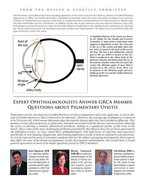
F R O M T H E H E A L T H & G E N E T I C S C O M M I T T E E<br />
There has been a great deal of discussion regarding pigmentary uveitis since it was first described as a disease of <strong>Golden</strong> <strong>Retriever</strong>s<br />
(Sapienza et al, 2000). The Health and Genetics Committee recommends yearly eye exams and online recording of eye exams for<br />
a lifetime in <strong>Golden</strong> <strong>Retriever</strong>s who have been bred. It is hoped that these recommendations will allow breeders to identify dogs<br />
who have had healthy eyes for a full lifetime. In addition, for the sake of early detection and more effective treatment, many ophthalmologists<br />
recommend annual eye exams for all <strong>Golden</strong> <strong>Retriever</strong>s beginning in early adulthood and continuing to an advanced<br />
age. The accompanying article was developed to provide members with expert opinions. The figure below is provided to help with<br />
some of the terms used in the article.<br />
A simplified diagram of the canine eye drawn<br />
by Dr. Hubbs for the Health and Genetics<br />
Committee to help owners understand what<br />
happens in pigmentary uveitis. The clear part<br />
of the eye is the cornea and light enters the<br />
eye and is focused on the back of the eye by<br />
the lens. The lens is just behind the colored<br />
part of the eye which is known as the iris.<br />
Fluid is produced in the ciliary body in the<br />
posterior chamber and drains from the eye in<br />
the anterior chamber where the iris meets the<br />
cornea (the filtration angle). If more fluid is<br />
produced in the ciliary body than can<br />
be drained at the filtration angle, pressure<br />
builds up in the eye and the <strong>Golden</strong> <strong>Retriever</strong><br />
develops glaucoma.<br />
Expert Ophthalmologists <strong>Answer</strong> GRCA Member<br />
<strong>Questions</strong> about <strong>Pigmentary</strong> <strong>Uveitis</strong><br />
<strong>Pigmentary</strong> uveitis, also known as <strong>Golden</strong> <strong>Retriever</strong> uveitis or pigmentary and cystic glaucoma, is an eye disease<br />
of <strong>Golden</strong> <strong>Retriever</strong>s that is believed to be inherited. However, the average age of diagnosis is reported<br />
to be 8.6 years old, which means that many dogs develop the disease after they have produced offspring. <strong>Pigmentary</strong><br />
uveitis often progresses to glaucoma, and pain associated with the disease can necessitate removing<br />
the eye(s) or placing a cosmetic intrascleral prosthesis, making it a significant quality of life issue in the<br />
breed. Since some of the most challenging problems presented by this disease have not yet been answered by<br />
the published science, we have asked three ophthalmologists with high levels of expertise and experience<br />
specifically in investigating, diagnosing, and treating pigmentary uveitis to help us respond to your questions.<br />
As is to be expected in areas of veterinary medicine that are the subject of ongoing investigation, these experts<br />
may differ on some of the details of pigmentary uveitis development and treatment; but overall there is broad<br />
general agreement on most of the major questions posed by GRCA members.<br />
John Sapienza, DVM<br />
is a veterinary ophthalmologist<br />
in private practice<br />
in Long Island, NY,<br />
and is the lead author of<br />
a pivotal scientific paper<br />
published in 2000 that<br />
characterized pigmentary<br />
uveitis.<br />
Wendy Townsend,<br />
DVM, MS is a veterinary<br />
ophthalmologist on the<br />
faculty of Purdue University<br />
in IN, and has<br />
had research grants<br />
investigating pigmentary<br />
uveitis for several<br />
years.<br />
Tom Sullivan, DVM is a<br />
veterinary ophthalmologist<br />
and <strong>Golden</strong> owner in<br />
private practice in Seattle,<br />
WA, where he has<br />
demonstrated a special<br />
commitment to early<br />
diagnosis and treatment<br />
of pigmentary uveitis.
Expert Ophthalmologists <strong>Answer</strong> <strong>Questions</strong>, continued<br />
1. Iris/ciliary body cysts sometimes appear before pigmentary<br />
uveitis can be definitively diagnosed, but not all <strong>Golden</strong>s<br />
with iris/ciliary body cysts will eventually get pigmentary<br />
uveitis. Is there a way to tell the difference between incidental<br />
cysts and those that may be associated with pigmentary<br />
uveitis, and if so, what are the differences?<br />
Sapienza: Incidental cysts tend to be free floating in the anterior<br />
chamber and often solitary. The cysts that are generally<br />
associated with the <strong>Golden</strong> <strong>Retriever</strong> (GR) uveitis are often iridociliary<br />
cysts (cysts located in the posterior chamber: the area<br />
between the back of the iris and the front of the lens). These iridociliary<br />
cysts are often multiple in nature, but can begin as<br />
one or two cysts. The pupil must be dilated to adequately see<br />
these iridociliary cysts.<br />
Townsend: I would agree with the comments shared by both<br />
Drs. Sapienza and Sullivan. I believe iridociliary cysts are a relatively<br />
common finding in <strong>Golden</strong> retrievers. In CERF clinics I<br />
have performed, 30% of the <strong>Golden</strong> <strong>Retriever</strong>s examined have<br />
had iridociliary cysts. Complete dilation of the pupil is critical<br />
because the iridociliary cysts are located behind the iris and<br />
without dilation are hidden from view.<br />
Sullivan: A: Iris/ciliary body cysts, as you’ve stated, can be<br />
seen as part of the uveitis syndrome, or as an unrelated entity.<br />
In the population of dogs that we see in our area, I would<br />
divide these into three groups:<br />
1. Dogs with free-floating cysts – singly or in low numbers.<br />
These likely arise on the back of the iris, but break free and<br />
float into the space between the iris and the inner surface of<br />
the cornea. These are often seen at 6 o’clock inside the eye<br />
because they sink with gravity. If the dog lies on his/her side<br />
for an extended period, they will roll to the new “down”<br />
side until the head is lifted. These usually continue to<br />
enlarge. Because that space – the anterior chamber – is<br />
dome shaped, continued growth results in these cysts<br />
becoming lodged between the iris and cornea, eventually<br />
moving into the pupillary space as that is where there is the<br />
most room. These free floating cysts, in my opinion, are not<br />
usually a result of uveitis.<br />
2. Dogs with one or two cysts attached to the ciliary body<br />
behind the iris way out at the periphery of the lens. These<br />
will sometimes grow to the point where they put pressure<br />
on the lens periphery, resulting in small equatorial cortical<br />
cataracts. These, too are usually not part of this syndrome,<br />
but could be the earliest stage of # 3 below.<br />
3. Dogs with multiple cysts growing on the back side of the iris<br />
such that when the pupil is dilated they can be seen emerging<br />
through the edge of the pupil. These in my area and<br />
opinion, are much more likely to be a part of the uveitis<br />
complex. Dr. Sapienza reported on blood filled ciliary body<br />
cysts in his paper. We rarely (if ever) see those, and in fact<br />
the cystic form of PU is the minority of our cases. (See photos<br />
below.)<br />
a. Are these differences reliable enough for breeders to feel<br />
“safe” breeding dogs whose cysts appear to be incidental?<br />
Sapienza: Yes and no. I have examined several GR dogs with<br />
solitary cysts in the anterior chamber that went on to develop<br />
severe GR uveitis. If the cysts are iridociliary in nature or there<br />
is radial pigment on the anterior capsule of the lens, then I<br />
would certainly say that there is the initial stage of GR uveitis.<br />
Townsend: The least concerning cyst would be the single, free<br />
floating cyst inside the anterior chamber. However that does<br />
not guarantee that they will not develop PU.<br />
Multiple cysts growing on the back side of the iris<br />
such that when the pupil is dilated they can be<br />
seen emerging through the edge of the pupil.<br />
(Sullivan, Q1, Group # 3)<br />
Cysts attached to the ciliary body behind the iris<br />
way out at the periphery of the lens. (Sullivan, Q1,<br />
Group # 2)
Expert Ophthalmologists <strong>Answer</strong> <strong>Questions</strong>, continued<br />
Sullivan: If there is a single cyst free floating inside the anterior<br />
chamber and no other evidence of PU, I wouldn’t be concerned<br />
that that cyst is a sign of PU. That does not mean that<br />
that dog will not develop PU independent of the cyst, so<br />
breeding is fine, but ongoing CERF exams are always warranted.<br />
b. Is there agreement among ophthalmologists regarding<br />
characteristics of incidental versus suspicious iris/ciliary<br />
body cysts so that if they were examining the same dog,<br />
they would reach the same conclusion?<br />
Sapienza: I believe so.<br />
Townsend: I think most are in agreement regarding the free<br />
floating cysts. Beyond that I’m not sure that there is a consensus.<br />
Sullivan: Good question – I have no idea – there is much more<br />
subjectivity to CERF evaluations and clinical impressions of<br />
what constitutes what disease than is ideal – especially in a<br />
disease, such as PU, that developed after the majority of the<br />
current ophthalmologists were trained. Different opinions and<br />
answers from different ophthalmologists are probably not<br />
uncommon.<br />
c. Is there medical/surgical treatment for the iris/ciliary body<br />
cysts that are associated with pigmentary uveitis?<br />
Sapienza: Often topical anti-inflammatory medications<br />
(namely, prednisolone actetate or dexamethasone) are started.<br />
There is no medication to make the cysts disappear.<br />
Townsend: There are no medications that will make the cysts<br />
disappear. There are surgical therapies to remove the cysts, but<br />
those are usually only used in patients with solitary free floating<br />
cysts that are large enough to obstruct vision.<br />
Sullivan: No medical treatment. The cysts can sometimes be<br />
treated via laser if they are heavily pigmented (the laser is<br />
absorbed by pigment – if cysts are relatively clear, doesn’t<br />
work), but more will likely occur. They can sometimes be aspirated<br />
under general anesthesia with a very small needle, but<br />
this can be tricky as you don’t want to puncture the lens, and<br />
the attached cysts are difficult to reach with a needle while<br />
avoiding the lens. Although it has been reported that these<br />
cysts are responsible for glaucoma development, they are not<br />
the only reason for the glaucoma (and in our “cystless” cases,<br />
obviously not even a contributing factor), so addressing the<br />
cysts medically or surgically may not be of any help in avoiding<br />
blindness.<br />
d. Is there a place on the CERF form where the examining<br />
ophthalmologist can provide his/her professional opinion<br />
as to whether iris/ciliary body cysts are either incidental or<br />
suspicious?<br />
Sapienza: Yes.<br />
Townsend: Not in the section where the cysts are marked. The<br />
ophthalmologist would have to write comments in the lower<br />
right hand corner of the form. However comments written<br />
there do not show up when an animal receives its CERF number.<br />
Therefore if a <strong>Golden</strong> retriever receives a CERF number<br />
and has category D1 marked, someone else viewing that CERF<br />
information cannot tell whether the cysts were thought to be<br />
incidental or suspicious.<br />
Sullivan: There are separate spots to mark PU and Iris/CB cysts.<br />
If there are other signs of PU, then “PU” would be marked. If<br />
there are cysts, but no other signs of PU then most ophthalmologists<br />
would likely just mark cysts. If you were suspicious<br />
that the cysts are part of early PU, then you can write that suspicion<br />
in the “comments” box in the lower right hand corner<br />
of the form.<br />
2. What percentage of dogs with pigmentary uveitis develops<br />
glaucoma?<br />
Sapienza: Difficult to say. Based on my article on GR uveitis,<br />
46% of GR dogs went on to develop glaucoma. If fibrin-like<br />
debris is present in the anterior chamber, glaucoma is a common<br />
secondary complication. Thirty-seven percent developed<br />
different stages of cataract formation (incipient to hypermature<br />
stages).<br />
Townsend: I believe this depends how early in the course of<br />
the disease treatment is started. In the patients that I see, 49%<br />
have developed glaucoma and most of those have had glaucoma<br />
at the very first visit. In fact the discomfort associated<br />
with the glaucoma was the reason they saw an ophthalmologist<br />
and the pigmentary uveitis was diagnosed. For patients<br />
participating in the pigmentary uveitis study the rate of glaucoma<br />
is lower at 30% as more of these patients are being diagnosed<br />
earlier at their annual CERF examinations.<br />
Sullivan: Depends upon how young the onset of disease and<br />
how early treatment is started. If treatment started very early, it<br />
is very rare for them to develop glaucoma in our cases (again,<br />
we don’t see the number of cysts that other areas apparently<br />
see). If not treated, glaucoma and cataracts are very likely if the<br />
patient lives long enough. In other words, if a 5 year old has<br />
significant changes from PU, chances are very high that he/she<br />
will get glaucoma by 8-9. If a dog has very early signs and<br />
treatment started, glaucoma less likely, but may develop<br />
vision-threatening cataracts several years later (we’ve done<br />
cataract surgery on a few of these dogs). If a dog develops early<br />
signs at 12 years of age, glaucoma is less likely just because<br />
they may not live long enough to develop secondary problems<br />
a few years down the road.<br />
3. What is your treatment protocol for dogs with very early<br />
pigmentary uveitis?<br />
Sapienza: Prednisolone acetate eye drops 1-3 times daily<br />
depending on the level of inflammation.<br />
Townsend: Either topical steroids or topical NSAIDs depending<br />
on the severity of the changes already present and monitor<br />
every couple of months<br />
Sullivan: In our cases, the inflammation does not respond well<br />
to steroids. Topical nonsteroidal anti-inflammatory drops<br />
(NSAIDs) work best. We use diclofenac, generally once daily.
Expert Ophthalmologists <strong>Answer</strong> <strong>Questions</strong>, continued<br />
Townsend: This is hard to say for sure as we don’t know how<br />
long the dogs that are diagnosed with more advanced disease<br />
were affected before they developed glaucoma, etc. However,<br />
patients seem to do better if it’s detected early so that we can<br />
prevent the scar tissue build up. We also don’t know how<br />
many older <strong>Golden</strong>s are affected with pigmentary uveitis, but<br />
never diagnosed because they are free of symptoms. I have<br />
been performing eye exams on senior <strong>Golden</strong>s in an effort to<br />
determine how prevalent pigmentary uveitis truly is in the senior<br />
population. On average I diagnose 1-2 cases of pigmentary<br />
uveitis for every 25-30 older <strong>Golden</strong>s examined. In each case<br />
the owners thought their older <strong>Golden</strong> had normal eyes as<br />
they had no symptoms.<br />
Photo of a <strong>Golden</strong> <strong>Retriever</strong> that has recovered from surgery to<br />
remove an eye with glaucoma due to pigmentary uveitis.<br />
The key issue to keep in mind is that inflammation inside the<br />
eye does two things: 1) it causes scar tissue development<br />
within the drain (fluid is always being produced inside the eye<br />
– it flows through the pupil into the front of the eye, then exits<br />
through the drain into the bloodstream), and 2) it reduces fluid<br />
production by interfering with the function of the fluid producing<br />
structure. An inflamed eye is a soft eye (this is why early<br />
on, PU eyes have very low pressures. As scar tissue builds up<br />
in the drain and contracts, it blocks drainage and increased<br />
pressure results). If we have an eye with moderate loss of<br />
drainage due to scar tissue (and we can’t see that on exam, you<br />
can surmise if the other signs of PU are moderate) then<br />
quelling the inflammation will turn fluid production back up to<br />
normal, and the damaged drain might no longer be able to<br />
cope with this normal fluid rate, leading to increased pressure<br />
inside the eye. This is how treating the inflammation with<br />
NSAIDs can lead to glaucoma. In dogs with early PU, these<br />
drops will usually halt the inflammation, preventing further<br />
damage. Because scar tissue contracts over time, glaucoma<br />
can still develop down the line even if ongoing inflammation<br />
is controlled. This is not usually the case in “early” cases, but<br />
is a concern in cases deemed moderate (moderate in our terminology<br />
means that inflammation has been smoldering for a<br />
moderate amount of time, so there is damage/scarring within<br />
the globe. Early would mean that the inflammation hasn’t been<br />
active for very long – it isn’t that some eyes are more inflamed<br />
than others as much as some have been inflamed for a longer<br />
period of time prior to diagnosis).<br />
a. In your experience, if pigmentary uveitis is diagnosed and<br />
treated in its very early stages, what percentage of dogs will<br />
remain clinically free of symptoms, versus those that<br />
progress to significant symptoms?<br />
Sapienza: My clinical suspicion is that 1/5 dogs go on to<br />
develop severe disease, but this truly depends on the level of<br />
anterior chamber inflammation (flare), the presence of fibrinlike<br />
debris in the anterior chamber, the number of iridociliary<br />
cysts, and the formation of cataracts.<br />
Sullivan: The vast majority will remain visual and comfortable<br />
lifelong if caught early and treated with diclofenac. Some – a<br />
very low percentage – will develop cataracts several years<br />
down the road from damage done to the lens back when the<br />
inflammation was active.<br />
4. At what age would you recommend beginning screening<br />
examinations for pet dogs that will not be bred, to give<br />
them the best chance for effective treatment if diagnosed?<br />
Sapienza: At three years of age.<br />
Townsend: I think two years of age is a good time to start to get<br />
a baseline and catch some of the rare cases that start at three<br />
to four years of age.<br />
Sullivan: We start screening at four years of age. I have seen<br />
two dogs with significant signs of active uveitis as early as two<br />
years of age, but that is very rare. Most start to develop definitive<br />
changes in the five- to seven-year range in our population.<br />
a. How frequently should pet <strong>Golden</strong> <strong>Retriever</strong>s be examined<br />
for pigmentary uveitis?<br />
Sapienza: If not clinical, once a year.<br />
Townsend: Yearly.<br />
Sullivan: We screen annually.<br />
b. Can general practice veterinarians detect early pigmentary<br />
uveitis if the owner specifically asks him/her to check for<br />
this?<br />
Sapienza: I do not believe so. A slit-lamp examination is<br />
imperative after the pupil has been dilated.<br />
Townsend: Better to see an ophthalmologist as our instruments<br />
allow us to detect very subtle changes that occur early in the<br />
disease process.<br />
Sullivan: Not reliably. It has taken many cases and several<br />
years of repeat examination for me to feel that I can see early<br />
PU. Many GP’s aren’t able to detect moderate or late PU – not<br />
because they are poor clinicians, just because they aren’t as<br />
familiar with ocular structures and the differences between<br />
normal and diseased tissues within an eye.
H&G; <strong>Pigmentary</strong> <strong>Uveitis</strong> FAQs, continued<br />
5. What is your best estimate of the number of <strong>Golden</strong>s with<br />
pigmentary uveitis that you have diagnosed and/or<br />
treated?<br />
Sapienza: Several hundred.<br />
Townsend: There are more than 200 dogs participating in my<br />
study with more that I know of.<br />
Sullivan: 400–500 (best guesstimate)<br />
a. Have you observed a trend in the number of affected dogs<br />
in recent years?<br />
Sapienza: Less so over the years, but still quite frequently seen.<br />
Townsend: They are being diagnosed at a younger age and earlier<br />
in the course of their disease<br />
Sullivan: Depends on definition of recent. When we perform<br />
screening clinics, the trend in new dogs without known ocular<br />
disease greater than or equal to four years of age is that 25% –<br />
33% have been affected. This has been stable over the past<br />
four years.<br />
6. Do other breeds get pigmentary uveitis?<br />
Sapienza: I had two Labrador cross-bred dogs with similar<br />
signs of “GR” uveitis.<br />
Townsend: No.<br />
Sullivan: Not that we’ve seen. Nor have I seen any <strong>Golden</strong><br />
crosses, i.e. “doodles” with it, although it is a new enough<br />
phenomenon that the 8-9 year old “typical presentation”<br />
hasn’t been reached for many.<br />
7. When diagnosing a <strong>Golden</strong> with pigmentary uveitis, how<br />
can you be sure that it’s not the form of uveitis that other<br />
dogs can get?<br />
Sapienza: The classical signs of GR uveitis are the pigment on<br />
the anterior lens capsule (often in a radial fashion), the presence<br />
of iridociliary cysts, posterior synechiae, fibrin-like debris<br />
in the anterior chamber, and then secondary complications<br />
like glaucoma and cataracts. In the GR breed, there is typically<br />
not a lot of anterior chamber inflammation (so called aqueous<br />
flare) as in the other types of uveitis seen in other breeds.<br />
Townsend: The classic appearance – pigment deposited on the<br />
anterior lens capsule in a radial fashion.<br />
Sullivan: Most cases of uveitis are sudden onset and aggressively<br />
inflamed. The <strong>Golden</strong> <strong>Retriever</strong> variety is very low grade<br />
inflammation (this is why owners don’t notice until secondary<br />
changes like glaucoma have developed) over prolonged periods<br />
of time. This leads to different symptoms/changes.<br />
8. What is the youngest age at which you’ve diagnosed pigmentary<br />
uveitis? What is the oldest? Is there an age at<br />
which a <strong>Golden</strong> can be determined to have no risk of getting<br />
the disease, or should eye exams continue for the dog’s<br />
lifetime?<br />
Sapienza: Youngest dog was 4.5 years old, oldest was 14.5<br />
years old at the time of examination.<br />
Townsend: Personally six years, but there are dogs that are 3-4<br />
years old in the study. I have also had a dog diagnosed at 13.5<br />
years of age, so there’s no safety zone.<br />
Sullivan: Youngest, 2.5 years of age. Oldest new onset 13 years<br />
of age. These are both very uncommon. I think in most cases it<br />
is fine to stop checking in the 11-year-old range as development<br />
later than this is unlikely, and a dog that develops it later<br />
would be unlikely to live long enough to have significant problems.<br />
9. Do you have any advice or recommendations about how<br />
breeders should evaluate the breeding prospects for<br />
<strong>Golden</strong> <strong>Retriever</strong>s that have a close relative such as a<br />
parent, aunt, uncle, grandparent, and/or sibling with pigmentary<br />
uveitis?<br />
Sapienza: My advice is to breed as far away from the affected<br />
animal as possible.<br />
Townsend: That is a hard question as we don’t know the inheritance<br />
of the condition.<br />
Sullivan: I would have an ophthalmologist familiar with the<br />
disease screen annually starting at four years of age. I would<br />
insist that pet owners do the same with any offspring. In our<br />
cases, at least, this appears to be a very treatable condition as<br />
long as it is caught and treatment started early enough. This is<br />
a much better health issue in terms of being able to manage as<br />
compared to severe allergic disease, heart disease, orthopedic<br />
disease, or cancer. This needs to be eliminated from the breed,<br />
but a genetic test is the best hope for that. Until that time, at<br />
least in our populations, it might be difficult to find lines free<br />
of this disease. Eliminating all related dogs from breeding<br />
might very well lead to unintentionally selecting for something<br />
else...<br />
Thank you very much for answering these questions from<br />
members of the <strong>Golden</strong> <strong>Retriever</strong> Club of America. Is there a<br />
final message that you would like to convey to our members<br />
regarding pigmentary uveitis in <strong>Golden</strong> <strong>Retriever</strong>s?<br />
Sapienza: This disorder has been around for years, but<br />
progress is being made in minimizing the frequency that we<br />
are seeing this syndrome. Judicious breeding and ethical decisions<br />
need to be continued to be implemented in order to<br />
eliminate this disorder in the GR breed.<br />
Townsend: I would like to thank everyone who has participated<br />
in my studies on pigmentary uveitis and the efforts that<br />
have been made to increase awareness of this condition. I<br />
think more dogs are being diagnosed earlier because of this<br />
increased awareness, which gives us a much better chance<br />
with therapy. ❖