Media Hafen (Harbor) nach Unbekannte Straße ... - AXIOM Solutions
Media Hafen (Harbor) nach Unbekannte Straße ... - AXIOM Solutions
Media Hafen (Harbor) nach Unbekannte Straße ... - AXIOM Solutions
Create successful ePaper yourself
Turn your PDF publications into a flip-book with our unique Google optimized e-Paper software.
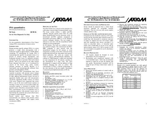
PSA quantitative<br />
PSA Enzyme Immunoassay<br />
96 Tests 88 98 56<br />
For In Vitro Diagnostic Use Only<br />
INTENDED USE<br />
For the quantitative determination of the Cancer<br />
Antigen PSA concentration in human serum.<br />
INTRODUCTION<br />
Human prostate-specific antigen (PSA) is a serine<br />
protease, a single chain glycoprotein with a<br />
molecular weight of approximately 34,000 daltons<br />
containing 7% carbohydrate by weight. PSA is<br />
immunologically specific for prostatic tissue, it is<br />
present in normal, benign hyperplastic, and<br />
malignant prostatic tissue, in metastatic prostatic<br />
carcinoma, and also in prostatic fluid and seminal<br />
plasma. PSA is not present in any other normal<br />
tissue obtained from men, nor is it produced by<br />
cancers of the breast, lung, colon, rectum,<br />
stomach, pancreas or thyroid. Besides, it is<br />
functionally and immunologically different from<br />
prostatic acid phosphatase (PAP).<br />
Elevated serum PSA concentrations have been<br />
reported in patients with prostate cancer, benign<br />
prostatic hypertrophy, or inflammatory conditions<br />
of other adjacent genitourinary tissues, but not in<br />
apparently healthy men, men with non-prostatic<br />
carcinoma, apparently healthy women, or women<br />
with cancer. Reports have suggested that serum<br />
PSA is one of the most useful tumor markers in<br />
oncology. It may serves as an accurate marker for<br />
assessing response to treatment in patients with<br />
prostatic cancer. Therefore, measurement of serum<br />
PSA concentrations can be an important tool in<br />
monitoring patients with prostatic cancer and in<br />
determining the potential and actual effectiveness<br />
of surgery or other therapies.<br />
Recent studies also indicate that PSA<br />
measurements can enhance early prostate cancer<br />
detection when combined with digital rectal<br />
examination (DRE).<br />
PRINCIPLE OF THE TEST<br />
The PSA ELISA test is based on the principle of a<br />
solid phase enzyme-linked immunosorbent assay.<br />
The assay system utilizes a rabbit anti-PSA<br />
antibody directed against intact PSA for solid<br />
phase immobilization (on the microtiter wells). A<br />
monoclonal anti-PSA antibody conjugated to<br />
horseradish peroxidase (HRP) is in the antibodyenzyme<br />
conjugate solution. The test sample is<br />
allowed to react first with the immobilized rabbit<br />
antibody at room temperature<br />
for 60 minutes. The wells are washed to remove<br />
any unbound antigen. The monoclonal anti-PSA-<br />
HRP conjugate is then reacted with the<br />
immobilized antigen for 60 minutes at room<br />
temperature resulting in the PSA molecules being<br />
sandwiched between the solid phase and enzymelinked<br />
antibodies. The wells are washed with<br />
water to remove unbound-labeled antibodies. A<br />
solution of TMB Reagent is added and incubated<br />
at room temperature for 20 minutes, resulting in<br />
the development of a blue color. The color<br />
development is stopped with the addition of Stop<br />
Solution changing the color to yellow. The<br />
concentration of PSA is directly proportional to<br />
the color intensity of the test sample. Absorbance<br />
is measured spectrophotometrically at 450 nm.<br />
REAGENTS<br />
Materials provided with the kits:<br />
• Rabbit anti-PSA coated microtiter plate with<br />
96 wells.<br />
• Zero Buffer, 7 ml.<br />
• Reference standard containing 0, 2, 4, 15, 60,<br />
and 120 ng/ml PSA, lyophilized. 1 set.<br />
• Enzyme Conjugate Reagent, 12 ml.<br />
• TMB Reagent (one step), 11 ml.<br />
• Stop Solution (1N HCl), 11 ml.<br />
Materials required but not provided:<br />
• Precision pipettes: 50 µl, 100 µl and 1.0 ml.<br />
• Disposable pipette tips.<br />
• Distilled water.<br />
• Vortex mixer or equivalent.<br />
• Absorbent paper or paper towel.<br />
• Graph paper.<br />
• A microtiter plate reader with a bandwidth of<br />
10 nm or less and an optical density range of<br />
0-2 OD or greater at 450 nm<br />
SPECIMEN COLLECTION AND PREPARATION<br />
Serum should be prepared from a whole blood<br />
specimen obtained by acceptable medical<br />
techniques. This kit is for use with serum samples<br />
without additives only.<br />
STORAGE OF TEST KIT AND INSTRUMENTATION<br />
Unopened test kits should be stored at 2-8°C upon<br />
receipt and the microtiter plate should be kept in a<br />
sealed bag with desiccants to minimize exposure<br />
to damp air. Opened test kits will remain stable<br />
until the expiration date shown, provided it is<br />
stored as described above. A microtiter plate<br />
reader with a bandwidth of 10 nm or less and an<br />
optical density range of 0-2 OD or greater at 450<br />
nm wavelength is acceptable for use in absorbance<br />
measurement.<br />
REAGENT PREPARATION<br />
1. All reagents should be brought to room<br />
temperature (18-25°C) before use.<br />
2. Reconstitute each lyophilized standard with<br />
1.0 ml distilled water. Allow the reconstituted<br />
material to stand for at least 20 minutes and<br />
mix gently. Reconstituted standards will be<br />
stable for up to 30 days when stored sealed at<br />
2-8°C<br />
ASSAY PROCEDURE<br />
1. Secure the desired number of coated wells in<br />
the holder.<br />
2. Dispense 50 µl of standards, specimens, and<br />
controls into appropriate wells.<br />
3. Dispense 50 µl of Zero Buffer into each well.<br />
4. Thoroughly mix for 30 seconds. It is very<br />
important to have a complete mixing in this<br />
setup.<br />
5. Incubate at room temperature (18-25°C) for 60<br />
minutes.<br />
6. Remove the incubation mixture by emptying<br />
plate contents into a waste container.<br />
7. Rinse and empty the microtiter wells 5 times<br />
with distilled or deionized water. (Please do<br />
not use tap water.)<br />
8. Strike the wells sharply onto absorbent paper<br />
or paper towels to remove all residual water<br />
droplets.<br />
9. Dispense 100 µl of Enzyme Conjugate<br />
Reagent into each well. Gently mix for 10<br />
seconds.<br />
10. Incubate at room temperature (18-25°C) for 60<br />
minutes.<br />
11. Remove the incubation mixture by emptying<br />
plate contents into a waste container.<br />
12. Rinse and empty the microtiter wells 5 times<br />
with distilled or deionized water. (Please do<br />
not use tap water.)<br />
13. Strike the wells sharply onto absorbent paper<br />
to remove residual water droplets.<br />
14. Dispense 100 µl of TMB Reagent into each<br />
well. Gently mix for 10 seconds.<br />
15. Incubate at room temperature for 20 minutes.<br />
16. Stop the reaction by adding 100 µl of Stop<br />
Solution to each well.<br />
17. Gently mix for 30 seconds. It is important to<br />
make sure that all the blue color changes to<br />
yellow color completely.<br />
18. Using a microtiter plate reader, read the optical<br />
density at 450nm within 15 minutes.<br />
CALCULATION OF RESULTS<br />
1. Calculate the average absorbance values<br />
(A450) for each set of reference standards,<br />
control, and samples.<br />
2. Construct a standard curve by plotting the<br />
mean absorbance obtained for each reference<br />
standard against its concentration in ng/ml on<br />
linear graph paper, with absorbance on the<br />
vertical (y) axis and concentration on the<br />
horizontal (x) axis.<br />
3. Using the mean absorbance value for each<br />
sample, determine the corresponding<br />
concentration of PSA in ng/ml from the<br />
standard curve.<br />
EXAMPLE OF STANDARD CURVE<br />
Results of a typical standard run with optical<br />
density readings at 450nm shown in the Y axis<br />
against PSA concentrations shown in the X axis.<br />
This standard curve is for the purpose of<br />
illustration only, and should not be used to<br />
calculate unknowns. Each user should obtain his<br />
or her own data and standard curve.<br />
PSA Absorbance (450<br />
(ng/ml) nm)<br />
0 0.141<br />
2 0.219<br />
4 0.320<br />
15 0.832<br />
60 2.300<br />
120 3.031<br />
10/05 BC/as 1<br />
10/05 BC/as 2
Absorbance (450 nm)<br />
4<br />
3<br />
2<br />
1<br />
0<br />
0 50 100 150<br />
Concentration PSA (ng/ml)<br />
EXPECTED VALUES AND SENSITIVITY<br />
Healthy males are expected to have PSA values<br />
below 4 ng/ml The minimum detectable<br />
concentration of PSA in this assay is estimated to<br />
be 1 ng/ml.<br />
LIMITATIONS OF THE PROCEDURE<br />
1. Reliable and reproducible results will be<br />
obtained when the assay procedure is carried<br />
out with a complete understanding of the<br />
package insert instructions and with adherence<br />
to good laboratory practice.<br />
2. The wash procedure is critical. Insufficient<br />
washing will result in poor precision and<br />
falsely elevated absorbance readings.<br />
3. Serum samples demonstrating gross lipemia,<br />
gross hemolysis, or turbidity should not be<br />
used with this test.<br />
4. The results obtained from the use of this kit<br />
should be used only as an adjunct to other<br />
diagnostic procedures and information<br />
available to the physician.<br />
REFERENCES<br />
1 Hara, M. and Kimura, H. Two prostate-specific antigens,<br />
gamma-seminoprotein and beta-microseminoprotein. J.<br />
Lab. Clin. Med. 113:541-548;1989.<br />
2 Yuan, J.J.; Coplen, D.E.; Petros, J.A.; Figenshau, R.S.;<br />
Ratliff, T.L.; Smith, D.S. and Catalona, W.J. Effects of<br />
rectal examination, prostatic massage, ultrasonography and<br />
needle biopsy on serum prostate specific antigen levels. J.<br />
Urol. 147:810-814; 1992.<br />
3 Wang, M.C.; Papsidero, L.D.; Kuriyama, M.; Valenzuela,<br />
L.A.; Murphy, G.P. and Chu, T.M. Prostatic antigen: a new<br />
potential marker for prostatic cancer. Prostate 2:89-93;<br />
1981.<br />
4 Stowell, L.I.; Sharman, I.E. and Hamel, K. An Enzyme-<br />
Linked Immunosorbent Assay ( ELISA ) for Prostatespecific<br />
antigen. Forensic Science Intern. 50:125-138;<br />
1991.<br />
5 Frankel, A.E.; Rouse, R.V.;Wang, M.C.; Chu, T.M. and<br />
Herzenberg, L.A. Monoclonal antibodies to a human<br />
prostate antigen. Canc. Res. 42:3714; 1982.<br />
6 Benson, M.C.; Whang, I.S.; Pantuck, A.; Ring, K.; Kaplan,<br />
S.A.; Olsson, C.A. and Cooner, W.H. Prostate specific<br />
antigen density: a means of distinguishing benign prostatic<br />
hypertrophy and prostate cancer. J. Urol. 147:815-816;<br />
1992.<br />
7 Gorman, C. The private pain of prostate cancer. Time<br />
10(5):77- 80; 1992.<br />
8 Walsh, P.C. Why make an early diagnosis of prostate<br />
cancer. J. Urol. 147:853-854; 1992.<br />
9<br />
Labrie, F.; Dupont, A.; Suburu, R.; Cusan, L.; Tremblay,<br />
M.; Gomez, J-L and Emond, J. Serum prostate specific<br />
antigen as pre-screening test for prostate cancer. J. Urol.<br />
147:846-852; 1992.<br />
10 McCarthy, R.C.; Jakubowski, H.V. and Markowitz, H.<br />
Human prostate acid phosphatase : purification,<br />
characterization, and optimization of conditions for<br />
radioimmunoassay. Clin. Chim. Acta. 132:287-293; 1983.<br />
11<br />
Heller, J.E. Prostatic acid phosphase: its current clinical<br />
status. J. Urol. 137:1091-1099; 1987.<br />
12 Filella, X.; Molina, R.; Umbert, J.J.B.; Bedini, J.L. and<br />
Ballesta, A.M. Clinical usefulness of prostate-specific<br />
antigen. Tumor Biol. 11:289-294; 1990.<br />
13<br />
Shin, W.J.; Gross, K.; Mitchell, B.; Collins, J.;<br />
Wierzbinski, B.; Magoun, S. and Ryo, U.Y. Prostate<br />
adenocarcinoma using Gleason scores correlates with<br />
prostate-specific antigen and prostate acid phosphatase<br />
measurements. J. Nat. Med. Assoc. 84:1049-1050; 1992.<br />
14<br />
Wirth, M.P. and Frohmuller, H.G. Prostate-specific<br />
antigen and prostate acid phosphatase in the detection of<br />
early prostate cancer and in the prediction of regional<br />
lymph node metastases. Eur. Urol. 21:263-268; 1992.<br />
15 Campbell, M.L. More cancer found with sensitive PSA<br />
assay. Urol. Times. 20:10; 1992.<br />
16 Vessella, R.L.; Noteboom, J. and Lange, P.H. Evaluation<br />
of the Abbott IMx Automated immunoassay of Prostate-<br />
Specific Antigen. Clin. Chem. 38:2044-2054; 1992.<br />
17 Brawer, M.K.; Chetner, M.P.; Beatie, J.; Buchner, D.M.;<br />
Vessella, R.L. and Lange, P. H. Screening for prostatic<br />
carcinoma with prostate specific antigen. J. Urol. 147:841-<br />
845; 1992.<br />
18 Benson, M.C. Whang, I.S.; Olsson, C.A.; McMahon, D.J.<br />
and Cooner, W.H. The use of prostate specific antigen<br />
density to enhance the predictive value of intermediate<br />
levels of serum prostate specific antigen. J. Urol.<br />
147:817-821; 1992.<br />
19 Oesterling, J.E. and Hanno, P.M. PSA still finding niches<br />
in cancer diagnosis. Urol. Times 20:13-18; 1992.<br />
20 Babaian, R.J.; Fritsche, H.A. and Evans, R.B. Prostatespecific<br />
antigen and the prostate gland volume: correlation<br />
and clinical application. J. Clin. Lab. Anal. 4:135-137;<br />
1990.<br />
10/05 BC/as 3


