Session 1 Hayward Platelet functional disorders and ... - NASCOLA
Session 1 Hayward Platelet functional disorders and ... - NASCOLA
Session 1 Hayward Platelet functional disorders and ... - NASCOLA

Create successful ePaper yourself
Turn your PDF publications into a flip-book with our unique Google optimized e-Paper software.
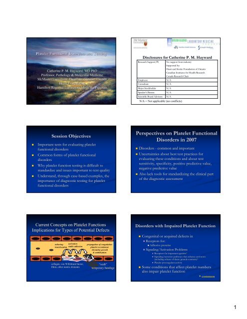
<strong>Platelet</strong> Functional Disorders <strong>and</strong> Testing<br />
Catherine P. M. <strong>Hayward</strong>, MD PhD<br />
Professor, Pathology & Molecular Medicine<br />
McMaster University, Hamilton, Ontario, Canada<br />
Head, Coagulation<br />
Hamilton Regional Laboratory Medicine Program<br />
Disclosures for Catherine P. M. <strong>Hayward</strong><br />
Research Support/PI<br />
Employee<br />
Consultant<br />
Major Stockholder<br />
Speaker’s s Bureau<br />
Scientific Board Advisory<br />
No support from industry<br />
Supported by:<br />
Heart <strong>and</strong> Stroke Foundation of Ontario<br />
Canadian Institutes for Health Research<br />
Canada Research Chair<br />
N/A<br />
N/A<br />
N/A<br />
N/A<br />
N/A<br />
N/A = Not applicable (no conflicts)<br />
<strong>Session</strong> Objectives<br />
• Important tests for evaluating platelet<br />
<strong>functional</strong> <strong>disorders</strong><br />
• Common forms of platelet <strong>functional</strong><br />
<strong>disorders</strong><br />
• Why platelet function testing is difficult to<br />
st<strong>and</strong>ardize <strong>and</strong> issues important to test quality<br />
• Underst<strong>and</strong>, through case-based examples, the<br />
importance of diagnostic testing for platelet<br />
<strong>functional</strong> <strong>disorders</strong><br />
Perspectives on <strong>Platelet</strong> Functional<br />
Disorders in 2007<br />
• Disorders - common <strong>and</strong> important<br />
• Uncertainties about best test practices for<br />
evaluating these conditions <strong>and</strong> about test<br />
sensitivity, specificity, positive predictive value,<br />
negative predictive value<br />
• Also lack tools for st<strong>and</strong>ardizing the clinical part<br />
of the diagnostic assessment<br />
Current Concepts on <strong>Platelet</strong> Functions<br />
Implications for Types of Potential Defects<br />
tethering activation<br />
translocation stable adhesion<br />
collagen, von Willebr<strong>and</strong> factor,<br />
fibrin, other matrix elements<br />
propagation of coagulation<br />
platelet recruitment<br />
thrombus growth<br />
& stabilization<br />
“scab”<br />
temporary b<strong>and</strong>age<br />
Disorders with Impaired <strong>Platelet</strong> Function<br />
• Congenital or acquired defects in<br />
• Receptors for:<br />
• Adhesive proteins<br />
• Signaling/Activation Problems<br />
• Receptors for important agonists*<br />
• Signaling/secretion pathways that enhance activation<br />
(including release of dense granule contents)*<br />
• <strong>Platelet</strong> procoagulant activity<br />
• Some conditions that affect platelet numbers<br />
also impair platelet function<br />
* common<br />
1
Adhesion<br />
Receptor<br />
Defects<br />
ADP (P2Y12)<br />
ADP (P2Y1)<br />
Thrombin<br />
Thromboxane<br />
PAF<br />
Collagen<br />
vWF<br />
GPIb<br />
BSS<br />
P<br />
Pleckstrin<br />
cAMP<br />
Gi<br />
AC<br />
PKC<br />
TxA2<br />
ATP<br />
TS<br />
DG<br />
PIP2<br />
PGG2/PGH2<br />
Gq<br />
PLC<br />
CO<br />
Arachidonic Acid<br />
IP3<br />
PLA2<br />
TK<br />
Phospholipids<br />
Ca<br />
MLC P<br />
MLC<br />
Ca<br />
vWD<br />
Fibrinogen<br />
Haemophilia 2006;12 (Suppl(<br />
3):128-136<br />
136<br />
VIIIa Ca<br />
IXa<br />
GPIIb-IIIa<br />
Aggregation<br />
Thrombasthenia<br />
Afibrinogenemia<br />
Secretion<br />
Disorders of Secretion/<br />
Signal Transduction<br />
IIa<br />
<strong>Platelet</strong> Coagulant<br />
Activities<br />
C<br />
Va a<br />
II<br />
Xa<br />
X<br />
Acquired Qualitative Defects<br />
• Drugs – antiplatelet agents are the most common<br />
• Uremia<br />
• Liver disease<br />
• Cushing’s s Syndrome<br />
• Cardiopulmonary bypass<br />
• Inhibitory antibodies<br />
• Bone marrow <strong>disorders</strong><br />
• Diverse – e.g. storage pool defects, membrane<br />
glycoprotein deficiencies<br />
<strong>Platelet</strong> Disorders<br />
Lack data from population surveys<br />
Secretion defects are more common than dense granule<br />
deficiency<br />
Dense granule deficiency is almost as common as von<br />
Willebr<strong>and</strong> disease<br />
about 3-5% 3<br />
of referred patients at our center<br />
Screening Tests for <strong>Platelet</strong> Functional Disorders<br />
• Bleeding time<br />
• Sensitivity limited, performance issues<br />
• Use – predicting response to DDAVP therapy?<br />
• Closure Time measured by PFA-100<br />
TM<br />
• Rapid, simple, test of shear-dependent platelet adhesion<br />
• Sensitivity<br />
• not perfect for screening<br />
• 24% to >90% sensitivity for congenital platelet <strong>disorders</strong><br />
• Poorer for studies prospectively evaluating platelet <strong>disorders</strong><br />
• Poorer for common platelet <strong>disorders</strong><br />
<strong>Hayward</strong>, Harrison, Cattaneo, Ortel <strong>and</strong> Rao; the <strong>Platelet</strong> Physiology ogy SSC of<br />
ISTH. <strong>Platelet</strong> function analyzer (PFA)-100 closure time in the evaluation<br />
of platelet <strong>disorders</strong> <strong>and</strong> platelet function. JTH 2006; 4: 312-9.<br />
PFA-100<br />
® Closure Times in Congenital <strong>Platelet</strong> Disorders<br />
JTH 2006; 4: 312-319.<br />
* - common <strong>disorders</strong><br />
‡ - associated with thrombocytopenia<br />
Glanzmann Thrombasthenia<br />
Aspirin-Like Defect<br />
ADP Receptor/Signaling Defect<br />
Dense Granule Deficiency*<br />
Hemansky Pudlak Syndrome<br />
Primary Secretion Defects*<br />
<strong>Platelet</strong> Procoagulant Defect<br />
Bernard-Soulier Syndrome‡<br />
<strong>Platelet</strong>-type type von Willebr<strong>and</strong> Disease ‡<br />
Gray <strong>Platelet</strong> Syndrome ‡<br />
Hereditary Macrothrombocytopenia Associated with Nonmuscle<br />
Myosin Heavy Chain IIa Syndromes ‡<br />
Undefined Autosomal Dominant Thrombocytopenia ‡<br />
CADP CT<br />
↑ ↑<br />
N<br />
↑-N<br />
N or ↑<br />
N to ↑<br />
N<br />
N<br />
↑ ↑<br />
↑↑<br />
↑<br />
N<br />
N<br />
CEPI CT<br />
↑ ↑<br />
↑<br />
N<br />
N or ↑<br />
N to ↑<br />
N or ↑<br />
N<br />
↑ ↑<br />
↑↑<br />
↑<br />
N or ↑<br />
N<br />
CT in Diagnostic Testing for <strong>Platelet</strong> Disorders<br />
• Potential advantages<br />
JTH 2006; 4: 312-319<br />
JTH 2005: 3;1309-11<br />
• Early clues about a defect if abnormal<br />
• Abnormal results may trigger a referral<br />
• More evidence is needed on its most appropriate<br />
use in clinical practice related to platelet <strong>disorders</strong><br />
• Sensitivity is better for VWD than for platelet <strong>disorders</strong><br />
• Diagnostic Screening: FURTHER TESTING NEEDED,<br />
REGARDLESS<br />
• Role in drug monitoring – needs further evaluation<br />
2
Drug<br />
CT with anti-platelet drugs<br />
inhibitors of α IIb β 3 abciximab,<br />
tirofiban, eptifibatide<br />
COX-1 1 inhibitors (aspirin <strong>and</strong><br />
other NSAIDs)<br />
Thienopyridines:<br />
Ticlopidine or clopidogrel<br />
Ticlopidine or clopidogrel<br />
plus aspirin<br />
JTH 2006; 4: 312-319<br />
319<br />
CADP CT<br />
P<br />
N<br />
N or P<br />
N or P<br />
CEPI CT<br />
P<br />
N or P<br />
N or P<br />
P<br />
Tests for Drug Resistance<br />
Assay<br />
Aggregation ‡<br />
VerifyNow® Aspirin<br />
& P2Y12 ‡<br />
(mod. aggreg. . with fibrinogen coated<br />
beads; RPFA)<br />
<strong>Platelet</strong>Works®<br />
(count platelets post-activation)<br />
PFA-100<br />
100® ‡<br />
(high shear adhesion test with<br />
activation: CEPI or CADP)<br />
Thromboxane Assays<br />
Serum (ex vivo generation)<br />
or urine (in vivo generation) ‡<br />
Impact (Cone <strong>and</strong> Plate device)<br />
Research Assays of <strong>Platelet</strong><br />
Activation (e.g. flow cytometry)<br />
VASP (vasodilator stimulated<br />
phosphoprotein – phosphorylation<br />
influenced by P2Y12)<br />
Advantages<br />
“Gold st<strong>and</strong>ard”<br />
Point-of<br />
of-Care, simple, rapid, semi-<br />
automated<br />
Simple, rapid, uses platelet counter<br />
Simple, Rapid, Semi-automated<br />
With both: samples can be stored<br />
for batch analyses<br />
More complex than Point-of<br />
of-Care<br />
Simple, Rapid, Semi-automated<br />
modifiable<br />
Endpoint for P2Y12 function<br />
Disadvantages<br />
All: need more data on relationship to outcomes, sensitivity <strong>and</strong><br />
specificity for resistance<br />
Tech challenging, time consuming, not st<strong>and</strong>ardized<br />
between labs, some variability, choice of procedures<br />
(light transmission, electrical impedance; use of platelet<br />
rich plasma, whole blood)<br />
relationship to outcomes?<br />
influenced by other variables (hematocrit(<br />
hematocrit, , platelet count,<br />
von Willebr<strong>and</strong> factor level)<br />
methods currently not in widespread use<br />
possible problems re specificity<br />
relationship to outcomes?<br />
Not in wide use, complex, time consuming<br />
Flow cytometry-based test; not in wide use<br />
Complexity of Diagnostic Testing for <strong>Platelet</strong> Disorders<br />
Include an assessment of/for:<br />
• <strong>Platelet</strong> number <strong>and</strong> size, platelet <strong>and</strong> leukocyte morphology<br />
• ~17% of referrals for testing are thrombocytopenic<br />
• Option – immunostaining for some conditions – e.g. MHY9 related <strong>disorders</strong><br />
• <strong>Platelet</strong> function, evaluated by aggregation tests<br />
• <strong>Platelet</strong> dense granule deficiency<br />
• Aggregation, BT, PFA-100<br />
100 CT - may be normal<br />
• <strong>Platelet</strong> secretion, evaluated by release of dense granule contents<br />
ts<br />
• ? More sensitive endpoint for defective function<br />
• Adhesion testing<br />
• apart from tests such as the PFA-100<br />
100 CT, this remains in research domain<br />
• Optional<br />
• Tests for procoagulant defects (appear rare, testing rarely done - ?Serum<br />
protein consumption to screen)<br />
• Others<br />
• transmission electron microscopy, glycoprotein analysis, thromboelastography<br />
(platelets contribute to properties of clots), etc<br />
What do we find with our st<strong>and</strong>ardized testing?<br />
Data for 391 Unselected Patients Prospectively Evaluated for<br />
von Willebr<strong>and</strong> Disease & <strong>Platelet</strong> Disorders<br />
platelet function abnormality &/or dense granule deficiency<br />
von Willebr<strong>and</strong> disease<br />
no laboratory abnormalities found<br />
abnormalities of uncertain signficance<br />
17%<br />
39%<br />
39%<br />
5%<br />
Hamilton Registry Data<br />
March 2002<br />
Setting<br />
General<br />
Diagnosis of <strong>Platelet</strong><br />
Dysfunction<br />
Monitoring of<br />
Antiplatelet Therapy<br />
Quality Assurance <strong>and</strong> <strong>Platelet</strong> Tests<br />
<strong>Hayward</strong> CPM, Eikelboom J. <strong>Platelet</strong> function testing: Quality assurance. a<br />
STH<br />
Characteristic<br />
Convenient<br />
Accurate & Precise<br />
St<strong>and</strong>ardized<br />
Sensitive<br />
Specific<br />
Population norms to<br />
guide interpretation<br />
Proven utility<br />
Specific<br />
Clinically relevant<br />
Modifiable<br />
Cost effective<br />
Additional comments<br />
Simple (no operator expertise required), rapid, inexpensive<br />
The test measures what it is supposed to measure. Reproducible,<br />
different observers agree on interpretation<br />
Test procedure is well described, st<strong>and</strong>ards are available,<br />
existing quality control program<br />
Negative test rules out disease<br />
Positive test rules in disease<br />
Test has been evaluated in full range of subjects (mild & severe,<br />
treated <strong>and</strong> untreated disease) <strong>and</strong> in subjects with other<br />
conditions that fall within the differential diagnosis<br />
Patients are better off as a result of undergoing the test<br />
Measures the effect of the drug on its target<br />
Results independently correlated with clinical outcome<br />
Altering antiplatelet treatment based on the results of the test<br />
improves clinical outcome<br />
Benefits of testing outweigh the direct <strong>and</strong> indirect costs of<br />
testing <strong>and</strong> follow up<br />
All Labs are Not the Same…..<br />
Variability Between Clinical Laboratories in<br />
Diagnostic Testing for Disorders of <strong>Platelet</strong><br />
Function<br />
Moffat et al, Thromb Haemost. 2005;93:549-53<br />
53<br />
• Goals<br />
• identify common practices <strong>and</strong> problems in the<br />
testing for <strong>disorders</strong> of platelet function<br />
• Enthusiastic participation!<br />
• 47 participating labs<br />
3
Aggregation methodologies<br />
37% of <strong>NASCOLA</strong> sites used >1 method<br />
<strong>NASCOLA</strong> Survey Data<br />
agonists used for clinical aggregation<br />
minority (15%) compared arachidonic acid & thromboxane analogue responses<br />
% sites<br />
100<br />
90<br />
80<br />
70<br />
60<br />
50<br />
40<br />
30<br />
20<br />
10<br />
0<br />
<strong>Platelet</strong> Rich Plasma Whole Blood Luminescence<br />
Collagen<br />
ADP<br />
Arachidonic Acid<br />
Ristocetin<br />
Epinephrine<br />
Thromboxane Analogue<br />
Thrombin<br />
TRAP<br />
Spontaneous<br />
ATP<br />
0 10 20 30 40 50 60 70 80 90 100<br />
% sites<br />
Survey 1 Survey 2<br />
Final Agonist Concentration for<br />
Testing <strong>Platelet</strong> Function by Aggregation<br />
Range<br />
Survey 1<br />
Median<br />
Range<br />
Survey 2<br />
Median<br />
Sources of reference intervals<br />
26% of sites used >1 method<br />
29% if sites had not determined their own reference range (12-250 250 tests/yr)<br />
Only 1 site did qualitative, without quantitative, interpretations<br />
ns<br />
Only 1 site formally evaluated data for normality in distribution<br />
See Moffat abstract this meeting<br />
Collagen<br />
ADP<br />
0.19 – 125 µg/mL<br />
0.5 – 1000 µM<br />
5 µg/mL<br />
5 µM<br />
0.62 – 190 µg/mL<br />
1 – 20 µM<br />
2.5 µg/mL<br />
5 µM<br />
Determined mean +/- 2 S.D.<br />
Epinephrine<br />
0.1 – 100 000 µM<br />
18 µM<br />
0.1 – 1000 µM<br />
10 µM<br />
Published literature<br />
Arachidonic<br />
Acid<br />
Ristocetin<br />
0.0005 – 1.7 mM<br />
0.25 – 1.5 mg/mL<br />
0.5 mM<br />
Low Dose:<br />
0.5 mg/mL<br />
High Dose:<br />
1.2 mg/mL<br />
0.0016 – 2.5 mM<br />
0.0012 – 2.0 mg/mL<br />
1.6 mM<br />
Low dose:<br />
0.5 mg/mL<br />
High dose:<br />
1.25 mg/mL<br />
Vendor of instrument or reagent<br />
qualitative interpretation<br />
0 20 40 60 80 100<br />
% sites<br />
Concerns Raised About <strong>Platelet</strong> Aggregation Testing<br />
there were many.............................<br />
• Labor intensive<br />
• Lack of evidence-based guidelines<br />
• Uncertainties – how to:<br />
• evaluate thrombocytopenic patients<br />
• Tam Abstract - this meeting<br />
• interpret epinephrine aggregation<br />
• Challenging to obtain reliable drug histories,<br />
uncertainties about the effects of different drugs<br />
• Influence of pre-analytical errors<br />
• proper sample procurement & transport<br />
Aggregation Testing – What is Best?<br />
• Agonist Concentrations<br />
• Medians – some conformity<br />
• Are these appropriate concentrations?<br />
• Review of published literature<br />
• The medians are probably good concentrations for testing<br />
• Useful strategies<br />
• e.g. comparing arachidonic acid/thromboxane responses<br />
• <strong>NASCOLA</strong> Study: 15% of labs used this comparison to sort out<br />
possible ASA/NSAID-like defects at the time of this survey<br />
4
Control (red, green) vs. Patient (P) with Secretion Defect<br />
P: also reduced aggregation with arachidonic acid <strong>and</strong> thromboxane e analogue<br />
ADP 2.5 <strong>and</strong> 5 μM<br />
Ristocetin 1.2 & 0.5 mg/mL<br />
Illustration of Aggregation Findings<br />
% aggregation with 4 μM ADP<br />
Collagen 1.2 <strong>and</strong> 5 mg/mL<br />
P<br />
P<br />
P<br />
P <strong>and</strong> C: no agg. with low dose, which<br />
screens for VWD type 2B & Plt-type<br />
Epinephrine 6 & 100 μM<br />
35.0<br />
30.0<br />
25.0<br />
20.0<br />
15.0<br />
10.0<br />
% subjects<br />
P<br />
P<br />
higher concentration of agonist: shown in red (C) & black (P)<br />
P<br />
patient<br />
control<br />
5 15 25 35 45 55 65 75 85 95 105 115<br />
% aggregation (interval mean)<br />
data is bimodal in distribution<br />
→ likely due to an influence of P2Y12 polymorphisms<br />
5.0<br />
0.0<br />
1 o<br />
2 o<br />
only 1 o<br />
Epinephrine<br />
Aggregation<br />
Variability in Aggregation Tests?<br />
data from repeat tests done on 115 patients in<br />
Hamilton<br />
abnormal on one or more occasions<br />
concordant results<br />
control<br />
Healthy Controls<br />
5 15 25 35 45 55 65 75 85 95 105<br />
% aggregation (interval mean)<br />
35.0<br />
30.0<br />
25.0<br />
20.0<br />
15.0<br />
10.0<br />
5.0<br />
0.0<br />
45.0<br />
40.0<br />
% subjects<br />
100<br />
90<br />
80<br />
70<br />
60<br />
% 50<br />
40<br />
30<br />
20<br />
10<br />
0<br />
ADP AA HD Rist all<br />
agonists<br />
Illustration: Usefulness of Aggregation Tests<br />
ADP 5 uM<br />
Collagen 5 ug/mL<br />
Collagen<br />
1.25 ug/mL<br />
Epinephrine 6 uM<br />
Arachidonic Acid 1.6 mM<br />
Thromboxane analogue 1 uM<br />
Ristocetin 0.5 mg/Ml<br />
Ristocetin 1.25 mg/mL<br />
Reference<br />
Interval<br />
% aggregn<br />
50-109<br />
85-101<br />
68-108<br />
5-37<br />
70-105<br />
72-108<br />
72-108<br />
0-7<br />
76-104<br />
Glanzmann<br />
Thrombasthenia<br />
0<br />
0<br />
0<br />
0<br />
0<br />
0<br />
0<br />
47<br />
Secretion<br />
Defect<br />
56<br />
83<br />
43<br />
15<br />
84<br />
21<br />
3<br />
62<br />
Dense Granule<br />
Deficiency<br />
(1/3 have<br />
normal results)<br />
71<br />
70<br />
12<br />
41<br />
47<br />
60<br />
8<br />
85<br />
Secretion absent or reduced with these agonists but normal with thrombin<br />
Thromboxane<br />
Generation<br />
Defect<br />
71<br />
62<br />
7<br />
36<br />
6<br />
94<br />
4<br />
90<br />
δ-granules: ~ 2-5/platelet2<br />
α-granules: ~ 80/platelet<br />
Diagnostic Evaluation of<br />
<strong>Platelet</strong> “Luggage” Defects<br />
Types of Luggage<br />
alpha (α)(<br />
– protein storage container<br />
delta (δ)–(<br />
electron dense*<br />
δ-granule deficiency:<br />
Fairly common<br />
~ 4% prevalence in our patients<br />
Aggregation, BT, CT may be normal<br />
α-granule deficiency:<br />
GRAY platelets<br />
- rare<br />
Clue – from evaluation of blood film<br />
combined αδ deficiency:<br />
Rarer than δ-granule deficiency<br />
5
Whole Mount<br />
Most popular method for assessing dense<br />
granule deficiency in North America<br />
Controls: average of 4 or more electron<br />
dense granules per platelet<br />
EDS<br />
Electron Dispersion Spectral Analysis<br />
analysis of the different dense<br />
granule constituents<br />
control<br />
P Ca<br />
αδ-storage pool defect<br />
P Ca<br />
Diagnostic Evaluation of <strong>Platelet</strong> Secretion<br />
• Secretion Defects<br />
• Paradox or knowledge translation gap<br />
• most common form of platelet disorder, yet secretion testing isn’t<br />
commonly done<br />
• Potential implications of NOT evaluating secretion?<br />
• Diagnostic label issue<br />
• Reduced detection of some platelet <strong>disorders</strong>?<br />
• Methods to evaluate secretion<br />
• Radioactive: e.g. serotonin release<br />
• Nonradioactive: : e.g. luminescence, other assays for<br />
nucleotides<br />
nM ATP Release<br />
Hamilton <strong>Platelet</strong> Secretion Testing<br />
Second Line Investigation - Luminescence Procedure<br />
~56% of patients are abnormal - half of these have normal aggregation studies<br />
bars - lower limit of reference range (determined using 48 controls)<br />
local testing done with 8 parameters, 6 agonists<br />
4.00<br />
3.50<br />
3.00<br />
2.50<br />
2.00<br />
1.50<br />
1.00<br />
0.50<br />
0.00<br />
0 0.5 1 1.5 2 2.5 3 3.5 4 4.5<br />
Thrombin ADP Collagen 1.25 Collagen 5<br />
Testing <strong>Platelet</strong> Function in Thrombocytopenic<br />
Patients<br />
17% of patients tested in Hamilton<br />
Data from 2002<br />
21%<br />
8% 4% 3% 2%1%<br />
<strong>Platelet</strong> function defect<br />
ITP<br />
asymptomatic<br />
liver disease<br />
known/probable MDS<br />
VWD<br />
other<br />
61%<br />
Reference<br />
Interval for samples<br />
with platelet count<br />
of 250 X 10 9 /L<br />
% aggregation<br />
Bernard Soulier<br />
Syndrome<br />
PRP: 29 X 10 9 /L<br />
(less than 5% GP<br />
IbIXV by flow)<br />
Control tested at<br />
same platelet count<br />
(PRP: 29 X 10 9 /L)<br />
Special Diagnostic Evaluations<br />
Illustration of Glycoprotein Analysis for Glanzmann Thrombasthenia<br />
ADP 5 uM<br />
50-109<br />
13<br />
37<br />
Collagen 5 ug/mL<br />
85-101<br />
21<br />
72<br />
Collagen 1.25 ug/mL<br />
68-108<br />
22<br />
8<br />
Epinephrine 6 uM<br />
5-37<br />
70-105<br />
20<br />
21<br />
Arachidonic Acid 1.6 mM<br />
72-108<br />
17<br />
62<br />
Thromboxane analog. 1 uM<br />
72-108<br />
18<br />
41<br />
Ristocetin 0.5 mg/Ml<br />
0-7<br />
0<br />
3<br />
Ristocetin 1.25 mg/mL<br />
76-104<br />
0<br />
79<br />
6
Testing for Rare Disorders - Quebec <strong>Platelet</strong> Disorder<br />
clues: family history, delayed bleeding responsive only to fibrinolytic inhibitors, absent<br />
epinephrine aggregation, reduced to low normal platelet counts<br />
<strong>Platelet</strong> u-PA u<br />
Western Blot<br />
known controls<br />
Quebec family members<br />
u-PA<br />
Q C 1 2 3 4 5 6<br />
J Thromb Haemost<br />
2006;4:1086-94<br />
Thromboelastography<br />
platelets contribute to clot strength<br />
blood samples recalcified, added TF & low concentration of t-PA<br />
lysis<br />
affected?<br />
√<br />
√<br />
√<br />
Control<br />
0 1200<br />
QPD<br />
0 1200<br />
Clots prepared with 0 or 1200 X 10 9 platelets/L<br />
J Thromb Haemost<br />
2006;4:1086-94<br />
Mystery Case – VWD screen<br />
55 year old male, severe bleeding after renal biopsy<br />
• First sample (referred in)<br />
• FVIIIC 2.43 U/mL<br />
(243 U/dL<br />
dL)<br />
• VWF:Ag 1.31 U/mL<br />
(131 U/dL<br />
dL)<br />
• VWF:RCo 0.29 U/mL<br />
(29 U/dL<br />
dL)<br />
• Interpretative comment: The von Willebr<strong>and</strong> factor ristocetin<br />
cofactor activity is significantly reduced. The discrepancy between een this<br />
value <strong>and</strong> the normal VWF antigen suggest a form of type 2 von<br />
Willebr<strong>and</strong> disease. An analyses of von Willebr<strong>and</strong> factor multimers<br />
would be helpful to further evaluate. Is there a family history of von<br />
Willebr<strong>and</strong> disease or a bleeding history that suggests acquired von<br />
Willebr<strong>and</strong> disease? Repeat testing, including ristocetin-induced induced platelet<br />
aggregation would be helpful to confirm <strong>and</strong> further evaluate the von<br />
Willebr<strong>and</strong> factor abnormalities.<br />
ADP 5 uM<br />
Collagen 5 ug/mL<br />
Collagen 1.25 ug/mL<br />
Epinephrine 6 uM<br />
Arachidonic Acid 1.6 mM<br />
Thromboxane analog. 1 uM<br />
Ristocetin 0.5 mg/Ml<br />
Ristocetin 1.25 mg/mL<br />
Further Investigations<br />
RI for samples with<br />
250 X 10 9 platelets/L<br />
% aggregation<br />
50-109<br />
85-101<br />
68-108<br />
5-37<br />
70-105<br />
72-108<br />
72-108<br />
0-7<br />
76-104<br />
Patient<br />
18<br />
45<br />
4<br />
12<br />
34<br />
32<br />
0<br />
3<br />
Control tested<br />
same day at same<br />
platelet count<br />
73<br />
91<br />
88<br />
86<br />
90<br />
89<br />
3<br />
91<br />
Patient CBC: Plt 64 X 109/L, MPV 8.5 fL<br />
Within RI for sample platelet count<br />
Additional Investigations<br />
• VWD screen done on day of aggregation testing<br />
• FVIIIC 1.34 U/mL<br />
• VWF:Ag 2.39 U/mL<br />
• VWF:RCo<br />
neat - 0.26; 1/2 - 0.63; 1/8 – 0.85 U/mL<br />
• Multimers Normal<br />
• Further RIPA testing (1.25 mg/mL<br />
mL) ) done after a 30 minute<br />
incubation of patient or control PPP, with control PRP (PPP<br />
added to adjust platelets from 440 down to 250 X 10 9 /L)<br />
• Patient Mixture: 2% aggregation<br />
• Control Mixture: 86% aggregation<br />
Acknowledgments:<br />
Colleagues <strong>and</strong> Collaborators<br />
Clinical <strong>and</strong> Research Lab Staff<br />
<strong>NASCOLA</strong> <strong>Platelet</strong> Study<br />
K. Moffat, M. Ledford-Kraemer,<br />
W. L. Nichols<br />
ISTH <strong>Platelet</strong> Physiology SSC<br />
PFA-100 Working Group<br />
• Diagnosis? Further tests that you would do?<br />
7