Introduction - Medical Education Online
Introduction - Medical Education Online
Introduction - Medical Education Online

You also want an ePaper? Increase the reach of your titles
YUMPU automatically turns print PDFs into web optimized ePapers that Google loves.
<strong>Introduction</strong>-mdm<br />
Abdominal pain<br />
In the Name of God<br />
<strong>Medical</strong> Decision Making<br />
for Common Disease<br />
Presentations<br />
BY<br />
Mitra Ahmad Soltani
<strong>Introduction</strong>-mdm<br />
Abdominal pain<br />
Table of Contents<br />
<strong>Introduction</strong>------------------------------------------------------------<br />
Priorities<br />
The process of making a decision<br />
Table I. Number and percent distribution of reasons of emergency department visits<br />
Table II. Number and percent distribution of emergency department visits with<br />
corresponding standard errors, by primary diagnosis<br />
Table III. Number and percent of drug mentions for the 20 most frequently<br />
occurring therapeutic drug classes at emergency department visits<br />
The Case<br />
Figure1- Approach to hemoptysis<br />
Figure 2- Approach to eosinophilia<br />
Figure 3- Approach to abdominal pain<br />
Figure-4: Approach to abdominal mass (organomegaly)<br />
Table IV- Yield and cost of individual common diagnostic tests performed for the<br />
clinical evaluation of patient’s illness and the case finding among 540 new,<br />
symptomatic primary care outpatients<br />
Table V- Comparing the three different ways to approach this case<br />
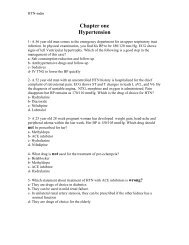
HTN------------------------------------------------------------------------<br />
Algorithm1-1- Approach to HTN<br />
Algorithm1-2: Drug choice in cases of chronic HTN<br />
Table1-1: Hypertension management<br />
Table1-2: HTN drugs
<strong>Introduction</strong>-mdm<br />
Abdominal pain<br />
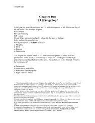
S3&S4--------------------------------------------------------------------<br />
Algorithm 2-1: Differential Diagnosis of S3 and S4<br />
Algorithm 2-2: Acute Pulmonary Edema Management<br />
Table 2-1: Treatment of different causes of S3 and S4 gallop<br />
Table2-2: Pulmonary edema drugs<br />
Arrhythmia---------------------------------------------------------<br />
Table3-2: Different ECG patterns with the counterpart laddergrams<br />
Table3-3: Answers<br />
Figure3-1: Algorithm to summarize the arrhythmia patterns<br />
Table3-4: Anti-arrhythmia drugs<br />
CAD-------------------------------------------------------------------<br />
Table4-1: IHD management<br />
Diagram4-1: ECG change after MI<br />
Diagram4-2: Enzyme change after MI<br />
Example #1: frontal plane leads with fully evolved inferior MI<br />
Example #2: Old inferior MI<br />
Example #3: Acute inferoposterior MI<br />
Example #4: Old posterolateral MI<br />
Example#5: Fully evolved anteroseptal MI<br />
Example#6: Acute anterior or anterolateral MI<br />
Example #7: Inferior MI + RBBB<br />
Example #8: Anteroseptal MI with RBBB<br />
Table4-2: Identifying Appropriate Patients For Thrombolytic Therapy<br />
Table4-3: Contraindications for thrombolytic drugs.<br />
Table4-4: Guidelines for the use of intravenous heparin with thrombolytic therapy.
<strong>Introduction</strong>-mdm<br />
Abdominal pain<br />
Table4-5: Heparin adjustment guideline<br />
Diagram4-3: Approach to Hyperlypidemia<br />
Table4-6: Determining Patient-Specific LDL Goals Through Risk Factors<br />
Table4-7: HMG-CoA Reductase Inhibitors (Statins)<br />
Table 4-8: Niacin (Nicotinic Acid)<br />
Table 4-9: Fibric Acid Derivatives (Fibrates)<br />
Table 4-10:Bile Acid Sequestrants<br />
Trauma------------------------------------------------------------<br />
Diagram5-1: Blunt abdominal trauma plus head trauma<br />
Diagram5-2: Cervical Spine trauma<br />
Diagram5-3: Fractured Pelvis<br />
Table5-1: Glasgow Coma Score-GCS<br />
Diagram5-4: Head Injury<br />
Diagram5-5: Penetration injury<br />
Table 5-2: Suitable Blood Replacement Regimes for Previously Healthy Adults<br />
Table 5-3: Clinical Signs of Shock<br />
Table 5-4: Protocol of certain trauma injuries management<br />
Empirical Antibiotic Therapy-------------------------------------------<br />
Table6-1: Genital Ulcer Antibiotic Therapy<br />
Table 6-2: Meningitis and Sepsis Empirical Therapy<br />
Table6-3: Endocarditis Empirical Therapy<br />
Table 6-4: Endocarditis prophylaxis<br />
Table6-5: Pneumonia Empirical therapy
<strong>Introduction</strong>-mdm<br />
Abdominal pain<br />
Table6-6: Empirical therapy of diarrhea.<br />
Table6-7: Central Nervous System Infections<br />
Table6-8: Gastrointestinal Infections<br />
Table6-9: Skin and Soft Tissue Infections<br />
Table6-10: Urinary tract infection<br />
Table6-11: Respiratory Tract Infections<br />
Altered Mental Status-----------------------------------------------<br />
Figure 7-1: Diagnosing the etiology of coma<br />
Figure7-2: Algorithm for the treatment of cerebrovascular accident (stroke) or<br />
suspected stroke<br />
Figure7-3: Algorithm for the management of acute poisoning.<br />
Table7-1: Poisoning Specific therapy<br />
Figure 7-4: Algorithm for the early management of meningococcal infection.<br />
Figure7-5: Early management of adults with an uncomplicated first generalized<br />
seizure<br />
Respiratory Aid--------------------------------------------------------<br />
Table8-1: Protocol of respiratory management<br />
Figure 8-1: Asthma management<br />
Figure 8-2: The diagnostic protocol of chest pain<br />
Figure 8-3: Algorithm of hypoxemia<br />
Figure 8-4: Etiology of low Vital Capacity<br />
Figure 8-5: Etiology of reduced chest movement<br />
Figure 8-6: Etiology of hypoxemia without hypercarbia
<strong>Introduction</strong>-mdm<br />
Abdominal pain<br />
Table 8-2: Mechanical Ventilation variables that need adjustment<br />
Figure8-7: Algorithm of weaning from mechanical ventilator<br />
Table8-3: Criteria for intubation &/or and mechanical ventilation<br />
Table8-4: Expected degrees of compensation in acid-base disorders<br />
Figure8-8: Steps to proceed with an ABG test.
<strong>Introduction</strong>-mdm<br />
Abdominal pain<br />
<strong>Introduction</strong><br />
Priorities<br />
When facing a patient, a beginner doctor encounters a recall of his/her prior education<br />
about the subject matter. Both clinically relevant and irrelevant details pop up<br />
simultaneously leading to a thought block. To avoid this, he/she should employ some<br />
kind of text organizers (like tables or charts) about most common issues of his/her area of<br />
practice, and make protocols for a fast reference.<br />
The aim of this handbook is to provide protocols of managing the emergency cases where<br />
the limited time allows just a single glance at a chart or a table.<br />
Based on CDC’s publication of the most prevalent diagnoses (table I,II), drugs (table III),<br />
and tests (table IV), this handbook is organized in seven chapters:<br />
• Abdominal pain (explained in this chapter of introduction)<br />
• Circulation (arrhythmia and Coronary Artery Disease)<br />
• Trauma<br />
• Empirical antibiotic therapy<br />
• Altered mental Status(CNS and poisoning)<br />
• Respiratory problems<br />
The most frequently used drugs are also discussed.<br />
The process of making a decision<br />
There is a growing awareness that physicians' decisions too often result in suboptimal<br />
outcomes, which can lead to adverse consequences for a patient. The question is what<br />
constitutes a good decision?<br />
There are two types of decisions: decisions based on fundamental references in any field<br />
(prescriptive), and decisions in experimental or real-world setting (descriptive).<br />
A physician takes into account certain set of facts when making a decision.<br />
Following list is an example:<br />
1. patients characteristics
<strong>Introduction</strong>-mdm<br />
Abdominal pain<br />
2. references(like ICD-10 which is designed to both reflect the practices of a<br />
physician as well as to shape them)<br />
3. technology<br />
4. monitoring and feedback<br />
5. group versus individual approach to a patient<br />
6. cost effectiveness<br />
7. considerations like predictive values of diagnostic tests he/she orders<br />
Each of these criteria has a level of uncertainty expressed in terms of probabilities; the<br />
likelihood of a given event to occur in a particular situation. Some probabilities are<br />
calculated based on references and some are calculated off hand by the physician in<br />
his/her unique setting. The first part; abdominal pain, is explained in words to explain<br />
algorithms and tables. This is meant to help the reader grasp an understanding of the style<br />
of material presentations in this handbook. Other chapters are not filled up by redundant<br />
explanations.
<strong>Introduction</strong>-mdm<br />
Abdominal pain<br />
Table I-Number and percent distribution of reasons of emergency department<br />
visits
<strong>Introduction</strong>-mdm<br />
Abdominal pain<br />
Table II. Number and percent distribution of emergency department visits with<br />
corresponding standard errors, by primary diagnosis
<strong>Introduction</strong>-mdm<br />
Abdominal pain<br />
Table III. Number and percent of drug mentions for the 20 most frequently<br />
occurring therapeutic drug classes at emergency department visits<br />
A case:<br />
A 38 year-old housewife is admitted for the Chief Complaint of diffuse abdominal<br />
pain with some accentuation in the LUQ. She states that the pain started some 5 years<br />
ago while it was diagnosed as colitits and was treated accordingly.<br />
Last month she had an episode of chocolate-color sputum and severe chills, chest pain<br />
and fever.<br />
Her Past History reveals that she has undergone two surgeries for a skin graft because<br />
of electrical shock injury and appendectomy. She also complains of palpitation and<br />
anxiety.
<strong>Introduction</strong>-mdm<br />
Abdominal pain<br />
Her Family History doesn’t show any particular event except for her mother who died<br />
of heart attack.<br />
In Review of Systems, she complains of weight loss, head-ache, change of skin color,<br />
sinusitis, chest pain, anorexia, nausea and vomiting, constipation, black stools,<br />
abdominal distention, muscular pain, weakness, claustrophobia, and excessive thirst.<br />
The findings of her Physical Examination are as follows:<br />
T=36.5 c<br />
P=80 bpm<br />
BP=100/60 mmHg<br />
BMI=27 kg/(m²)<br />
She has falling hair, a palpable tender spleen with some fluctuation. The muscular<br />
force is 4/5. The rest of the examination reveals no positive findings.<br />
Lab Results shows a normal stool exam and urine analysis.<br />
ESR=25<br />
CBC is normal except for a 10% Eos.<br />
QUESTION:<br />
Which approach is the most effective approach to reach a diagnosis?<br />
a- Based on the woman’s statement: chocolate-color sputum, abdominal pain<br />
b- Based on physical exam: splenomegaly<br />
c- Based on the lab results: eosinophilia .<br />
In figure one an algorithm of hemoptysis can be found. Figure two shows the<br />
approach to eosinophilia and figure three is for organomegaly in the context of<br />
abdominal pain approach. The yield and cost-effectiveness of common diagnostic<br />
tests are presented in table-IV.
<strong>Introduction</strong>-mdm<br />
Abdominal pain<br />
Figure1- Approach to hemoptysis
<strong>Introduction</strong>-mdm<br />
Abdominal pain<br />
Figure 2- Approach to eosinophilia
<strong>Introduction</strong>-mdm<br />
Abdominal pain<br />
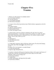
Abdominal pain:<br />
• RUQ/LUQ pain: ECG & CXR (to Rule out DX1)---->( If NL)<br />
Sonography (DX2)(figure3) ---> (NL) Amylase, Lipase,<br />
Aminotransferases (DX3)--->(NL) ERCP/CT(DX4)<br />
• Epigastric: ECG &CXR(DX1)--->(NL) UGI SERIES/Endoscopy(DX5)<br />
--> (NL)Sonography(DX6)---->(NL)DX7<br />
• Generalized:Abdominal XR(DX8)-->(NL)CT Scan(9)---><br />
(NL) Angiography (DX10) --->(NL)DX11<br />
• PERIUMBILICAL: Sonography (DX12)---> (NL)Contrast Enema (DX13)<br />
---> (NL) DX14<br />
• RLQ/LLQ: Digital Rectal Exam +Gravindex(♀) +CBC(DX15)--->(NL)<br />
Contrast Enema(DX16)--->(NL)IVP(DX17)--->(NL) DX18<br />
• Pelvic: DRE+ Gravindex +CBC(DX19)--->(NL)Bladder<br />
Catheterization(DX20)--->(NL) Sigmoidoscopy(DX21)<br />
---> (NL) IBS<br />
---------------------------------------------------------------------------------------------------<br />
Figure 3- Approach to abdominal pain<br />
DX1=MI, pericarditis, pleuritis, basilar pneumonia, pleural effusion<br />
DX2= perforated vicerea, stone, organomegaly, intestinal infarct or obstruction<br />
DX3= Acute pancreatitis, small intestine obstruction, cholecystitis, cholelithiasis,<br />
perforated PU, viral hepatitis, perihepatitis, liver abscess, liver parenchymal<br />
disease<br />
DX4=Biliary disease, CBD strictures, carcinoma<br />
DX5=PUD, reflux, tumor, gastritis<br />
DX6=cholecystitis<br />
DX7= acute pancreatitis, abdominal henia, DES<br />
DX8=perforation (of appendicitis, PU, cholecystitis, carcinoma, bowel ischemia,<br />
diverticulum), peritonitis, kidney stone, obstruction
<strong>Introduction</strong>-mdm<br />
Abdominal pain<br />
DX9= ascites, pancreatitis, perforated abscess, aneurism<br />
DX10=bowels ischemia<br />
DX11=porphyria, lead, uremia, DKA<br />
DX12=aneurism, infarct, obstruction<br />
DX13=appendicitis, diverculitis<br />
DX14=hyperperistalsis, diverticulitis<br />
DX15=ectopic pregnancy, PID, ovary disease, rectal carcinoma, prostatitis<br />
DX16=appendicitis, diverticulitis, IBD, ischemia, cancer, obstruction<br />
DX17=Ureter carcinoma, stone<br />
DX18=herpes zoster, IBS<br />
DX19=prostatitis, rectal carcinoma, proctitis, PID, EP, endometriosis, uterine<br />
rupture, acute cervicitis, endometritis<br />
DX20= Bulged bladder,<br />
DX21= sigmoid carcinoma, crohn disease, ulcerative colitis, diverticulitis
<strong>Introduction</strong>-mdm<br />
Abdominal pain<br />
FIGURE-4: Approach to abdominal mass (organomegaly)
<strong>Introduction</strong>-mdm<br />
Abdominal pain<br />
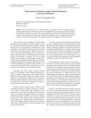
Contribution of test 1 to UR-generation<br />
Cost 2 /Test yielding a UR<br />
Dipstick urinalysis 3 0.044 6270<br />
CRP 3 0.252 116<br />
ESR 3 0.177 580<br />
Hemoglobin 3 0.084 679<br />
RBC indices 34 0.072 789<br />
WBC 3 0.204 278<br />
Total protein 3 0.022 1027<br />
Albumin 3 0.013 1732<br />
A/G 3 0.118 381<br />
Urine sediment 0.037 7731<br />
Sialic acid 0.219 542<br />
Platelet count 0.035 1622<br />
LDC 0.231 205<br />
Serum protein fraction 0.192 962<br />
profile<br />
Total cholesterol 0.076 369<br />
Glucose 0.019 1723<br />
AST 0.089 280<br />
ALT 0.086 293<br />
LD 0.062 368<br />
ALP 0.035 708<br />
GGT 0.038 628<br />
Cholinesterase 0.063 442<br />
Serum urea nitrogen 0.014 1814<br />
Creatinine 0.008 4399<br />
Uric acid 0.006 4612<br />
Fecal occult blood 5 (n 0.057 1894<br />
= 53)<br />
Chest x-ray 5 (n = 198) 0.136 6430<br />
Abdominal x-ray 5 (n = 0.353 2484<br />
17)<br />
ECG 5 (n = 79) 0.177 3909<br />
CBC 6 0.099 576<br />
CBC + LDC 0.125 440<br />
Chemistry profile 7 0.070 522<br />
Table IV. Yield and cost of individual common diagnostic tests performed for the clinical<br />
evaluation of patient’s illness and the case finding among 540 new, symptomatic primary care<br />
outpatients. (UR=useful Results)<br />
(1 Contribution of the test is calculated as follows: the number of tests yielding a UR or contributing to case finding/total number<br />
of tests performed.<br />
2 Costs are indicated in Yen (¥)<br />
3 Test components of the ELT(Essential Laboratory Tests ) panel.<br />
4 RBC, red blood cell; WBC, white blood cell count.<br />
5 Optional test items ordered if necessary. Values in parentheses indicate the number of patients in whom each of these optional<br />
tests was performed. Fecal occult blood was considered as Stool Exam for ova.<br />
6 Yield and cost of simultaneous measurement of hemoglobin + RBC indices + white blood cell count + platelet count on an<br />
automated blood cell counter<br />
7 Yield and cost of simultaneous analysis of 16 test items, including chemistry tests, CRP, and sialic acid, on an automated multi<br />
channel analyzer. )
<strong>Introduction</strong>-mdm<br />
Abdominal pain<br />
Table-V summarizes these figures.<br />
Problem list<br />
eosinophilia<br />
splenomegaly<br />
hemoptysis<br />
The first<br />
diagnostic<br />
test<br />
Stool<br />
exam<br />
Abdominal<br />
x ray or US<br />
Chest x<br />
ray<br />
Cost/Test<br />
yielding a<br />
UR<br />
1894 Yen<br />
(3570 Rls)<br />
2484 Yen<br />
(24000 Rls)<br />
6430 Yen<br />
(29000 Rls)<br />
Steps taken to Any<br />
Level of Contrib<br />
“no further<br />
limitations(timemoney-patient’s<br />
Of the of test<br />
invasiveness ution<br />
investigation”<br />
compliancereliance<br />
on<br />
external<br />
resources<br />
diagnostic<br />
tests<br />
to URgenerat<br />
ion<br />
2 depends NI-I 0.057<br />
3 depends NI 0.353<br />
4 depends NI -I 0.136<br />
Table V-comparing the three different ways to approach this case.(UR=Useful<br />
Result /NI=noninvasive/I=invasive)<br />
One point to be mentioned is that costs are indicated in Yen (¥) and can be converted<br />
to US dollars at a rate of $1.00 = ¥115.00= 9100 Iran Rials on Jan, 26th 2006. The<br />
computation, however, should be adjusted according to the country. For example, the<br />
cost of a chest x-ray is 6430 Yen or 55.91 $ or 508780 Rials. Yet, the actual cost is<br />
29000 in Iran. As table-V shows, the approach to splenomegaly by an abdominal x-<br />
ray has the highest contribution of test to useful results (calculated as : the number of<br />
tests yielding a useful result or contributing to case finding/total number of tests<br />
performed). Its cost is moderate, it’s non-invasive and it takes fewer steps to the “no<br />
further investigation is needed”.<br />
So the case was managed by splenomegaly approach.The abdominal imaging (x-ray<br />
followed by ultrasound) revealed multiple cystic lesions (30x25mm) in the right lobe<br />
of the liver and splenic cysts (one as big as 125x90 mm) in the spleen.<br />
She had a surgery and the cysts proved to be hydatid.<br />
This could explain the anaphylactoid reaction, chocolate-color sputum and<br />
eosinophilia.
<strong>Introduction</strong>-mdm<br />
Abdominal pain<br />
As deducted, what we find useful in real-world setting decision making is schematic<br />
comparisons and not isolated information of texts. That is the main reason the bulk of<br />
this book is written in figures, tables, algorithms and cases.<br />
Each chapter begins with some questions about some medical cases. The cases are<br />
discussed by schematic presentations. The drugs are presented by the dosage,<br />
contraindications, and prices in a table for comparison. The chapter ends with<br />
answers to questions and suggested reading.<br />
Acknowledgment:<br />
I should like to thank Professor David J. Solomon the editor of <strong>Medical</strong> <strong>Education</strong><br />
<strong>Online</strong> for his help in making this manuscript more eligible for archiving,<br />
Professor Lawrence Martin for the permission to use some of his ABG and<br />
mechanical ventilation problems in Respiratory Aid section,<br />
Professor Bruce Argyle for his permission to use MicroEKG Computer Program<br />
Manual.MadScientist Software of Alpine, Utah for arrhythmia management in<br />
Arrhythmia section,<br />
And Dr. Yuzuru Takemura for the permission to use his article on “yield and cost<br />
of common diagnostic tests” in the <strong>Introduction</strong> section.<br />
It should also be mentioned that ECG recordings are from Professor Frank<br />
Yanowitz’s ECG learning center , and sections on drugs and procedures are<br />
unchanged citations from the related references to avoid misunderstanding.<br />
Mitra Ahmad Soltani,<br />
MD, MS in Midwifery, MA in TEFL<br />
Azad University-Tehran School of Medicine
<strong>Introduction</strong>-mdm<br />
Abdominal pain<br />
References:<br />
1- Ayalew Tefferi, Mayo Foundation for <strong>Medical</strong> <strong>Education</strong> And Research.<br />
(2005).Blood Eosinophilia: A New Paradigm In Disease Classification, Diagnosis,<br />
And Treatment.;80:75-83<br />
2- Bidwell ,Jacob L. Pachner , Robert W. American Family Physician. (2005)<br />
Hemoptysis: Diagnosis And Management. University Of Wisconsin <strong>Medical</strong><br />
School, Milwaukee, Wisconsin.Vol. 72/No. 7<br />
www.aafp.org/afp/20051001/1253.html<br />
3- Braunwald Eugene, et al. Harrison's Principles of Internal Medicine. 16th<br />
edition. McGrawHill; 2005<br />
4- CDC.Advance Data No. 340 . March 18, 2004<br />
5- Patel ,Vimla L. Kaufman ,David R And Arocha ,Jose F. Journal of Biomedical<br />
Informatics (2002), Emerging Paradigms Of Cognition In <strong>Medical</strong> Decision<br />
Making, Columbia University, New York, USA<br />
6-Ringertz,Hans. State of The Art Imaging Of Abdominal Masses In Childhood,<br />
Http://Www.Star-Program.Com/Data--Star-Program/Upload /Star_Abstracts_<br />
752_Ringertz1.Pdf<br />
7- Takemura,Yuzuru. Haku Ishida, Yuji Inoue And Beck J. Robert. Clinical<br />
Chemistry. (2002) .Yield And Cost Of Individual Common Diagnostic Tests In<br />
New Primary Care Outpatients In Japan.;48:42-54.<br />
www.clinchem.org/cgi/content/full/48/1/42
<strong>Introduction</strong>-mdm<br />
Abdominal pain