New Jersey Neuroscience Institute at JFK Medical Center
New Jersey Neuroscience Institute at JFK Medical Center
New Jersey Neuroscience Institute at JFK Medical Center
Create successful ePaper yourself
Turn your PDF publications into a flip-book with our unique Google optimized e-Paper software.
J O U R N A L O F T H E<br />
<strong>New</strong> <strong>Jersey</strong><br />
<strong>Neuroscience</strong> <strong>Institute</strong><br />
JNJNI • V o l u m e 5 I s s u e 2 • D e c e m b e r 2010<br />
UNPARALLELED DEPTH. UNRIVALED EXCELLENCE.<br />
J F K M E D I C A L C E N T E R
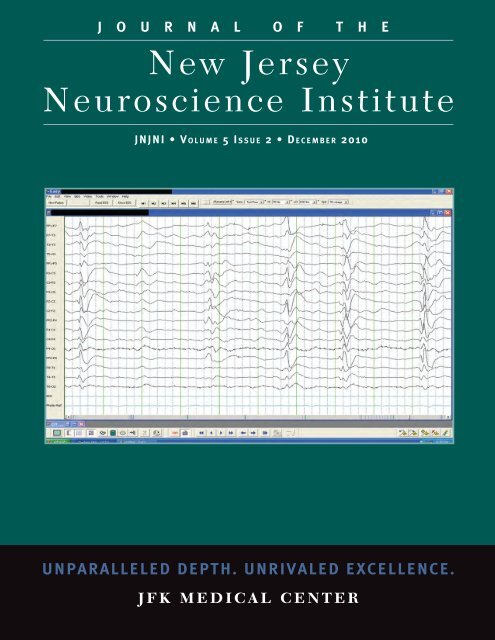
Cover Image: For details see page 18, Figure 3
Table of Contents<br />
<strong>New</strong> <strong>Jersey</strong> <strong>Neuroscience</strong> <strong>Institute</strong> <strong>at</strong> <strong>JFK</strong> <strong>Medical</strong> <strong>Center</strong>.. . . . . . . . . . . . . . . . . . . . . . . . . . . . . . . . . . . . . . . . 2<br />
Aim and Scope.. . . . . . . . . . . . . . . . . . . . . . . . . . . . . . . . . . . . . . . . . . . . . . . . . . . . . . . . . . . . . . . . . . . . . . . . . . . . 3<br />
Editors’ Corner.. . . . . . . . . . . . . . . . . . . . . . . . . . . . . . . . . . . . . . . . . . . . . . . . . . . . . . . . . . . . . . . . . . . . . . . . . . . . 4<br />
Sudhansu Chokroverty, MD, FRCP, FACP; Martin Gizzi, MD, PhD<br />
Management of Low Back Pain . . . . . . . . . . . . . . . . . . . . . . . . . . . . . . . . . . . . . . . . . . . . . . . . . . . . . . . . . . . . . . . 6<br />
Ronald D. Karnaugh, MD; Sandeep Vaid, MD<br />
An Unusual Present<strong>at</strong>ion of Creutzfeldt Jacob Disease and an Example of<br />
How Hickam’s Dictum and Ockham’s Razor Can Both Be Right .. . . . . . . . . . . . . . . . . . . . . . . . . . . . . . . . . . 17<br />
Eli S. Neiman, DO; Amtul Farheen, MD; Nancy Gadallah, DO; Thomas Steineke, MD, PhD; Peter<br />
Parsells, PA-C; Michael Rosenberg, MD<br />
Hirayama Disease/Monomelic Amyotrophy: A Case Report . . . . . . . . . . . . . . . . . . . . . . . . . . . . . . . . . . . . . . 20<br />
Amtul Farheen, MD<br />
Cardiovascular Comorbidities in Rapid Eye Movement Sleep Predominant Obstructive<br />
Sleep Apnea (REM-pOSA): A Pilot Study.. . . . . . . . . . . . . . . . . . . . . . . . . . . . . . . . . . . . . . . . . . . . . . . . . . . . . . 22<br />
Justin Pi, MD; Sushanth Bh<strong>at</strong>, MD; Irving Smith, DO; Besher Kabak, MD; Divya Gupta, MD; Sudhansu<br />
Chokroverty, MD, FRCP, FACP<br />
A Historical Overview of Legal Insanity . . . . . . . . . . . . . . . . . . . . . . . . . . . . . . . . . . . . . . . . . . . . . . . . . . . . . . . 24<br />
Daniel P. Greenfield, MD, MPH, MS; Jack A. Gottschalk, JD, MA, MSM<br />
Wh<strong>at</strong>’s <strong>New</strong> in <strong>Neuroscience</strong>?.. . . . . . . . . . . . . . . . . . . . . . . . . . . . . . . . . . . . . . . . . . . . . . . . . . . . . . . . . . . . . . . 31<br />
Sudhansu Chokroverty, MD, FRCP, FACP<br />
Instructions to the Authors .. . . . . . . . . . . . . . . . . . . . . . . . . . . . . . . . . . . . . . . . . . . . . . . . . . . . . . . . . . . . . . . . . 32<br />
p a g e o n e
<strong>New</strong> <strong>Jersey</strong> <strong>Neuroscience</strong> <strong>Institute</strong> <strong>at</strong> <strong>JFK</strong> <strong>Medical</strong> <strong>Center</strong><br />
<strong>New</strong> <strong>Jersey</strong> <strong>Neuroscience</strong> <strong>Institute</strong> (NJNI) <strong>at</strong> <strong>JFK</strong> <strong>Medical</strong> <strong>Center</strong> is a comprehensive facility designed<br />
exclusively for the diagnosis, tre<strong>at</strong>ment, and research of complex neurological and neurosurgical disorders in<br />
adults and children. Services offered <strong>at</strong> the <strong>Institute</strong> include programs in minimally invasive and reconstructive<br />
spine surgery, peripheral nerve surgery, brain tumors, dizziness and balance disorders, epilepsy, sleep, memory<br />
problems/dementia, cerebral palsy, stroke, spasticity, movement disorders, and neuromuscular disorders. As<br />
a department of Seton Hall University’s (SHU) School of Gradu<strong>at</strong>e <strong>Medical</strong> Educ<strong>at</strong>ion, NJNI serves as the<br />
clinical setting for residency training in neurology and fellowship training in clinical neurophysiology and sleep<br />
medicine. For more inform<strong>at</strong>ion on the <strong>New</strong> <strong>Jersey</strong> <strong>Neuroscience</strong> <strong>Institute</strong>, call 732-321-7950 or visit the<br />
facility online <strong>at</strong> www.njneuro.org.<br />
p a g e t w o<br />
T h e N e w J e r s e y N e u r o s c i e n c e I n s t i t u t e a t J F K M e d i c a l C e n t e r
AIM and SCOPE<br />
The Journal of the <strong>New</strong> <strong>Jersey</strong> <strong>Neuroscience</strong> <strong>Institute</strong> (JNJNI) focuses on topics of interest to clinical scientists<br />
covering all subspecialty disciplines of neuroscience as practiced in the <strong>Institute</strong> and makes clinical inform<strong>at</strong>ion<br />
accessible to all practitioners. The fundamental goal is to promote good health throughout the community by<br />
educ<strong>at</strong>ing practitioners and investig<strong>at</strong>ing the causes and cures of neurological and neurosurgical ailments.<br />
JNJNI publishes the following types of articles: editorials, reviews, original research articles, historical<br />
articles, controversies, case reports, wh<strong>at</strong>’s new in neuroscience, images in neuroscience, letters to the editors,<br />
and news and announcements.<br />
Editors<br />
Sudhansu Chokroverty, MD, FRCP, FACP<br />
Martin Gizzi, MD, PhD<br />
Editorial Advisory Board<br />
Stephen Bloomfield, MD<br />
Raji Grewal, MD<br />
Gay Holstein, PhD<br />
Thomas Steineke, MD, PhD<br />
Michael Rosenberg, MD<br />
Editorial Assistant:<br />
Annabella Drennan<br />
Director of Public Rel<strong>at</strong>ions and Marketing:<br />
Steven Weiss<br />
Publishing Office:<br />
<strong>New</strong> <strong>Jersey</strong> <strong>Neuroscience</strong> <strong>Institute</strong> <strong>at</strong> <strong>JFK</strong><br />
p a g e t h r e e
Editors’ Corner<br />
JNJNI, first published in 2007, encourages submissions of clinically relevant articles to enhance the quality of<br />
p<strong>at</strong>ient care and excellence of the journal. We begin this issue by trying to rejuvin<strong>at</strong>e you, the readers and the<br />
contributors. This is your journal and its success belongs to all who are dedic<strong>at</strong>ed to raising the standard of the<br />
journal and, indirectly, p<strong>at</strong>ient care. We hope to <strong>at</strong>tract quality articles from clinicians and researchers both<br />
within and outside the <strong>Institute</strong> in future issues. This is a call for action.<br />
In this winter issue we are publishing five articles in addition to our ongoing section “Wh<strong>at</strong>’s <strong>New</strong> in<br />
<strong>Neuroscience</strong>?”<br />
The first article is a timely review of low back pain management. This is probably the commonest condition<br />
in neuroscience practice, and it requires meticulous <strong>at</strong>tention. Karnaugh and Vaid write an excellent review<br />
of management issues, strictly adhering to practical points and emphasizing the clinical approach. Their<br />
fundamental point is th<strong>at</strong> management must depend on astute clinical judgment and assessment—not on<br />
labor<strong>at</strong>ory investig<strong>at</strong>ions (e.g., MRI, EMG/NCV, etc.)—in order to provide the best quality tre<strong>at</strong>ment, r<strong>at</strong>her<br />
than subjecting p<strong>at</strong>ients to a b<strong>at</strong>tery of tests and numerous unnecessary drugs, some of which are very toxic and<br />
addictive.<br />
The second article is a case report with a logical approach to a complic<strong>at</strong>ed and unusual case of chiari<br />
malform<strong>at</strong>ion with brainstem compression and Creutzfeldt-Jacob disease by Neiman and coinvestig<strong>at</strong>ors. The<br />
authors have shown th<strong>at</strong> through a structured approach, r<strong>at</strong>her than haphazard investig<strong>at</strong>ions, one can make a<br />
diagnosis even in a complex and rare case.<br />
The third article by Farheen describes an interesting case of monomelic amyotrophy which often poses a<br />
clinical dilemma in terms of differential diagnosis between the rel<strong>at</strong>ively benign and more serious conditions<br />
like amyotrophic l<strong>at</strong>eral sclerosis.<br />
The fourth article deals with an uncommon condition, namely rapid eye movement (REM) sleep predominant<br />
obstructive sleep apnea (REM-pOSA), which may have serious long-term adverse consequences (as in OSAS)<br />
particularly on the cardiovascular system. Many sleep specialists, including neurologists, are not always aware<br />
of this.<br />
p a g e f o u r<br />
T h e N e w J e r s e y N e u r o s c i e n c e I n s t i t u t e a t J F K M e d i c a l C e n t e r
The fifth article appearing under the “Historical Section” gives a fascin<strong>at</strong>ing overview of legal insanity as a<br />
legal defense, starting from ancient to modern jurisprudence, citing many historically significant examples.<br />
The final section “Wh<strong>at</strong>’s <strong>New</strong> in <strong>Neuroscience</strong>?” deals with two articles. Dijk and Tijseen gave a nice<br />
overview of therapeutic management of p<strong>at</strong>ients with myoclonus based on clinical, etiological and an<strong>at</strong>omical<br />
classific<strong>at</strong>ion of myoclonus. The second article by Dubois and collabor<strong>at</strong>ors brought to our <strong>at</strong>tention the<br />
revised new definition of Alzheimer’s disease clinical spectrum based on recent advances in the use of reliable<br />
biomarkers instead of the time-honored McKhann criteria. This will stimul<strong>at</strong>e further research for potential<br />
drug discovery to intercede in the p<strong>at</strong>hogenic cascade of the disease.<br />
We hope these articles will stimul<strong>at</strong>e readers and contributors to look for a copy of JNJNI’s last issue of 2010<br />
and encourage new submissions for the spring 2011 issue.<br />
Sudhansu Chokroverty, MD, FRCP, FACP<br />
Martin Gizzi, MD, PhD<br />
Editors-in-Chief, JNJNI<br />
p a g e f i v e
R e v i e w<br />
Management of Low Back Pain<br />
Ronald D. Karnaugh, MD; Sandeep Vaid, MD<br />
Abstract<br />
Low back pain (LBP) is a common complaint th<strong>at</strong><br />
prompts many p<strong>at</strong>ients to visit their primary care<br />
provider and causes a significant socioeconomic burden<br />
on the U.S. healthcare system. The management<br />
of LBP requires the clinician to be aggressive with<br />
the diagnosis, yet conserv<strong>at</strong>ive with the medical<br />
management. A comprehensive p<strong>at</strong>ient evalu<strong>at</strong>ion<br />
is required to identify the pain gener<strong>at</strong>or and help<br />
guide clinicians toward an appropri<strong>at</strong>e tre<strong>at</strong>ment<br />
plan. Furthermore, imaging and diagnostic studies<br />
should be performed as an extension of the history and<br />
physical examin<strong>at</strong>ion to help identify an underlying<br />
p<strong>at</strong>hology and to guide tre<strong>at</strong>ment. Most importantly,<br />
the clinician should be aware of red flags which may<br />
indic<strong>at</strong>e the presence of a more serious p<strong>at</strong>hology<br />
requiring immedi<strong>at</strong>e intervention. Implement<strong>at</strong>ion of a<br />
multidisciplinary conserv<strong>at</strong>ive care approach consisting<br />
of reassurance, p<strong>at</strong>ient educ<strong>at</strong>ion, recognizing<br />
underlying psychosocial problems, NSAIDs, physical<br />
therapy, and injection procedures can prevent acute<br />
LBP from progressing into chronic LBP.<br />
Introduction<br />
Spine Care is the fastest growing sector of outp<strong>at</strong>ient<br />
practice for neurologists, physi<strong>at</strong>rists and primary care<br />
providers. An estim<strong>at</strong>ed 25% of adults in the U.S.<br />
report <strong>at</strong> least one episode of low back pain (LBP) in<br />
the previous 3 months. LBP is reported to be the fifth<br />
most common reason th<strong>at</strong> p<strong>at</strong>ients visit their primary<br />
care provider in the U.S. 1<br />
The management of LBP requires a comprehensive<br />
approach with the goal of allevi<strong>at</strong>ing pain, restoring<br />
function, and improving overall quality of life. The<br />
clinician must formul<strong>at</strong>e an accur<strong>at</strong>e diagnosis in<br />
order to implement a targeted tre<strong>at</strong>ment plan. To<br />
achieve this goal, the clinician must be able to obtain a<br />
detailed history and perform a comprehensive physical<br />
examin<strong>at</strong>ion. The results from the evalu<strong>at</strong>ion will<br />
then determine the appropri<strong>at</strong>e tre<strong>at</strong>ment plan and<br />
whether further diagnostic testing is warranted . 2<br />
History<br />
LBP is common and generally a benign, self limiting<br />
disease. During the initial present<strong>at</strong>ion, the neurologist<br />
should rule out any life thre<strong>at</strong>ening causes for LBP<br />
and be aware of red flags (see Table 1). For example,<br />
cauda equina syndrome (which may present as saddle<br />
sensory disturbances, bladder and bowel dysfunction,<br />
and variable lower extremity motor and sensory loss),<br />
malignancy (night pain th<strong>at</strong> awakens the p<strong>at</strong>ient from<br />
sleep and unexplained weight loss) or spinal infections<br />
including discitis, osteomyelitis, and epidural abscess<br />
(with associ<strong>at</strong>ed febrile illness) all require emergent<br />
workup and intervention. 3<br />
The clinician should make<br />
note of symptom onset in order to differenti<strong>at</strong>e acute<br />
LBP from chronic LBP. The subjective p<strong>at</strong>ient history<br />
should include the quality of pain, timing, frequency,<br />
severity, referral p<strong>at</strong>tern, aggrav<strong>at</strong>ing and allevi<strong>at</strong>ing<br />
factors and any other associ<strong>at</strong>ed symptoms. 4<br />
The use<br />
of a pain assessment chart (such as a body chart) can<br />
be helpful (see Figure 1) in localizing the pain while<br />
identifying areas of radi<strong>at</strong>ion...It is important to note th<strong>at</strong><br />
by the time a p<strong>at</strong>ient presents to the neurology clinic for<br />
p a g e s i x<br />
T h e N e w J e r s e y N e u r o s c i e n c e I n s t i t u t e a t J F K M e d i c a l C e n t e r
low back pain, they have most likely tried conserv<strong>at</strong>ive<br />
methods of tre<strong>at</strong>ment. These tre<strong>at</strong>ments may include<br />
over the counter medic<strong>at</strong>ions for pain, chiropractic<br />
manipul<strong>at</strong>ion, physical therapy, and epidural steroid<br />
injections. Identific<strong>at</strong>ion of previous effective and<br />
non-effective medic<strong>at</strong>ions or modalities will aid in<br />
the differential diagnosis. Detailed questioning of the<br />
p<strong>at</strong>ient about aggrav<strong>at</strong>ing and allevi<strong>at</strong>ing factors can<br />
assist in localizing the probable pain gener<strong>at</strong>or. Pain<br />
associ<strong>at</strong>ed with prolonged sitting, forward flexion, pain<br />
radi<strong>at</strong>ing down the leg, coughing, sneezing, and bowel<br />
movement may be discogenic in n<strong>at</strong>ure. Pain th<strong>at</strong> is<br />
exacerb<strong>at</strong>ed with prolonged walking, standing, spinal<br />
hyperextension or l<strong>at</strong>eral bending may be secondary to<br />
facet joint dysfunction. 5.. P<strong>at</strong>ients may also complain of<br />
pain <strong>at</strong> the tailbone with prolonged sitting, standing up<br />
from sitting position, and bowel movement, which may<br />
be due to coccydynia. Coccydynia can be secondary to<br />
trauma, fracture, and malignancy to the coccyx bone.<br />
The clinician should, however, keep in mind th<strong>at</strong> these<br />
pain gener<strong>at</strong>ors have overlapping referral p<strong>at</strong>terns and<br />
the physical examin<strong>at</strong>ion will further localize the source<br />
of pain.<br />
Physical examin<strong>at</strong>ion<br />
In conjunction with the detailed history, a<br />
comprehensive physical examin<strong>at</strong>ion will assist in<br />
narrowing the diagnosis. The examin<strong>at</strong>ion of the<br />
Figure 1. Figure 1.<br />
Please Complete The Pain Drawing<br />
WHERE IS YOUR PAIN? Please mark on<br />
the drawing where you feel pain right now<br />
and use the key below the figures.<br />
Pins & Needles - oooo<br />
Stabbing - ////<br />
Burning - xxxx<br />
Deep Ache - zzzz<br />
p a g e s e v e n
R e v i e w<br />
lumbar spine should focus on the evalu<strong>at</strong>ion of the<br />
musculoskeletal, neurological, and vascular systems.<br />
Focus should be placed on several key elements<br />
including visual inspection, palp<strong>at</strong>ion, range of motion,<br />
neurological, and functional assessment.<br />
Pain associ<strong>at</strong>ed with flexion can be secondary to<br />
p<strong>at</strong>hologies of the intervertebral disc, vertebral body,<br />
interspinous ligament, or paraspinal muscul<strong>at</strong>ure. .<br />
This will help narrow the differential diagnosis<br />
to discogenic p<strong>at</strong>hology, compression fracture,<br />
interaspinous ligmantus sprain, or paraspinal muscle<br />
strain. Typical p<strong>at</strong>ient complaints with such diagnoses<br />
include pain with sitting, bending or lifting. The pain<br />
will improve in supine or prone position or while<br />
standing or walking. On examin<strong>at</strong>ion forward flexion<br />
will increase the pain intensity and the p<strong>at</strong>ient may<br />
complain of radicular symptoms in the leg. Special<br />
tests which provoke symptoms such as a straight-leg<br />
raise (SLR), slump test, or femoral nerve stretch test<br />
will further clarify if the pain gener<strong>at</strong>or is discogenic in<br />
n<strong>at</strong>ure (see Table 2). 6<br />
LBP associ<strong>at</strong>ed with back extension is usually<br />
associ<strong>at</strong>ed with posterior element p<strong>at</strong>hology such<br />
as pars interarticularis defect, facet arthrop<strong>at</strong>hy,<br />
foraminal stenosis, or l<strong>at</strong>eral recess stenosis. P<strong>at</strong>ients<br />
will complain of pain with standing, walking, running,<br />
and extension. The pain is generally relieved when<br />
flexing the back, sitting or lying with some flexion, but<br />
the pain is typically worse when lying prone. P<strong>at</strong>ients<br />
may complain of some leg symptoms, but usually the<br />
pain does not radi<strong>at</strong>e below the knee unless significant<br />
stenosis is present. On examin<strong>at</strong>ion, there is increased<br />
back pain with extension and l<strong>at</strong>eral bending but<br />
no significant pain on forward flexion. 6<br />
Tenderness<br />
to palp<strong>at</strong>ion in the region of lower lumbar facet<br />
joint is noted and this can be localized close to the<br />
sacroiliac (SI) joint. SLR is usually neg<strong>at</strong>ive; however,<br />
hamstrings are usually tight. Possible gener<strong>at</strong>ors for<br />
back pain associ<strong>at</strong>ed with hyperextension include<br />
spondylosis, scoliosis, spondylolysis, spondylolisthesis,<br />
spinal stenosis, and facet joint syndromes.<br />
Confirming the diagnosis<br />
Diagnostic studies should be used as an extension<br />
of the history and physical examin<strong>at</strong>ion and not as a<br />
substitute. They should not be ordered indiscrimin<strong>at</strong>ely<br />
due to number of false positive results noted in<br />
Table 1.<br />
Life Thre<strong>at</strong>ening and Urgent Conditions<br />
MEDICAL<br />
Infection: osteomyelitis, epidural abscess,<br />
discitis<br />
Hem<strong>at</strong>ologic: primary or metast<strong>at</strong>ic cancer,<br />
multiple myeloma, myelodysplasia<br />
Retroperitoneal p<strong>at</strong>hology: pyelonephritis,<br />
renal calculus<br />
Benign tumors<br />
Aortic aneurysm<br />
Abdominal p<strong>at</strong>hology: pancre<strong>at</strong>itis,<br />
perfor<strong>at</strong>ed viscera<br />
MUSCULOSKELETAL<br />
Lumbar or sacral nerve root compression<br />
Disc herni<strong>at</strong>ion, cauda equina syndrome,<br />
spinal stenosis<br />
Vertebral fracture<br />
Sacroiliac joint sprain<br />
Arthritic conditions: osteoarthritis,<br />
rheum<strong>at</strong>ologic conditions<br />
Lumbar muscle strain<br />
Modified from Deyo et al. 3<br />
p a g e e i g h t<br />
T h e N e w J e r s e y N e u r o s c i e n c e I n s t i t u t e a t J F K M e d i c a l C e n t e r
asymptom<strong>at</strong>ic subjects. Diagnostic studies such as<br />
plain radiographs of the lumbar spine are not routinely<br />
needed in the evalu<strong>at</strong>ion of most episodes of LBP.<br />
Abnormalities on X-ray do not correl<strong>at</strong>e with degree<br />
of p<strong>at</strong>ient symptoms, but X-rays are helpful in the<br />
initial evalu<strong>at</strong>ion to rule out a more serious condition<br />
in p<strong>at</strong>ients with red flags such as tumors, osteomyelitis,<br />
and retroperitoneal p<strong>at</strong>hology including small and<br />
large bowel obstruction and bowel perfor<strong>at</strong>ion. 3<br />
Bone Scans<br />
In general, bone scans are not useful in evalu<strong>at</strong>ing acute<br />
LBP. However, they can be very helpful in confirming<br />
the diagnosis of an acute spondylolysis (stress fracture<br />
of pars interarticularis) especially with single photon<br />
emission computed tomography (SPECT) imaging.<br />
Furthermore, they may be useful when the history<br />
and examin<strong>at</strong>ion indic<strong>at</strong>e the possibility of infection<br />
or tumor. 7<br />
Table 2. Provac<strong>at</strong>ive maneuvers for the diagnosis of lumbar disk herni<strong>at</strong>ion.<br />
MANEUVER<br />
Straight-leg raise<br />
Cross straight-leg raise<br />
Slump test<br />
Femoral nerve stretch test<br />
PROCEDURE<br />
With the p<strong>at</strong>ient lying supine, the leg is raised with<br />
the knee extended; elev<strong>at</strong>ion of the leg is stopped<br />
when the p<strong>at</strong>ient begins to feel pain; the result is<br />
positive when the angle is between 30º and 70º<br />
and the pain is reproduced down the posterior<br />
thigh to below the knee.<br />
Same as above, with pain elicited on raising the<br />
contral<strong>at</strong>eral leg.<br />
The p<strong>at</strong>ient is se<strong>at</strong>ed with legs together and knees<br />
against the examin<strong>at</strong>ion table; the p<strong>at</strong>ient slumps<br />
forward as far as possible and the examiner applies<br />
firm pressure to bow his or her back while keeping<br />
the sacrum vertical; the p<strong>at</strong>ient is then asked to flex<br />
the head, and pressure is added to neck flexion;<br />
the examiner then extends the knee and adds<br />
dorsiflexion to the ankle; the test result is positive<br />
when there is reproduction of symptoms.<br />
With the p<strong>at</strong>ient prone, the examiner places<br />
his palm <strong>at</strong> the popliteal fossa as the knee is<br />
dorsiflexed; the test result is positive when there is<br />
reproduction of symptoms in the anterior thigh or<br />
back or both; this test is used to make a diagnosis<br />
of high lumbar disk herni<strong>at</strong>ions.<br />
Modified from Solomon J et al. Physical examin<strong>at</strong>ion of the lumbar spine. In: Malanga GA, Nadler SF, editors: Musculoskeletal Physical<br />
Examin<strong>at</strong>ion. Philadelphia: Elsevier Mosby, 2006; p. 210-213. Reproduced with permission from GA Malanga.<br />
p a g e n i n e
R e v i e w<br />
Magnetic Resonance Imaging (MRI)<br />
MRI has been shown to have excellent sensitivity in<br />
the diagnosis of lumbar disc herni<strong>at</strong>ion. This modality<br />
should be reserved for p<strong>at</strong>ients who present with<br />
persistent pain for more than 6 to 8 weeks or with<br />
neurologic deficits such as progressive weakness,<br />
sensory deficit, or a dropped reflex. Other consider<strong>at</strong>ions<br />
include cauda equina symptoms, high risk p<strong>at</strong>ients<br />
for cancer, infection, or inflamm<strong>at</strong>ory disorders. 8<br />
It<br />
should be noted, however, th<strong>at</strong> a significant number<br />
of false positive results are found in the asymptom<strong>at</strong>ic<br />
popul<strong>at</strong>ion. 9<br />
In p<strong>at</strong>ients presenting with pain th<strong>at</strong><br />
radi<strong>at</strong>es in a specific derm<strong>at</strong>omal distribution along<br />
with other neurological findings of weakness, sensory<br />
changes, or asymmetrical reflex secondary to a<br />
radiculop<strong>at</strong>hy due to disc herni<strong>at</strong>ion, an MRI study of<br />
lumbar spine is necessary to help guide tre<strong>at</strong>ment such<br />
as an injection procedure. In the p<strong>at</strong>ient presenting<br />
with LBP, the addition of gadolinium is not necessary<br />
in the majority of p<strong>at</strong>ients. 9<br />
Computerized tomography (CT) imaging<br />
CT scans are a good modality for imaging osseous<br />
structures; however, they are inferior to MRI for<br />
detecting disc herni<strong>at</strong>ion. P<strong>at</strong>ients who are unable to<br />
undergo MRI scanning due to permanent pacemaker<br />
or metallic implants are imaged using CT. A CT scan<br />
of the spine can detect spinal stenosis, facet arthrosis,<br />
herni<strong>at</strong>ed nucleus pulposus (HNP), spondylolysis,<br />
osteoporosis, and neoplasms. 10 CT scans of the spine<br />
in conjunction with myelography are a gre<strong>at</strong> modality<br />
for p<strong>at</strong>ients with spinal stenosis who are contempl<strong>at</strong>ing<br />
surgery.<br />
Electrodiagnosis<br />
Electrodiagnosis, which includes electromyography<br />
(EMG) and nerve conduction studies (NCS),<br />
should be considered as an extension of the physical<br />
examin<strong>at</strong>ion. It is helpful among p<strong>at</strong>ients with limb<br />
pain where the diagnosis remains unclear. The study<br />
can be helpful in ruling out other causes of sensory and<br />
motor complic<strong>at</strong>ions such as peripheral neurop<strong>at</strong>hy,<br />
mononeurop<strong>at</strong>hy, and motor neuron disease. EMG<br />
studies can be used in p<strong>at</strong>ients with significant<br />
weakness secondary to neuropraxia and pain inhibition<br />
from significant axonal injury. The use of EMG is not<br />
necessary in p<strong>at</strong>ients with isol<strong>at</strong>ed low back symptoms.<br />
If diagnosis of radiculop<strong>at</strong>hy is unequivocal after a<br />
detailed history and physical examin<strong>at</strong>ion, the addition<br />
of EMG and NCS does not improve the tre<strong>at</strong>ment<br />
outcome and thus is not required. 11<br />
General tre<strong>at</strong>ment principles<br />
LBP tre<strong>at</strong>ment should begin once the diagnosis is<br />
made and the red flags have been ruled out. LBP<br />
management requires a multifaceted approach with<br />
the goal of minimizing pain, normalizing activities of<br />
daily livings, and improving overall quality of life. The<br />
n<strong>at</strong>ural history of LBP is to improve with or without<br />
tre<strong>at</strong>ment, but certain tre<strong>at</strong>ments can hasten the<br />
process and are worthwhile. Successful management<br />
requires the p<strong>at</strong>ient’s particip<strong>at</strong>ion in the tre<strong>at</strong>ment<br />
plan and understanding the cause of the LBP. The<br />
clinician must explain the working diagnosis and<br />
review the basic an<strong>at</strong>omy and biomechanics th<strong>at</strong> led to<br />
the LBP. The tre<strong>at</strong>ment plan should be reviewed and<br />
r<strong>at</strong>ionale for diagnostic studies should be identified. It<br />
is important to note th<strong>at</strong> the vast majority of p<strong>at</strong>ients<br />
with LBP can be tre<strong>at</strong>ed through conserv<strong>at</strong>ive methods<br />
with reassurance. However, p<strong>at</strong>ients presenting to the<br />
neurologist or physi<strong>at</strong>rist may have tried conserv<strong>at</strong>ive<br />
p a g e t e n<br />
T h e N e w J e r s e y N e u r o s c i e n c e I n s t i t u t e a t J F K M e d i c a l C e n t e r
methods without success. These p<strong>at</strong>ients may be<br />
candid<strong>at</strong>es for more aggressive tre<strong>at</strong>ment modalities<br />
such as stronger non-steroidal anti-inflamm<strong>at</strong>ory drugs<br />
(NSAIDs), opioid analgesics, oral corticosteroids,<br />
antidepressants, antiepileptic medic<strong>at</strong>ion, injection<br />
therapy, and finally surgical intervention. Again, the<br />
physician must have the correct diagnosis in order to<br />
implement the correct tre<strong>at</strong>ment plan.<br />
Life style modific<strong>at</strong>ion<br />
For most p<strong>at</strong>ients, LBP can be tre<strong>at</strong>ed with lifestyle<br />
modific<strong>at</strong>ion and educ<strong>at</strong>ion. P<strong>at</strong>ients should be<br />
instructed on proper posture and biomechanics while<br />
performing household and occup<strong>at</strong>ional activities.<br />
The American Obesity Associ<strong>at</strong>ion has reported th<strong>at</strong><br />
one-third of Americans classified as obese suffer from<br />
musculoskeletal and joint pain. Due to the excess<br />
weight the spine can become misaligned and stressed<br />
unevenly. Weight reduction has been proven to control<br />
pain among this popul<strong>at</strong>ion. Studies have shown th<strong>at</strong><br />
smoking is also a caus<strong>at</strong>ive factor of LBP. A recent<br />
public<strong>at</strong>ion by Shiri and associ<strong>at</strong>es concluded th<strong>at</strong><br />
current smokers and individuals who smoked in the<br />
past have a higher incidence of LBP when compared<br />
to individuals who never smoked. 12<br />
The risk of back<br />
pain associ<strong>at</strong>ed with smoking was modest among adults<br />
and was noted to be even gre<strong>at</strong>er in the adolescent<br />
popul<strong>at</strong>ion. Lastly, one must also consider assessing<br />
psychosocial factors which will undermine the results<br />
of tre<strong>at</strong>ment, i.e., by tre<strong>at</strong>ing depression in conjunction<br />
with tre<strong>at</strong>ing the p<strong>at</strong>ient’s underlying pain gener<strong>at</strong>or.<br />
Bed rest<br />
Some benefits can be gained from bed rest secondary<br />
to the reduction in intradiscal pressure while lying in<br />
the supine position. But bed rest has many detrimental<br />
effects on bone, connective tissue, muscles, and<br />
cardiovascular fitness. For non-radicular LBP, 2 days<br />
of bed rest have been shown to be as effective as 7<br />
days. 13<br />
For radicular symptoms, however, limited<br />
bed rest in conjunction with standing and walking (as<br />
toler<strong>at</strong>ed) is beneficial and ideal. 14<br />
P<strong>at</strong>ients should be<br />
educ<strong>at</strong>ed on positions th<strong>at</strong> may exacerb<strong>at</strong>e their back<br />
pain symptoms. P<strong>at</strong>ients with discogenic pain should<br />
be instructed to avoid prolonged sitting, bending and<br />
lifting and educ<strong>at</strong>ed on proper work st<strong>at</strong>ion ergonomics<br />
to minimize back and neck pain due to poor posture.<br />
Exercise<br />
The goal of an exercise program in p<strong>at</strong>ients with LBP is<br />
to control pain, strengthen muscles and restore proper<br />
motion of the spine and trunk. Studies have shown<br />
th<strong>at</strong> p<strong>at</strong>ients with LBP have a reduction in aerobic<br />
fitness th<strong>at</strong> may contribute to exacerb<strong>at</strong>ion of pain. 15<br />
Conversely, evidence has shown th<strong>at</strong> <strong>at</strong>hletes with high<br />
cardiovascular fitness rarely have LBP. 16<br />
Improving<br />
cardiovascular fitness in addition to an active exercise<br />
program is a reasonable tre<strong>at</strong>ment modality; p<strong>at</strong>ients<br />
should be encouraged to stay active in order to avoid<br />
deconditioning.<br />
Physical therapy<br />
There is conflicting liter<strong>at</strong>ure on the effects of<br />
strengthening exercises in acute LBP, but their effects<br />
on p<strong>at</strong>ients with chronic LBP have the best outcome.<br />
Various exercise modalities are used when strength<br />
training such as flexion, extension, and dynamic lumbar<br />
stabiliz<strong>at</strong>ion exercise. Deb<strong>at</strong>e surrounds the merits<br />
of flexion versus extension type exercise. Flexion<br />
exercise appears to be more reasonable in p<strong>at</strong>ients<br />
with posterior element problems; 17 extension exercise<br />
has been effective in p<strong>at</strong>ients with discogenic LBP. 18<br />
A dynamic lumbar stabiliz<strong>at</strong>ion program has been<br />
shown to be most effective for a multitude of low<br />
p a g e e l e v e n
R e v i e w<br />
back problems. 19 The program emphasizes maintaining<br />
a neutral spine position coupled with progressive<br />
strengthening exercise of the trunk muscles, i.e.,<br />
back extensors, abdominals, and gluteal muscles. The<br />
goal is to develop muscular support of the trunk to<br />
diminish stress on the bones, discs, ligaments, etc. 19<br />
This tre<strong>at</strong>ment modality requires close supervision,<br />
direction and a hands-on approach by the tre<strong>at</strong>ing<br />
physical therapist.<br />
Manipul<strong>at</strong>ion/Mobiliz<strong>at</strong>ion<br />
With tre<strong>at</strong>ing acute LBP, several studies suggest th<strong>at</strong><br />
manipul<strong>at</strong>ion during the first 3 weeks decreases painful<br />
episodes, but this is controversial. Nevertheless, the<br />
benefits are primarily reduction in symptoms in the<br />
acute phase with no evidence of long term benefits.<br />
Only p<strong>at</strong>ients with LBP appear to respond; those with<br />
radicular pain do not show improvement in their pain.<br />
Medic<strong>at</strong>ions<br />
Medic<strong>at</strong>ions from many different classes have been<br />
used to tre<strong>at</strong> LBP and each has unique risks and<br />
benefits in the tre<strong>at</strong>ment. When prescribing these<br />
medic<strong>at</strong>ions, the clinician should have knowledge of<br />
the indic<strong>at</strong>ion and contraindic<strong>at</strong>ion.<br />
Acetaminophen<br />
Acetaminophen is an inexpensive over the counter<br />
medic<strong>at</strong>ion and generally a safe analgesic. It is<br />
effective for mild to moder<strong>at</strong>e pain but has no effect<br />
on inflamm<strong>at</strong>ion or muscle spasm. This medic<strong>at</strong>ion<br />
is generally not considered a first-line medic<strong>at</strong>ion for<br />
LBP and used if the p<strong>at</strong>ient has contraindic<strong>at</strong>ion to<br />
other medic<strong>at</strong>ions.<br />
Nonsteroidal anti-inflamm<strong>at</strong>ory drugs (NSAIDs)<br />
NSAIDs are reasonable medic<strong>at</strong>ions for pain relief<br />
and have anti-inflamm<strong>at</strong>ory effects. NSAIDs are<br />
most effective during the first week of exacerb<strong>at</strong>ion<br />
and the anti-inflamm<strong>at</strong>ory doses are significantly<br />
different from the analgesic dose. Many times, the<br />
doses prescribed are too small and far too short to<br />
produce an anti-inflamm<strong>at</strong>ory effect. 20 There are high<br />
risks associ<strong>at</strong>ed with NSAIDs, particularly in elderly<br />
p<strong>at</strong>ients and p<strong>at</strong>ients with hypertension, diabetes, and<br />
gastrointestinal ulcer; therefore prolonged use (gre<strong>at</strong>er<br />
than 4 to 6 weeks) should be avoided.<br />
Muscle relaxants<br />
Muscle relaxants have the unique effect of inducing<br />
muscle relax<strong>at</strong>ion and often sed<strong>at</strong>ion; they primarily<br />
affect the central nervous system (CNS) and have<br />
associ<strong>at</strong>ed effects on the neuromuscular system. They<br />
are often prescribed during episodes of acute LBP<br />
and have been shown to be beneficial when used with<br />
NSAIDs. Muscle relaxants should be used <strong>at</strong> night to<br />
take advantage of their sed<strong>at</strong>ing effect and to minimize<br />
daytime sed<strong>at</strong>ion.<br />
Opioid analgesics<br />
Use of opioid analgesics in acute LBP should be<br />
limited and considered only in p<strong>at</strong>ients with pain th<strong>at</strong><br />
is unresponsive to NSAIDs and muscle relaxants.<br />
When prescribed for chronic LBP, they should be<br />
written on an appropri<strong>at</strong>e dosing schedule and not on<br />
a PRN basis. 21 If opioid medic<strong>at</strong>ions are used, the risks<br />
and benefits should be considered due to potential<br />
addiction or misuse in p<strong>at</strong>ients with abuse history.<br />
p a g e t w e l v e<br />
T h e N e w J e r s e y N e u r o s c i e n c e I n s t i t u t e a t J F K M e d i c a l C e n t e r
Oral corticosteroids<br />
Theoretically these agents are useful in p<strong>at</strong>ients with<br />
radiculop<strong>at</strong>hy due to disc herni<strong>at</strong>ion. But there are<br />
no controlled studies to support their use, and only<br />
anecdotal clinical success is reported.<br />
Antidepressant medic<strong>at</strong>ion<br />
Antidepressant medic<strong>at</strong>ions have been shown to be<br />
helpful in the tre<strong>at</strong>ment of chronic LBP. These<br />
medic<strong>at</strong>ions can typically take up to 4 weeks to be<br />
effective and studies suggest th<strong>at</strong> the effects are<br />
not dependent on changes in depression scores. 22<br />
Antidepressants are usually not indic<strong>at</strong>ed in the<br />
tre<strong>at</strong>ment of acute LBP.<br />
Conserv<strong>at</strong>ive modalities<br />
Transcutaneous electrical stimul<strong>at</strong>ion (TENS)<br />
TENS is a modality used to tre<strong>at</strong> a variety of pain<br />
conditions and generally used in the tre<strong>at</strong>ment of<br />
chronic pain conditions. These modalities are generally<br />
not indic<strong>at</strong>ed in the tre<strong>at</strong>ment of acute LBP and the<br />
success r<strong>at</strong>es vary gre<strong>at</strong>ly. The mechanism of pain<br />
reduction is secondary reduction in nerve conduction,<br />
muscle contractility and its counter-irritant effects. 23<br />
Use of electrical stimul<strong>at</strong>ion should be limited to<br />
the initial phase of tre<strong>at</strong>ment to facilit<strong>at</strong>e an active .<br />
exercise program.<br />
Ultrasound<br />
Ultrasound is a deep he<strong>at</strong>ing modality and most<br />
effective in he<strong>at</strong>ing deeper musculoskeletal tissue. It<br />
improves connective tissue distensibility th<strong>at</strong> facilit<strong>at</strong>es<br />
stretching of contracted tissue. This modality is often<br />
misused and abused due to its use in acute injuries<br />
and especially in acute radiculop<strong>at</strong>hy. Ultrasound as a<br />
therapeutic modality should be used to facilit<strong>at</strong>e soft<br />
tissue mobiliz<strong>at</strong>ion and to improve range of motion.<br />
Superficial he<strong>at</strong><br />
He<strong>at</strong> modality is helpful in decreasing stiffness in<br />
smaller more superficial joints. He<strong>at</strong>ing effects occur<br />
<strong>at</strong> a level of 1 to 2 cm and can decrease pain and<br />
muscle spasm. This modality should be used as an<br />
adjunct to facilit<strong>at</strong>e an active exercise program.<br />
Cryotherapy<br />
Cryotherapy is a more effective tissue penetr<strong>at</strong>ion<br />
modality which is able to decrease muscle temper<strong>at</strong>ure<br />
by as much as 3 to 7°C. This causes vasoconstriction<br />
which in turn results in a reduction of metabolism,<br />
edema, and local inflamm<strong>at</strong>ion. There is a decrease<br />
in nerve conduction velocity and reduction in muscle<br />
spasm. Cryotherapy tre<strong>at</strong>ment should be employed<br />
15 to 20 minutes, 3 to 4 times a day during the acute<br />
injury. 24<br />
Therapeutic injections<br />
Therapeutic injections are helpful in confirming<br />
a diagnosis after a careful history and physical<br />
examin<strong>at</strong>ion. This interventional modality should not<br />
be used in isol<strong>at</strong>ion and the number should be<br />
limited to th<strong>at</strong> which allows for an active exercise<br />
program. Therapeutic injections can range from in<br />
office trigger point injections to minimally invasive<br />
epidural steroid and facet joint injections performed<br />
under fluoroscopic guidance.<br />
Trigger point injection<br />
Myofascial trigger points are felt to be hyperirritable<br />
foci in muscle associ<strong>at</strong>ed with taut muscle bands.<br />
They are diagnosed by palp<strong>at</strong>ion and production of<br />
local pain th<strong>at</strong> is referred away from the site of the<br />
tender muscle. The injection of these foci should be<br />
reserved for p<strong>at</strong>ients who have not responded to other<br />
tre<strong>at</strong>ment modality after 4 to 6 weeks. There is no<br />
p a g e t h i r t e e n
R e v i e w<br />
evidence to support the use of corticosteroids in these<br />
injection techniques. 25 More than 1 injection may be<br />
required but gre<strong>at</strong>er than 3 in the same trigger point is<br />
not usually necessary.<br />
Epidural Corticosteroid Injection (ESI)<br />
The r<strong>at</strong>ionale for the use of epidural injections has<br />
improved with the evidence of inflamm<strong>at</strong>ory medi<strong>at</strong>ors<br />
in p<strong>at</strong>ients with radiculop<strong>at</strong>hy. The physician must<br />
correl<strong>at</strong>e the p<strong>at</strong>ient’s history, physical examin<strong>at</strong>ion<br />
findings, and imaging studies to determine th<strong>at</strong><br />
the appropri<strong>at</strong>e procedure is indic<strong>at</strong>ed for further<br />
diagnostic and therapeutic value. The goal of ESI is<br />
to reduce the pain and inflamm<strong>at</strong>ion so the p<strong>at</strong>ient<br />
can progress to an active exercise program. 26 There is<br />
release of inflamm<strong>at</strong>ory medi<strong>at</strong>ors in p<strong>at</strong>ients with disc<br />
herni<strong>at</strong>ion and acute radiculop<strong>at</strong>hy which leads to pain<br />
and immobility. In our clinical practice, all p<strong>at</strong>ients<br />
who present with radiculop<strong>at</strong>hy (except cauda equnia<br />
syndrome) receive a trial of ESI before any surgical<br />
referral is made. Scientific evidence regarding efficacy<br />
is mixed, but overall the short term results have been<br />
very positive in p<strong>at</strong>ients with acute radiculop<strong>at</strong>hy<br />
from disc herni<strong>at</strong>ion. In a prospective case series<br />
which examined the outcomes of p<strong>at</strong>ients with lumbar<br />
HNP and radiculop<strong>at</strong>hy who received transforaminal<br />
epidural steroid injections (TF ESI), Lutz et al 27 found<br />
th<strong>at</strong> 75.4% of the p<strong>at</strong>ients had a successful longterm<br />
outcome, as determined by their post-injection<br />
pain scores and ability to return to previous levels<br />
of functional activities. In summary, TF ESI is the<br />
tre<strong>at</strong>ment of choice for unil<strong>at</strong>eral radicular pain (see<br />
Figure 2).<br />
In our clinical practice, many p<strong>at</strong>ients with spinal<br />
stenosis have benefited from caudal ESI. Fluoroscopic<br />
guidance is imper<strong>at</strong>ive for proper placement and<br />
administr<strong>at</strong>ion of the medic<strong>at</strong>ion. Repe<strong>at</strong> injection<br />
should be based on pre-tre<strong>at</strong>ment goals and the<br />
therapeutic response. It is not necessary for most<br />
p<strong>at</strong>ients to undergo a set number or series of injections.<br />
A. B. C. D.<br />
Figure 2. A 44-year old male with a 3 month history of severe LBP with radi<strong>at</strong>ion to right leg; physical examin<strong>at</strong>ion findings and<br />
imaging studies (lumbar MRI above) correl<strong>at</strong>ed with diagnosis of right L5 radiculop<strong>at</strong>hy secondary to L4-5 disc extrusion. He<br />
underwent a right L5 Transforaminal Epidural Steroid Injection (TF ESI) and <strong>at</strong> follow up remains asymptom<strong>at</strong>ic. A: Sagittal<br />
view shows evidence of a large L4-5 disc extrusion. B: Axial view confirms a large right L4-5 paracentral disc extrusion (measuring<br />
approxim<strong>at</strong>ely 8mm in size) which narrows the right l<strong>at</strong>eral recess and causes a mass-effect on the traversing L5 nerve root in the<br />
spinal canal. C: L<strong>at</strong>eral view of a fluoroscopically guided, contrast enhanced right L5 TF ESI with needle tip placement into the<br />
superol<strong>at</strong>eral neuroforamen. D: AP view with needle tip placement under the 6 o’clock position of the L5 pedicle with contrast dye<br />
flow outlining the exiting L5 nerve and into the proximal epidural space.<br />
p a g e f o u r t e e n<br />
T h e N e w J e r s e y N e u r o s c i e n c e I n s t i t u t e a t J F K M e d i c a l C e n t e r
If no improvement in pain is noted after two injections,<br />
then a third injection is not indic<strong>at</strong>ed. P<strong>at</strong>ients should<br />
be followed up after the injection to monitor pain level<br />
and reassess neurological st<strong>at</strong>us.<br />
Facet injections<br />
Facet joints are a potential source of LBP gener<strong>at</strong>ion<br />
and the facet injection is used in the diagnosis of facet<br />
medi<strong>at</strong>ed pain after correl<strong>at</strong>ing the history and physical<br />
examin<strong>at</strong>ion findings with imaging. The facet injection<br />
should be reserved for p<strong>at</strong>ients with examin<strong>at</strong>ion<br />
findings consistent with facet pain who have not<br />
responded to tre<strong>at</strong>ment in the first 4 to 6 weeks. 28<br />
Even though facet joints can be a source of pain, the<br />
therapeutic benefits of the injections are controversial.<br />
Conclusion<br />
Management of LBP requires the clinician to be<br />
aggressive in the diagnosis and conserv<strong>at</strong>ive in the<br />
management. The neurologist must be knowledgeable<br />
in the an<strong>at</strong>omy and the biomechanics of the spine<br />
to assist in the diagnosis of LBP. Obtaining a<br />
comprehensive history and performing a thorough<br />
physical examin<strong>at</strong>ion will lead to differential diagnosis<br />
which can be further confirmed by imaging studies,<br />
if needed. The implement<strong>at</strong>ion of the tre<strong>at</strong>ment plan<br />
requires th<strong>at</strong> the proper diagnosis for LBP has been<br />
made. Understand the N<strong>at</strong>ural History and intervene<br />
when you can change it with the least invasive options<br />
first. The clinician should keep in mind th<strong>at</strong> most<br />
p<strong>at</strong>ients with LBP will improve over time and th<strong>at</strong> they<br />
should be active and not become bed bound. Tre<strong>at</strong>ment<br />
comprises of reassurance, educ<strong>at</strong>ion with life style<br />
modific<strong>at</strong>ions, and addressing underlying psychosocial<br />
issues. Liter<strong>at</strong>ure has shown th<strong>at</strong> also starting a short<br />
course of NSAIDs and an active exercise program<br />
<strong>at</strong> the initial onset of LBP will allow the p<strong>at</strong>ient to<br />
return to normal activity and prevent the back pain<br />
from becoming chronic. LBP in some p<strong>at</strong>ients will<br />
become chronic in n<strong>at</strong>ure and these p<strong>at</strong>ients will<br />
benefit from a dynamic lumbar stabiliz<strong>at</strong>ion program<br />
and core muscul<strong>at</strong>ure strengthening to help control<br />
and prevent further back pain and deconditioning. For<br />
p<strong>at</strong>ients with back pain secondary to mechanical and<br />
degener<strong>at</strong>ive p<strong>at</strong>hology, minimally invasive approaches<br />
with injections will help control and allevi<strong>at</strong>e LBP with<br />
or without radiculop<strong>at</strong>hy. Currently in our clinical<br />
practice, all p<strong>at</strong>ients without cauda equina receive<br />
a trial of conserv<strong>at</strong>ive tre<strong>at</strong>ment and if they do not<br />
respond then a surgical referral is made. We will<br />
continue to tre<strong>at</strong> the p<strong>at</strong>ient, and not the MRI<br />
findings. Our goal is to assess and tre<strong>at</strong> the p<strong>at</strong>ient with<br />
acute LBP appropri<strong>at</strong>ely and hence prevent cre<strong>at</strong>ing<br />
the p<strong>at</strong>ient with chronic LBP.<br />
Key References<br />
1. Hart LG, Deyo RA, Cherkin DC. Physician office visits for low.<br />
back pain: frequency, clinical evalu<strong>at</strong>ion, and tre<strong>at</strong>ment p<strong>at</strong>terns.<br />
from a US n<strong>at</strong>ional survey. Spine (Phila Pa 1976). 1995;20:11-19.<br />
2. Borenstein DG, Wiesel SW, Boden SD. Low Back Pain: <strong>Medical</strong><br />
Diagnosis and Comprehensive Management. 2nd ed.<br />
Philadelphia, Pa: WB Saunder Co: 1995: 109-135.<br />
3. Deyo RA, Rainville J, Kent DL. Wh<strong>at</strong> can the history.<br />
and physical examin<strong>at</strong>ion tell us about low back pain? JAMA.<br />
1992;268:760-765<br />
4. Nadler S, Stitik T. Occup<strong>at</strong>ional low back pain: history and.<br />
physical examin<strong>at</strong>ion. Occup Med. 1998;13:61-81.<br />
5. Schwarzer AC, Aprill CN, Derby R, Fortin J, Kine G, Bogduk.<br />
N. Clinical fe<strong>at</strong>ures of p<strong>at</strong>ients with pain stemming from the.<br />
lumbar zygapophysial joints: is the lumbar facet syndrome a.<br />
clinical entity? Spine. 1994;19:1132-1137.<br />
6. Solomon J, Malanga GA, Nadler SF, editors: Musculoskeletal.<br />
Physical Examin<strong>at</strong>ion. Philadelphia: Elsevier Mosby, 2006;.<br />
p. 192-193<br />
7. Greenspan A. Imaging techniques. In: Orthopedic Radiology: A<br />
Practical Approach. 2nd ed. <strong>New</strong> York: Gower <strong>Medical</strong><br />
Publishers; 1992:2.1-2.11.<br />
p a g e f i f t e e n
R e v i e w<br />
8. Boden SD, Davis DO, DINA TS, P<strong>at</strong>ronas NJ, Wiesel SW,.<br />
Abnormal magnetic-resonance scans of the lumbar spine in.<br />
asymptom<strong>at</strong>ic subjects: a prospective investig<strong>at</strong>ion. J Bone Joint<br />
Surg Am. 1990;72:403-408<br />
9. Jensen MC, Brant-Zawadzki MN, Obuchowski N, Modic MT,.<br />
Malkasian D, Ross JS. Magnetic resonance imaging of.<br />
the lumbar spine in people without back pain. N Engl J Med.<br />
1994;331:69-73.<br />
10. Thornbury JR, Fryback DG, Turski PA, ET AL. Disc-caused.<br />
nerve compression in p<strong>at</strong>ient with acute low-back pain: diagnosis.<br />
with MR, CT myelography, and plain CT [published correction.<br />
appears in RADIOLOGY. 1993;187:880]. Radiology.<br />
1993;186:731-738.<br />
11. Devlin VJ. Spine Secrets: Question and Answers Reveal the<br />
Secrets to Successful Diagnosis and Tre<strong>at</strong>ment Of Spinal<br />
Disorders. Philadelphia, Pa: Hanley and Belfus, INC: 2003: 119.<br />
12. Shiri et al. The associ<strong>at</strong>ion between smoking and low back pain: a.<br />
meta-analysis. Am J Med. 2010 Jan;123(1):87.e7-35. DOI: .<br />
10.1016/j.amjmed.2009.05.028<br />
13. Deyo RA, Diehl AK, Rosenthal M: How many days of bed rest.<br />
for acute low back pain? N Eng J Med 1986;315:1064-1070.<br />
14. Hagen KB, Hilde G, Jamtvedt G, Winnem M. Bed rest for.<br />
acute low back pain and sci<strong>at</strong>ica. Cochrane D<strong>at</strong>abase Sys Rev.<br />
2004;(4):CD001254. Article withdrawn June 16, 2010.<br />
15. Hayden JA, van Tulder MW, Tomlinson G. System<strong>at</strong>ic review:.<br />
str<strong>at</strong>egies for using exercise therapy to improve outcomes in.<br />
chronic low back pain. Ann Intern Med. 2005;142:776-785.<br />
16. Casazza BA, Young JL, Herring SA: The role of exercise in the.<br />
prevention and management of acute low back pain. Occup Med<br />
1998; 13:47-60<br />
17. Donchin M, Woolf O, Kaplan L, Floman Y. Secondary.<br />
prevention of low-back pain: a clinical trial. Spine. 1990;<br />
15:1317-1320.<br />
18. Stankovic R, Johnell O. Conserv<strong>at</strong>ive tre<strong>at</strong>ment of acute low-back.<br />
pain. A prospective randomized trial: McKenzie method of.<br />
tre<strong>at</strong>ment versus p<strong>at</strong>ient educ<strong>at</strong>ion in “mini back school”.<br />
Spine 1990;15:120–123.<br />
19. Saal JA. Dynamic muscular stabiliz<strong>at</strong>ion in the nonoper<strong>at</strong>ive.<br />
tre<strong>at</strong>ment of lumbar pain syndrome. Orthop Rev. 1990;<br />
19:691-700.<br />
20. Van Tulder MW, Scholten RJ, Koes BW, Deyo RA. Nonsteroidal.<br />
anti-inflamm<strong>at</strong>ory drugs for low back pain: a system<strong>at</strong>ic review.<br />
within the framework of the Cochrane Collabor<strong>at</strong>ion Back.<br />
Review Group. Spine (Phila Pa 1976). 2000;25:2501-2513.<br />
21. Chou R, Clark E, Helfand M. Compar<strong>at</strong>ive efficacy and safety.<br />
of long-acting oral opioids for chronic non-cancer pain: a.<br />
system<strong>at</strong>ic review. J Pain Symptom Manage. 2003;26:1026-1048.<br />
22. Malanga GA, Dennis RL. Use of medic<strong>at</strong>ions in the tre<strong>at</strong>ment of.<br />
acute low back pain. Clin Occup Environ Med. 2006;<br />
5:643-653, vii.<br />
23. Mannheimer JS. Electrode placements for transcutaneous.<br />
electrical nerve stimul<strong>at</strong>ion . Phys Ther. 1978;58:1455-1462<br />
24. Drez D, Faust D, Evans JP. Cryotherapy and nerve palsy. .<br />
AM J Sports Med. 1981;9:256-257<br />
25. Garvey TA, Marks MR, Wiesel SW. A prospective, randomized,.<br />
double-blind evalu<strong>at</strong>ion of trigger-point injection therapy for low.<br />
back pain. Spine. 1989;14:962-964.<br />
26. Rosen CD, Kahanovitz N, Bernstein R, Viola K. A retrospective.<br />
analysis of the efficacy of epidural steroid injections. .<br />
Clin Orthop.1988;228:270-272.<br />
27. Lutz GE,Vad VB,Wisneski RJ. Fluoroscopic Transforaminal.<br />
Lumbar Epidural Steroid Injections: an outcome study. Arch.<br />
Phys Med Rehabil 1998;79:1362-1366.<br />
28. Bogduk N. Evidence-informed management of chronic low back.<br />
pain with facet injections and radiofrequency neurotomy. .<br />
Spine J. 2008;8:56-64.<br />
p a g e s i x t e e n<br />
T h e N e w J e r s e y N e u r o s c i e n c e I n s t i t u t e a t J F K M e d i c a l C e n t e r
An Unusual Present<strong>at</strong>ion of Creutzfeldt Jacob Disease and<br />
An Example of How Hickam’s Dictum and Ockham’s Razor Can<br />
Both Be Right<br />
Eli S. Neiman, DO; Amtul Farheen, MD; Nancy Gadallah, DO; Thomas Steineke, MD, PhD; Peter Parsells, PA-C;<br />
Michael Rosenberg, MD<br />
Introduction<br />
P<strong>at</strong>ients can have more than one neurological problem,<br />
and sorting out acute from chronic disease can be<br />
challenging. We report a middle-aged p<strong>at</strong>ient who<br />
presented with <strong>at</strong>axia, right hemipareis, and abnormal<br />
nystagmus. Magnetic resonance imaging (MRI)<br />
showed a Chiari and an arachnoid cyst with brainstem<br />
compression th<strong>at</strong> appeared to explain his abnormal<br />
examin<strong>at</strong>ion. Shortly after admission he was noted to<br />
have intermittent abnormal behaviors and confusion.<br />
History from family revealed significant acute and<br />
chronic psychi<strong>at</strong>ric problems th<strong>at</strong> appeared to explain<br />
his abnormal mental st<strong>at</strong>us; this delayed the diagnosis<br />
of intermittent complex partial seizures. All of these<br />
problems resulted in a delay of the final diagnosis<br />
of Creutzfeldt Jacob disease, which in retrospect<br />
explained the entire new physical examin<strong>at</strong>ion, seizures<br />
and mental st<strong>at</strong>us changes.<br />
Case<br />
A 48-year-old man with mild mental retard<strong>at</strong>ion and<br />
history of controlled hypertension was escorted to<br />
the Emergency Department by the local police after<br />
being stopped for driving err<strong>at</strong>ically and being slow<br />
to respond to questioning. MRI of the brain showed<br />
tonsillar herni<strong>at</strong>ion with large left posterior fossa<br />
arachnoid cyst. The p<strong>at</strong>ient was transferred to our<br />
facility for a neurosurgical evalu<strong>at</strong>ion concerning<br />
possible herni<strong>at</strong>ion.<br />
Figure 1. Saggital FLAIR: Arachnoid cyst and Chiari type 1<br />
malform<strong>at</strong>ion.<br />
The family st<strong>at</strong>ed th<strong>at</strong> he always had “learning<br />
problems,” but for the past month he had become<br />
progressively more forgetful, with increasing difficulty<br />
in understanding simple commands and a gait<br />
disturbance. Behavioral changes were also noted <strong>at</strong> his<br />
place of work. On examin<strong>at</strong>ion the p<strong>at</strong>ient was tearful<br />
and confused. Mental st<strong>at</strong>us examin<strong>at</strong>ion revealed<br />
disorient<strong>at</strong>ion to place, persever<strong>at</strong>ion, and difficulty<br />
understanding simple commands. Cranial nerve<br />
examin<strong>at</strong>ion revealed a primary position right be<strong>at</strong>ing<br />
nystagmus. He had a mild right-sided hemiparesis with<br />
intact sens<strong>at</strong>ion to all modalities. His gait was <strong>at</strong>axic and<br />
there was a left-sided dysmetria.<br />
MRI revealed a Type I Arnold-Chiari malform<strong>at</strong>ion<br />
and a large left l<strong>at</strong>eral posterior fossa arachnoid cyst with<br />
compression of the brainstem and tonsillar herni<strong>at</strong>ion<br />
(Fig 1.)<br />
An external ventricular drain (EVD) was<br />
p a g e s e v e n t e e n
C a s e R e p o r t<br />
Figure 2. Initial EEG with right hemispheric PLEDs 1.5-2<br />
Hz discharges maximally seen right frontal.<br />
Figure 3. Burst suppression p<strong>at</strong>tern with generalized<br />
periodic epilelptiform discharges seen every 1-2 seconds.<br />
placed into the posterior fossa arachnoid cyst for<br />
decompression and to safely obtain a cerebrospinal<br />
fluid (CSF) sample without worsening the herni<strong>at</strong>ion<br />
(Fig 2). No clinical improvement was noticed after<br />
decompression. CSF protein, glucose and white blood<br />
cell (WBC) count were unremarkable (RBC 707, WBC<br />
4, protein 19.8, glucose 91.7), and CSF cultures for<br />
viruses, bacteria, and fungi were neg<strong>at</strong>ive.<br />
The p<strong>at</strong>ient became progressively more agit<strong>at</strong>ed and<br />
confused. His change in mental st<strong>at</strong>us prompted an<br />
electroencephalography (EEG), and CSF from EVD<br />
was sent. EEG showed 1 to 2 Hz periodic l<strong>at</strong>eralized<br />
epileptiform discharges (PLEDs) predominantly in<br />
the right hemisphere seen maximally over the right<br />
frontal head region (Fig. 3). Electrographic seizures<br />
were also seen with right frontal onset and spread to<br />
the contral<strong>at</strong>eral hemisphere. The p<strong>at</strong>ient developed<br />
nonconvulsive st<strong>at</strong>us. The seizures were difficult to<br />
control and were tre<strong>at</strong>ed aggressively with various<br />
anticonvulsants including levetiracetam, phenytoin,<br />
valpro<strong>at</strong>e, and lacosamide. The p<strong>at</strong>ient’s seizures<br />
became more frequent, requiring intub<strong>at</strong>ion, and<br />
propofol drip and lorazepam drip were also used to<br />
stop nonconvulsive seizure activity. The EEG p<strong>at</strong>tern<br />
evolved over weeks to a burst suppression p<strong>at</strong>tern with<br />
continuous 1 Hz generalized periodic epileptiform<br />
discharges seen every 1 to 2 seconds (Fig 4).<br />
A subsequent MRI showed diffusion weighted<br />
imaging abnormality within the basal ganglia (caud<strong>at</strong>e<br />
and putamen) and cortical ribbon diffusion restriction<br />
Figure 4. External ventricular drain placed in arachnoid<br />
cyst for CSF removal and brain stem decompression.<br />
Figure 5. Final DWI: with cortical ribbon sign and more<br />
prominent basal ganglia diffusion abnormality.<br />
p a g e e i g h t e e n<br />
T h e N e w J e r s e y N e u r o s c i e n c e I n s t i t u t e a t J F K M e d i c a l C e n t e r
seen most prominently in the right frontal head region<br />
and in the right intrahemispheric region as often<br />
classically seen in CJD (Fig 5). He failed weaning<br />
trials for extub<strong>at</strong>ion and the family withdrew care, per<br />
the p<strong>at</strong>ient’s wishes in his living will. The p<strong>at</strong>ient was<br />
discharged to hospice where he subsequently died days<br />
l<strong>at</strong>er and had an autopsy.<br />
The CSF sample sent for protein 14-3-3 was positive.<br />
ELISA immunoassay for tau protein was also positive<br />
in an amount of 10090pg/ml in CSF (decision point<br />
1200pg/ml). 1 Brain tissue was sent to the N<strong>at</strong>ional Prion<br />
Disease P<strong>at</strong>hology Surveillance <strong>Center</strong> in Cleveland,<br />
Ohio. Western Blot analysis on frozen sections<br />
revealed the presence of abnormal protease resistant<br />
prion protein (PrPSc) often identified as PrP 27-30.<br />
Immunostaining with 3F4, the monoclonal antibody to<br />
the prion protein, revealed granular deposits as seen<br />
in prion diseases. The cause of de<strong>at</strong>h was sporadic<br />
Creutzfeldt-Jakob disease (sCJD) MM1 according to<br />
the classific<strong>at</strong>ion proposed by Parchi et al. 2<br />
Discussion<br />
When considering a p<strong>at</strong>ient’s possible diagnosis there<br />
are often discussions about whether or not all of<br />
the p<strong>at</strong>ient’s problems can be explained by a single<br />
diagnosis or if in fact there may be several. In medicine<br />
it has become axiom<strong>at</strong>ic th<strong>at</strong> a single diagnosis, if<br />
possible, is usually the correct one. This philosophy<br />
has long been <strong>at</strong>tributed to a 14th century theologian,<br />
F<strong>at</strong>her William of Ockham, who said, “plurality should<br />
not be posited without necessity.” 3<br />
(Most medical<br />
students and residents of this century have been taught<br />
the importance of trying to come to a single diagnosis of<br />
their p<strong>at</strong>ient’s problem and have heard this philosophy<br />
<strong>at</strong>tributed to Ockham. It is called Ockham’s razor:<br />
diagnoses are “shaved off” the list, leaving the only true<br />
diagnosis.<br />
This approach, however, did not transl<strong>at</strong>e in our<br />
p<strong>at</strong>ient. The chiari and arachnoid cyst appeared to<br />
explain the physical examin<strong>at</strong>ion findings, and the<br />
psychi<strong>at</strong>ric history similarly seemed comp<strong>at</strong>ible with<br />
his change in mental st<strong>at</strong>us; even the intermittent<br />
complex partial seizures seemed to be unrel<strong>at</strong>ed...Thus<br />
we felt th<strong>at</strong> Ockham’s razor had given way to Hickam’s<br />
dictum. John Hickam, a physician <strong>at</strong> Duke and then<br />
Indiana University, st<strong>at</strong>ed (paraphrased) “a man can<br />
have as many diseases as he darn well pleases.” 4<br />
Over<br />
the next several days, as the p<strong>at</strong>ient continued to<br />
rapidly deterior<strong>at</strong>e and an EEG confirmed PLEDS and<br />
nonconvulsive st<strong>at</strong>us, it became clear th<strong>at</strong> both dictums<br />
were true. CJD was a single disease th<strong>at</strong> best explained<br />
all fe<strong>at</strong>ures of the p<strong>at</strong>ient’s subacute course as expected<br />
by Ockham’s razor. Nonetheless, the p<strong>at</strong>ient did have<br />
multiple other problems confirming Hickam’s dictum.<br />
Our case shows th<strong>at</strong> these ideas are not necessarily<br />
mutually exclusive. Although it has become axiom<strong>at</strong>ic<br />
th<strong>at</strong> we try to apply Ockham’s razor to the differential<br />
diagnosis of our p<strong>at</strong>ients, we must be careful as some<br />
have both “fleas and lice.”<br />
References<br />
1. Otto, O. Wiltfang, L. Cepek, M. et al. Tau Protein and 14-3-3.<br />
protein in the differential diagnosis of Creutzfeldt-Jakob disease..<br />
Neurology 2002; 58: 192-197.<br />
2. Parchi, P, Giese, A, et al. Classific<strong>at</strong>ion of sporadic Creutzfeld.<br />
Jakob disease based on molecular and phenotyptic analysis of 300.<br />
subjects. Annals of Neurology 1999; 46: 224-233.<br />
3. “Ockham’s razor”. Encyclopædia Britannica. Encyclopædia.<br />
Britannica Online. 2010. http://www.britannica.com/EBchecked.<br />
topic/424706/Ockhams-razor.<br />
4. Trobe, Jon<strong>at</strong>han D, Noble J. David, MD, Reminisces. Journal of.<br />
Neuro-Ophthalmology: 2002; 22: 240-246<br />
p a g e n i n e t e e n
C a s e R e p o r t<br />
Hirayama disease/Monomelic Amyotrophy: A case report<br />
Amtul Farheen, MD; Aiesha Ahmed, MD<br />
Introduction<br />
Monomelic amyotrophy (MMA) is a rare disorder<br />
presenting with <strong>at</strong>rophy and weakness restricted to<br />
one limb. It is restricted to lower motor neurons. The<br />
benign n<strong>at</strong>ure of MMA helps distinguish it from other<br />
lower motor neuron disorders like amyotrophic l<strong>at</strong>eral<br />
sclerosis (ALS).<br />
Case description<br />
A 26-year-old right-handed Indian man initially noted<br />
mild left hand weakness while playing volleyball.<br />
Over the next 7 years he developed slowly progressive<br />
weakness and <strong>at</strong>rophy of the entire left upper extremity.<br />
He denied pain, numbness, diplopia, dysphagia, ptosis,<br />
muscle cramps, fascicul<strong>at</strong>ions, headache or neck pain.<br />
There was no history of febrile illness, poliomyelitis,<br />
and exposure to toxins or heavy metals. He had no<br />
significant past medical or surgical history, and the<br />
family history was unremarkable. On examin<strong>at</strong>ion he<br />
was alert and oriented to time, place and person. His<br />
cranial nerve and sensory examin<strong>at</strong>ion was normal.<br />
Motor examin<strong>at</strong>ion revealed <strong>at</strong>rophy of entire left<br />
upper extremity and weakness (power of 4/5) of left<br />
deltoid, biceps, triceps, wrist flexio and extensor, and<br />
hand muscles. The rest of the motor examin<strong>at</strong>ion was<br />
normal. Coordin<strong>at</strong>ion and gait testing was unremarkable.<br />
Serum electrolytes, urea, cre<strong>at</strong>inine, liver function test,<br />
thyroid panel, erythrocyte sediment<strong>at</strong>ion r<strong>at</strong>e (ESR),<br />
and cre<strong>at</strong>ine phosphokinase were normal. Human<br />
immunodeficiency virus (HIV) test was neg<strong>at</strong>ive.<br />
Magnetic resonance imaging (MRI) of the cervical<br />
spine showed vertically oriented signal alter<strong>at</strong>ion in left<br />
mid anterior spinal cord from C 3-C7, suggestive of cord<br />
<strong>at</strong>rophy on the left and small disc herni<strong>at</strong>ion <strong>at</strong> C5-C6<br />
on the right. Electrodiagnostic testing revealed normal<br />
nerve conduction studies and positive sharp waves<br />
and fibrill<strong>at</strong>ion potentials on needle electromyography<br />
(EMG) testing in almost all of the muscles in the left<br />
upper extremity. Fascicul<strong>at</strong>ion potentials and motor<br />
unit action potentials of increased amplitude and<br />
dur<strong>at</strong>ion with reduced recruitment were also noted in<br />
most of the left upper limb muscles tested. The needle<br />
EMG of the other limbs was normal. Genetic testing<br />
for spinal muscular <strong>at</strong>rophy (SMA) was neg<strong>at</strong>ive. The<br />
p<strong>at</strong>ient was diagnosed with Monomelic amyotrophy<br />
(MMA).<br />
Discussion<br />
Benign Monomelic Amyotrophy is a rare condition in<br />
which neurogenic <strong>at</strong>rophy is restricted to one limb. 1 An<br />
upper limb involvement is referred to as Brachial<br />
Monomelic amyotrophy( BMMA) or Hirayama disease, 2<br />
and lower limb as crural monomelic amyotrophy. 3<br />
MMA is a rare disorder predominantly affecting young<br />
men in the second and third decades. 4<br />
Most of the<br />
cases reported are Japanese, Indian or Malaysian. 1<br />
MMA has insidious onset. Sporadic occurrence of<br />
wasting and weakness confined to one limb, initial slow<br />
progression for 2 to 4 years followed by a st<strong>at</strong>ionary<br />
course, and absence of spreading to other limbs are<br />
characteristic fe<strong>at</strong>ures. There is lack of involvement<br />
of the cranial nerves, cerebrum, brain stem or sensory<br />
p a g e t w e n t y<br />
T h e N e w J e r s e y N e u r o s c i e n c e I n s t i t u t e a t J F K M e d i c a l C e n t e r
nervous system. 5 In upper limb involvement wasting and<br />
<strong>at</strong>rophy of the muscles of medial aspect of the forearm<br />
and the small muscles of the hand are noted. 5 Sparing<br />
of brachioradialis muscle is noteworthy in p<strong>at</strong>ients. 5,6<br />
In the lower extremities <strong>at</strong>rophy is mainly restricted to<br />
quadriceps; however, diffuse wasting of entire lower<br />
extremity has also been reported. 5 Irregular, jerky<br />
coarse tremors not peculiar to this disease have been<br />
reported and design<strong>at</strong>ed minipolymyoclonus. 5,7 Another<br />
characteristic fe<strong>at</strong>ure is aggrav<strong>at</strong>ion of symptoms on<br />
exposure to cold—cold paresis—and it is possibly due<br />
to an autonomic disturbance or sensitivity of <strong>at</strong>rophic<br />
muscles to cold. 5 Interestingly, even after a dur<strong>at</strong>ion<br />
of illness ranging from 5 to 15 years there is no<br />
clinical evidence of it spreading to other limbs. 5.. EMG<br />
studies are characterized by potentials of increased<br />
amplitude and polyphasic potentials consistent with the<br />
underlying p<strong>at</strong>hophysiologic process of denerv<strong>at</strong>ion<br />
and reinnerv<strong>at</strong>ion. 1<br />
Imaging of cervical and lumbar<br />
cord has shown variable results: focal unil<strong>at</strong>eral<br />
<strong>at</strong>rophy or anterior horn cells. 1 Computed tomography<br />
(CT) and MRI of skeletal muscles can offer useful<br />
additional inform<strong>at</strong>ion on muscle involvement. 1,8,9,10 The<br />
differential diagnosis of MMA includes distal spinal<br />
muscular <strong>at</strong>rophy, amyotrophic l<strong>at</strong>eral sclerosis, chronic<br />
focal myositis, l<strong>at</strong>e progression of poliomyelitis, post<br />
polio syndrome, and multiple motor neurop<strong>at</strong>hies. 1 In<br />
conclusion, we should consider the diagnosis of MMA<br />
in cases of slowly progressive unil<strong>at</strong>eral amyotrophy<br />
restricted to one limb followed by stabiliz<strong>at</strong>ion and with<br />
neurogenic changes in the EMG. 1<br />
References<br />
1. De Frietas Marcos R. G., Nascimento Osvaldo J.M., Benign.<br />
Monomelic Amyotrophy: A study of 21 cases; Arq Neuropsiqui<strong>at</strong>r.<br />
2000; 58(3-B):808-813<br />
2. Hirayama K, Toyukura Y, Tsubaki T.; Juvenile muscular <strong>at</strong>rophy.<br />
of unil<strong>at</strong>eral upper extremity: A new clinical entity. Psychi<strong>at</strong>r.<br />
Neurol Jpn;1959;61:2190-7.<br />
3. Gourie-Devi M. Monomelic Amyotrophy of upper or lower limbs..<br />
In:Wisen AA, Shaw PJ, editors. Handbook of Clinical Neurology..<br />
Elsevier, BV;2007.p.207-27.<br />
4. Pal Pramod K, Atchayaram Nalini, Goel Gaurav, et al. Central.<br />
Motor Conduction in brachial monomelic amyotrophy. Neurol.<br />
India; 2008;56:438-443<br />
5. Gourie-Devi M., Suresh T.,Shankar S. Monomelic Amyotrophy;.<br />
Arch Neurol;41:388-394.<br />
6. Hirayama K: Juvenile Nonprogressive muscular <strong>at</strong>rophylocalised in.<br />
the hand and forearm:Observ<strong>at</strong>ions in 38 cases. RinshoShinkeigaku.<br />
1972;12:313-324.<br />
7. Spiro A: Minipolymyoclonus: A neglected sign in childhood spinal.<br />
muscular <strong>at</strong>rophy. Neurology 1970;20:1124-1126.<br />
8. De Visser M, de Visser BWO, Verbeeten B Jr. Electromyographic.<br />
and computed tomographic findings in five p<strong>at</strong>ients with.<br />
monomelic spinal muscular <strong>at</strong>rophy. Eur Neurol;1988;28:135-138<br />
9. Di Muzio A, Pizzi CD, Lugaresi A, et alBenign Monomelic.<br />
amyotrophy of lower limb: a rare entity with a characteristic.<br />
muscular CT. J Neurol Sci; 1994;126:153-151.<br />
10. Hamano T, Mutoh T, Hirayama M, et al. MRI findings of benign.<br />
monomelic amyotrophy of lower limb. J Neurol Sci 1999;<br />
165:184-187.<br />
p a g e t w e n t y - o n e
O r i g i n a l R e s e a r c h<br />
Cardiovascular Comorbidities in Rapid Eye Movement Sleep<br />
Predominant Obstructive Sleep Apnea (REM-pOSA): A Pilot Study<br />
Justin Pi, MD; Sushanth Bh<strong>at</strong>, MD; Irving Smith, DO; Besher Kabak, MD; Divya Gupta, MD;<br />
Sudhansu Chokroverty, MD, FRCP, FACP<br />
Abstract<br />
We compared cardiovascular comorbidities in<br />
p<strong>at</strong>ients with REM-pOSA and those with OSA in<br />
general. We retrospectively reviewed 200 consecutive<br />
polysomnograms to identify REM-pOSA and selected<br />
15 p<strong>at</strong>ients. Five p<strong>at</strong>ients with REM-pOSA had a history<br />
of hypertension, 2 had diabetes, 4 had hyperlipidemia,<br />
and 3 complained of palpit<strong>at</strong>ions. These numbers were<br />
5, 2, 5 and 2 p<strong>at</strong>ients, respectively, in p<strong>at</strong>ients with OSA<br />
in general. Thus, there was no significant difference<br />
between p<strong>at</strong>ients with REM-pOSA and those with<br />
OSA in general with regard to hypertension, diabetes,<br />
or hyperlipidemia. Nevertheless, palpit<strong>at</strong>ions were 1.5<br />
times more prevalent in the REM-pOSA group. The<br />
findings suggest th<strong>at</strong> REM-pOSA may represent an<br />
early and perhaps milder stage of OSA, and vigorous<br />
control of comorbid risk factors may be beneficial in<br />
this p<strong>at</strong>ient popul<strong>at</strong>ion.<br />
Introduction<br />
The n<strong>at</strong>ural history of REM-pOSA is ill defined.<br />
Our aim was to study cardiovascular comorbidities<br />
in p<strong>at</strong>ients with REM-pOSA to determine whether<br />
p<strong>at</strong>ients with this condition had a significantly different<br />
clinical profile from those with OSA in general. We<br />
also looked <strong>at</strong> body mass index (BMI) and demographic<br />
fe<strong>at</strong>ures, drawing from p<strong>at</strong>ients referred to a single<br />
labor<strong>at</strong>ory.<br />
Methods<br />
We retrospectively reviewed 200 adult polysomnograms<br />
performed <strong>at</strong> <strong>JFK</strong> <strong>Medical</strong> <strong>Center</strong> to identify .<br />
REM-pOSA. A study was included if REM comprised<br />
≥ 10% of total sleep time, the overall apnea-hypopnea<br />
index (AHI) was ≥ 5 and the r<strong>at</strong>io of the AHI in REM<br />
(AHIREM) and in NREM (AHI NREM) )> 2...Sleepiness,<br />
sleep maintenance and cardiovascular comorbidity<br />
inform<strong>at</strong>ion was obtained through p<strong>at</strong>ient interviews.<br />
Cardiovascular comorbidity (including the presence of<br />
hypertension, diabetes and hypercholesterolemia) and<br />
demographic d<strong>at</strong>a were obtained by contacting p<strong>at</strong>ients<br />
via telephone or during sleep clinic visits.<br />
Results<br />
We included 15 p<strong>at</strong>ients in the case group, with a<br />
mean age of 42.1 (SD +/- 8.7), a mean BMI of 32.6<br />
(SD +/- 7.9) and a 0.33 female/male r<strong>at</strong>io. Mean AHI<br />
was 15.9/h (SD 8.0); mean AHI in REM sleep 45.5/h<br />
(SD 18.1) and mean REM-AHI/NREM-AHI 3.1 (SD<br />
1.1). This was compared to a comparison group of 15<br />
p<strong>at</strong>ients with the following characteristics: mean age of<br />
56.6 (SD +/- 11.9), a mean BMI of 28.3 (SD +/- 3.4),<br />
and a 0.3 female/male r<strong>at</strong>io. Mean AHI was 37.4/h<br />
(SD 20.7), mean AHI in REM sleep 48.2/h (SD 20.2),<br />
and mean REM-AHI/NREM-AHI 1.4. Thirty-three<br />
percent of p<strong>at</strong>ients had a history of hypertension, and<br />
20% complained of palpit<strong>at</strong>ions.<br />
p a g e t w e n t y - t w o<br />
T h e N e w J e r s e y N e u r o s c i e n c e I n s t i t u t e a t J F K M e d i c a l C e n t e r
Discussion<br />
Our study showed th<strong>at</strong> p<strong>at</strong>ients with REM-pOSA<br />
tended to be younger than those with OSA in general;<br />
however, the mean BMI was noted to be higher. This<br />
appears to suggest th<strong>at</strong> REM-pOSA may represent an<br />
earlier stage of the disease. In addition, the prevalence<br />
of hypertension, diabetes, and hyperlipidemia were<br />
similar in p<strong>at</strong>ients with REM-pOSA and those with<br />
OSA in general. Hence, early tre<strong>at</strong>ment may help<br />
prevent adverse cardiovascular outcomes.<br />
Nevertheless, the number of p<strong>at</strong>ients in this<br />
study is too small to draw any st<strong>at</strong>istically significant<br />
conclusions, and further long-term prospective studies,<br />
using more objective measures for cardiovascular risk<br />
factors, such as hypertension, seem to be warranted to<br />
valid<strong>at</strong>e these findings.<br />
Key References<br />
1. Hassan A Chami, Carol M Baldwin, et al. Sleepiness, Quality of.<br />
Life and Sleep Maintenance in REM vs. NREM Sleep Disordered.<br />
Bre<strong>at</strong>hing. Am J. Respir Crit. Care Med. 2010<br />
2. Loureiro CC, Durmmond M, Winck JC, et al. Clinical and.<br />
polysomnographic characteristic of p<strong>at</strong>ients with REM sleep.<br />
disordered bre<strong>at</strong>hing. Rev Port Pneumo. 2009 Sep-Oct;.<br />
15(5):847-57<br />
3. Haba-Rubio J, Janssens JP, Roch<strong>at</strong> T, et al. Rapid eye movement.<br />
rel<strong>at</strong>ed disorder bre<strong>at</strong>hing: clinical and polysomnographic fe<strong>at</strong>ures. .<br />
Chest. 2005 Nov; 128(5): 3350-7.<br />
4. Koo BB, P<strong>at</strong>el SR, Strohl, K, et al. Rapid eye movement-rel<strong>at</strong>ed.<br />
sleep-disordered bre<strong>at</strong>hing: influence of age and gender. Chest.<br />
2008 Dec; 134(6):1156-61<br />
5. Punjabi NM, Bandeen-Roche K, et al. The associ<strong>at</strong>ion between.<br />
daytime sleepiness and sleep-disordered bre<strong>at</strong>hing in NREM and.<br />
REM sleep. Sleep. 2002 May 1; 25(3):307-14<br />
6. Kass JE, Akers SM, Bartter TC, et al. Rapid-eye-movement.<br />
specific sleep-disordered bre<strong>at</strong>hing: a possible cause of excessive.<br />
daytime sleepiness. Am J Respir Crit Care Med. 1996 Jul;.<br />
154(1):167-9<br />
7. O’Connor C, Thornley KS, et al. Gender differences in.<br />
polysomnographic fe<strong>at</strong>ures of obstructive sleep apnea. Am J.<br />
Respir Crit Care Med. 2000 May; 161(5): 1465-72<br />
8. George CF, West P, Kryger MH. Oxygen<strong>at</strong>ion and bre<strong>at</strong>hing.<br />
p<strong>at</strong>tern during phasic and tonic REM in p<strong>at</strong>ients with chronic.<br />
obstructive pulmonary disease. Sleep. 1987 Jun; 10(3):234-43<br />
9. Muraki M, Kitaguchi S, et al. Apnoea-hypopnoea index during.<br />
rapid eye movement and non-rapid eye movement sleep in.<br />
obstructive sleep apnoea. J Int Med Res. 2008 Sep-Oct;.<br />
36(5):906-13<br />
Table 1. Demographic and Clinical D<strong>at</strong>a<br />
Demographic Factors<br />
Age years (SD)<br />
Female n (%)<br />
BMI<br />
Comorbidities (%)<br />
AHI<br />
REM/NREM AHI<br />
Risk for OSA<br />
Hypertension<br />
Diabetes<br />
Hypercholesterolemia<br />
Palpit<strong>at</strong>ions<br />
Cases (n=15)<br />
42.1 (8.7)<br />
33.3<br />
32.6 (7.9)<br />
15.9 (8.0)<br />
3.1 (1.1)<br />
60<br />
27<br />
15<br />
33.3<br />
20<br />
Comparisons (n=15)<br />
56.6 (11.9)<br />
33.3<br />
27.8 (3.4)<br />
37.4 (20.7)<br />
1.4 (0.3)<br />
33.3<br />
26<br />
13<br />
33.3<br />
13<br />
As shown in Table 1, other comorbidities were also comparable in the two groups. These included diabetes (present in 15% of the cases<br />
and 13% of the comparisons) and hypercholesterolemia (present in 33% of both groups). However, p<strong>at</strong>ients with REM-pOSA tended<br />
to be younger (mean age 42.1 years) compared to those with OSA in general (mean age 56.6 years). The mean BMI was noted to be<br />
higher in p<strong>at</strong>ients with REM-pOSA (mean value 32.6) compared to the controls (mean BMI 27.8/hr).<br />
p a g e t w e n t y - t h r e e
H i s t o r i c a l S e c t i o n<br />
A Historical Overview of Legal Insanity<br />
Daniel P. Greenfield, MD, MPH, MS; Jack A. Gottschalk, JD, MA, MSM<br />
Abstract<br />
After reviewing the history and evalu<strong>at</strong>ion of the<br />
concept of “Legal Insanity,” this article presents 15<br />
well-known individuals, from the 17th century to the<br />
present, illustr<strong>at</strong>ive of the concept. We conclude with<br />
a discussion of the clinical, evalu<strong>at</strong>ive, and ethical role<br />
of the neurosciences in bridging the formidable gap<br />
between clinical sciences and the law.<br />
Keywords: Insanity Defense; NGI/NGRI; legal proofs;<br />
scientific proof; criminal/clinical responsibility<br />
“In Anglo-American law, a criminal defendant must<br />
be sane to be found guilty of the offense(s) with which<br />
he or she is charged. If th<strong>at</strong> individual is not sane,<br />
he or she must be adjudic<strong>at</strong>ed not guilty by reason of<br />
insanity.” 1<br />
Although the legal principle enunci<strong>at</strong>ed above applies<br />
to a recent (2009) Superior Court decision in <strong>New</strong><br />
<strong>Jersey</strong>, its underlying principle is very old in the history<br />
of the law. D<strong>at</strong>ing back to Biblical times and first<br />
formally articul<strong>at</strong>ed in the “Wilde Beeste Test” in 18th<br />
century England, the concept of a legal “Insanity test”<br />
for criminal responsibility has evolved a gre<strong>at</strong> deal over<br />
the years. The concept presently consists of 5 variants<br />
in the United St<strong>at</strong>es and other jurisdictions based on<br />
English common law. 2<br />
To explore the concept of “Legal Insanity” historically<br />
and to discuss its present applic<strong>at</strong>ions to criminal law,<br />
this article will (1) review the historical evolution of<br />
the concept of “Legal Insanity,” leading to its current<br />
variants and applic<strong>at</strong>ions; (2) present, summarize, and<br />
discuss the illustr<strong>at</strong>ive stories of 15 selected well-known<br />
individuals for whom “Legal Insanity” was or may have<br />
been an issue in their legal defense, whether they were<br />
ultim<strong>at</strong>ely adjudic<strong>at</strong>ed “legally insane”* or not; and (3)<br />
discuss issues and dilemmas inherent to the concept of<br />
“Legal Insanity,” as well as altern<strong>at</strong>ive approaches to<br />
the concept, as used in some jurisdictions.<br />
Background and History of the<br />
NGI/NGRI* Defense<br />
Public interest in the legal insanity defense is based<br />
on curiosity, often fueled by equal proportions of<br />
ignorance and anger, as well as on a firm belief th<strong>at</strong> the<br />
justice system is flawed. A common public view is th<strong>at</strong><br />
many criminal defendants either <strong>at</strong>tempt to use the<br />
insanity defense or are successful in th<strong>at</strong> <strong>at</strong>tempt. This<br />
is not true: The insanity defense is rarely <strong>at</strong>tempted<br />
and rarely successful. The NGRI defendant rarely<br />
walks happily and freely out of the court. R<strong>at</strong>her,<br />
th<strong>at</strong> exculp<strong>at</strong>ed individual is generally committed to a<br />
secure institution–often a st<strong>at</strong>e hospital–for periods of<br />
preventive institutionaliz<strong>at</strong>ion th<strong>at</strong> can be lifelong.<br />
In examining the history of “Legal Insanity” and the<br />
“Insanity Defense,” we can only guess how ancient<br />
civiliz<strong>at</strong>ions managed th<strong>at</strong> thorny issue. The ancient<br />
Greeks showed some compassion toward people who<br />
were violent toward others without reason. The Roman<br />
successors to the Greeks regarded mental illness as a<br />
sign of religious superiority in some circumstances. 3<br />
*In this article, the acronyms “NGI” and “NGRI” (“not guilty by reason of insanity”) will be used to describe this legal determin<strong>at</strong>ion.<br />
p a g e t w e n t y - f o u r<br />
T h e N e w J e r s e y N e u r o s c i e n c e I n s t i t u t e a t J F K M e d i c a l C e n t e r
Legal tre<strong>at</strong>ment of the “criminally insane” did not<br />
advance until medieval England. In 1265, 50 years<br />
after the signing of the Magna Carta, England’s Lord<br />
Bracton wrote th<strong>at</strong> “...an insane person is one who<br />
does not know wh<strong>at</strong> he is doing, is lacking in mind and<br />
reason and is not far removed from the brutes.” 3<br />
The connection made with a type of “brute” as a<br />
basis for “Legal Insanity” continued for many years.<br />
In 1671, another English jurist, Lord Hale, described<br />
a mentally deranged individual as “laboring under<br />
melancholy distempers.” 3<br />
In 1724, about 50 years<br />
after Lord Hale’s characteriz<strong>at</strong>ion, Lord Tracey wrote<br />
th<strong>at</strong> to be exempted from punishment as a madman,<br />
a criminal defendant must be “totally deprived of<br />
his understanding and memory and doth not know .<br />
wh<strong>at</strong> he is doing, no more than an infant, a brute or a<br />
wild beast.” 3<br />
This insanity exemption did not apply <strong>at</strong> th<strong>at</strong> time<br />
across the Atlantic Ocean, in the Massachusetts<br />
Bay Colony. In th<strong>at</strong> jurisdiction, in 1638, a woman<br />
named Dorothy Talbye, with “trouble of mind,” was<br />
thrown out of the church in Salem because of various<br />
actions, including assaulting her husband. She sank<br />
further into despondency and murdered her 3-yearold<br />
daughter because she said th<strong>at</strong> S<strong>at</strong>an had told her<br />
to do it. The Bay Colony used the English common .<br />
law th<strong>at</strong> called for the de<strong>at</strong>h penalty in cases of .<br />
murder, and Massachusetts law had no exemption based<br />
on any concept of insanity. The Bible was the ultim<strong>at</strong>e<br />
source of punishment: It offered no altern<strong>at</strong>ives to<br />
the de<strong>at</strong>h penalty. Dorothy Talbye was hanged in<br />
early 1639. Two years l<strong>at</strong>er, in 1641, however, the<br />
Massachusetts “Body of Liberties” was enacted, which<br />
provided th<strong>at</strong> various classes of individuals (including<br />
“children, idiots, and distracted persons...”) have such<br />
allowances and dispens<strong>at</strong>ions in any cause “...as religion<br />
and reason require.” 4<br />
During those early years of English jurisprudence,<br />
<strong>at</strong>tempts were made to develop standards th<strong>at</strong> could<br />
be used to separ<strong>at</strong>e an ordinary murderer from .<br />
one who is, using Justice Tracy’s word, a “madman.”<br />
But other legal jurisdictions during the Enlightenment<br />
were not as understanding or advanced in the .<br />
humane and fair applic<strong>at</strong>ion of such knowledge as was<br />
England. The case of Robert François Damiens in<br />
France is an example.<br />
Damiens was probably mentally disturbed. He<br />
<strong>at</strong>tempted a clumsy assassin<strong>at</strong>ion of Louis XV on<br />
January 5, 1757. He was immedi<strong>at</strong>ely arrested, and<br />
then tortured for two months by French authorities<br />
in an effort to determine his mental st<strong>at</strong>e <strong>at</strong> the time<br />
of the assassin<strong>at</strong>ion <strong>at</strong>tempt. Finally, on March 28,<br />
1757, his flesh was torn away in a public square by<br />
red-hot pincers before he was drawn and quartered by<br />
six horses. This was the last such execution to occur in<br />
France. 6<br />
The problem of lacking a legal standard to .<br />
distinguish between insane and non-insane offenders<br />
persisted well into the 19th century. Many cases<br />
illustr<strong>at</strong>e this problem; one of the most notable is th<strong>at</strong><br />
of Richard Lawrence.<br />
Lawrence was a rel<strong>at</strong>ively unknown American who<br />
thought he was King Richard III of England. On<br />
January 30, 1835, while Lawrence was apparently in<br />
a psychotic and delusional st<strong>at</strong>e of mind, he confronted<br />
President Andrew Jackson and fired two pistols <strong>at</strong> him.<br />
The guns failed to work properly and Jackson knocked<br />
the would-be assassin to the ground with his heavy cane.<br />
Lawrence claimed not only th<strong>at</strong> he was a dead king, but<br />
th<strong>at</strong> President Jackson was his clerk, with whom he had<br />
had a lengthy dispute and who he felt “deserved to be<br />
punished.” Lawrence was examined by two physicians,<br />
deemed to be insane, and institutionalized indefinitely. 6<br />
The Lawrence case and its subsequent legal decision<br />
p a g e t w e n t y - f i v e
H i s t o r i c a l S e c t i o n<br />
was the first in the United St<strong>at</strong>es to have a significant<br />
impact on the jurisprudential problem of how to deal<br />
with alleged perpetr<strong>at</strong>ors of violence who were not of<br />
sound mind.<br />
Another such case took place several years l<strong>at</strong>er on<br />
a London street, on January 20, 1843. On th<strong>at</strong> day,<br />
a Scottish woodcutter named Daniel M’Naghten was<br />
suffering from his long and strongly held paranoid<br />
psychotic delusion th<strong>at</strong> the English Prime Minister, Sir<br />
Robert Peel, wanted to kill him. He approached Peel<br />
and tried to shoot him <strong>at</strong> close range. He missed Peel<br />
and instead killed Peel’s secretary, Edward Drummond.<br />
A trial was held.<br />
<strong>Medical</strong> testimony was produced by the defense<br />
th<strong>at</strong> was successful in proving to the court th<strong>at</strong> the<br />
defendant was psychotic and therefore not responsible<br />
for his actions. The acquittal cre<strong>at</strong>ed a firestorm<br />
throughout Victorian England, from the halls of<br />
Buckingham Palace to the public-<strong>at</strong>-large. In the wake<br />
of the M’Naghten decision, the British House of Lords<br />
ordered the Lords of the Queen’s Bench to cre<strong>at</strong>e a<br />
set of legal guidelines for future cases in which the<br />
insanity defense was raised. L<strong>at</strong>er, in the same year<br />
the trial was held (1843), the Queen’s Bench issued the<br />
“M’Naghten Rule,” which st<strong>at</strong>ed:<br />
“...To establish a defense on the ground of insanity,<br />
it must be clearly proved th<strong>at</strong>, <strong>at</strong> the time of the<br />
committing of the act, the party accused was laboring<br />
under such a defect of reason, from disease of the mind,<br />
as not to know the n<strong>at</strong>ure and quality of the act he was<br />
doing; or if he did know it, th<strong>at</strong> he did not know (wh<strong>at</strong>)<br />
he was doing was wrong.” 7<br />
Table 1 lists M’Naghten and other variants of “Legal<br />
Insanity” in Anglo-American jurisprudence. The<br />
M’Naghten Rule was l<strong>at</strong>er adopted in an American<br />
jurisdiction (<strong>New</strong> Hampshire, in 1871). Nevertheless,<br />
before th<strong>at</strong>, a lurid “insanity” case had occurred in<br />
Washington, DC.<br />
On February 27, 1859, US Represent<strong>at</strong>ive Daniel<br />
Sickles (Democr<strong>at</strong>-<strong>New</strong> York) encountered US District<br />
Attorney Philip Barton Key (the son of Francis Scott<br />
Key who wrote the poem Defence of Fort McHenry,<br />
from which the lyrics for The Star-Spangled Banner<br />
were taken) in broad daylight in Lafayette Park in<br />
Washington, DC, across the street from the White<br />
House. Sickles correctly believed th<strong>at</strong> Key was having<br />
an affair with his wife. He took a revolver out of his<br />
pocket and shot Key, killing him instantly.<br />
The subsequent criminal trial of Represent<strong>at</strong>ive<br />
Sickles fe<strong>at</strong>ured an “all-star” defense team, headed by<br />
Edwin Stanton (l<strong>at</strong>er President Lincoln’s Secretary of<br />
War), and based its defense on the idea th<strong>at</strong> Sickles was<br />
Table 1.<br />
Variants of “Legal Insanity” in Anglo-American Jurisprudence<br />
M’Naghten test<br />
Durham Rule (“Product Test”)<br />
Brawner Rule<br />
Insanity Defense Reform Act (“IDRA”)<br />
Model Penal Code (American Law <strong>Institute</strong>)<br />
1843<br />
1954-1972<br />
1972<br />
1984<br />
1984<br />
p a g e t w e n t y - s i x<br />
T h e N e w J e r s e y N e u r o s c i e n c e I n s t i t u t e a t J F K M e d i c a l C e n t e r
suffering from temporary insanity brought on by his<br />
knowledge of his affair. An all-male jury deliber<strong>at</strong>ed<br />
for 2 hours and acquitted Sickles. This was the first<br />
American case in which insanity was used as a criminal<br />
defense.<br />
After the M’Naghten Rule came to America,<br />
it was applied in a Presidential assassin<strong>at</strong>ion case.<br />
Charles Guiteau, a frustr<strong>at</strong>ed office seeker and bizarre<br />
individual, was charged in 1881 with the assassin<strong>at</strong>ion of<br />
President James Garfield...The shooting was carried out<br />
on July 2, 1881, in Union St<strong>at</strong>ion in Washington, DC.<br />
In retrospect, Guiteau was probably psychotic,<br />
paranoid, and delusional. Factors of his having preplanned<br />
the shooting underscored Guiteau’s level of<br />
premedit<strong>at</strong>ion of the act. An insanity defense was not<br />
offered, and Guiteau was found guilty of murder and<br />
hanged on June 30, 1882.<br />
Although Guiteau was insane and was executed, the<br />
applic<strong>at</strong>ion of the law in cases of mental illness was not<br />
even-handed. On June 25, 1906, the clearly paranoid<br />
schizophrenic (and heir to a $50 million fortune), Harry<br />
Thaw, shot and killed the noted architect, Sanford<br />
White, <strong>at</strong> a <strong>New</strong> York City restaurant before <strong>at</strong> least<br />
100 witnesses. The “Trial of the Century” fe<strong>at</strong>ured the<br />
expert testimony of some prominent “alienists” (mental<br />
health professionals, especially psychologists, <strong>at</strong> the<br />
time). These experts asserted th<strong>at</strong> Thaw had suffered<br />
a “brainstorm” brought on by his belief th<strong>at</strong> White<br />
had deflowered Thaw’s girlfriend, Evelyn Nesbitt, the<br />
“It-Girl” of Victorian America. After one trial in which<br />
the jury could not agree on a verdict, a second one was<br />
held in which Thaw was acquitted on the grounds of<br />
insanity. Thaw then spent several years in a mental<br />
institution, and then declared cured of his mental<br />
illness.<br />
Other noteworthy early 20th century insanity cases<br />
included th<strong>at</strong> of John Schrank. Schrank <strong>at</strong>tempted to<br />
kill President Theodore Roosevelt in 1912. Schrank was<br />
subsequently examined by physicians and committed<br />
to a mental hospital indefinitely before a trial took<br />
place. His commitment turned out to be permanent. 10<br />
For years after the M’Naghten Rule was promulg<strong>at</strong>ed<br />
in this country, courts had tried to modify or abandon<br />
it altogether. Other defenses were adopted, including<br />
the concept of an “irresistible impulse.” Th<strong>at</strong> defense,<br />
in turn, was based on the idea th<strong>at</strong> an individual may<br />
have “known” (cognitively) th<strong>at</strong> an act was illegal, but<br />
because of a mental impairment, he or she had lost<br />
control over his or her actions. Ohio was the first st<strong>at</strong>e<br />
to adopt such a defense in 1834, and in 1994, Lorena<br />
Bobbit successfully interposed th<strong>at</strong> defense when<br />
charged with amput<strong>at</strong>ing her husband’s penis.<br />
Probably the most serious challenge to the M’Naghten<br />
Rule occurred in 1954 when the US Court of Appeals<br />
for the District of Columbia adopted the Durham<br />
Rule, or “Product Test.” Th<strong>at</strong> Rule was based on the<br />
idea th<strong>at</strong> “an accused is not criminally responsible if<br />
his unlawful act was the product [emphasis added] of<br />
mental disease or defect.” However, in 1972, 2 the same<br />
court th<strong>at</strong> had cre<strong>at</strong>ed the Durham Rule rejected it<br />
and accepted the Brawner Rule. This effectively put<br />
gre<strong>at</strong>er faith in a jury—and less in expert testimony—to<br />
determine the question of insanity.<br />
In more recent years, a number of cases has evoked<br />
public anger and dismay when the insanity defense has<br />
been successfully employed. The Hinckley and Y<strong>at</strong>es<br />
cases are examples.<br />
On March 30, 1981, John Hinckley, Jr.—with a<br />
history of mental illness—<strong>at</strong>tempted to shoot and<br />
assassin<strong>at</strong>e President Ronald Reagan. He failed to hit<br />
the President directly but wounded the President’s<br />
press secretary, James Brady, a Secret Service agent,<br />
and a police officer.. . The subsequent trial took<br />
place in the District of Columbia, and Federal law<br />
p a g e t w e n t y - s e v e n
H i s t o r i c a l S e c t i o n<br />
therefore applied. Specifically, the Brawner Rule 2<br />
contained elements of M’Naghten and the theory of<br />
irresistible impulse, and required th<strong>at</strong> an individual<br />
who is suffering from a mental disease or defect is<br />
not responsible for his criminal actions if he lacks a<br />
“substantial capacity”* to appreci<strong>at</strong>e the criminality<br />
or to conform his conduct to the law. Hinckley’s<br />
defense successfully used this defense <strong>at</strong> trial, resulting<br />
in Hinckley’s civil commitment to a mental hospital<br />
(St. Elizabeth’s Hospital in the District of Columbia),<br />
where he remains hospitalized to this day.<br />
In the wake of Hinckley’s NGI verdict, the wave<br />
of public dismay—if not outright disgust—with the<br />
decision was reminiscent of th<strong>at</strong> which had occurred<br />
in Gre<strong>at</strong> Britain after the M’Naghten decision over<br />
100 years before. In direct reaction to the Hinckley<br />
decision, however, Congress passed the Insanity<br />
Defense Reform Act (“IDRA”) in 1984. The IDRA<br />
Table 2. Prominent NGI/NGRI & Rel<strong>at</strong>ed Cases<br />
1. NGI/NGRI pre-M’Naghten<br />
2. Convicted: NGI/NGRI not considered<br />
3. Two Trials: Hung jury, then NGI/NGRI<br />
4. Twelve-year psychi<strong>at</strong>ric hospitaliz<strong>at</strong>ion (civil commitment) <strong>at</strong> St. Elizabeth’s (Federal) Hospital, Washington, DC<br />
5. Refused insanity plea<br />
6. Convicted in 2002; reversed, retried, and found NGI/NGRI in 2006.<br />
p a g e t w e n t y - e i g h t<br />
T h e N e w J e r s e y N e u r o s c i e n c e I n s t i t u t e a t J F K M e d i c a l C e n t e r
eiter<strong>at</strong>es the M’Naghten standard, except th<strong>at</strong> it also<br />
required th<strong>at</strong> a defendant suffer from a “severe mental<br />
defect,” and it placed the burden of proving th<strong>at</strong> defect<br />
or illness on the defendant by a different standard, viz.,<br />
th<strong>at</strong> of “clear and convincing evidence.” 11 . .<br />
Several other well-known individuals might have<br />
had, or did have, a “Legal Insanity” psychi<strong>at</strong>ric defense.<br />
Although infamous and quite possibly psychotic, “Jack<br />
the Ripper” (1888) was and is sufficiently unknown to<br />
permit legal scholarship to decide whether or not he<br />
would have been considered “Legally Insane” (by the<br />
50-year-old M’Naghten standard, <strong>at</strong> the time) of his<br />
crimes. Leopold and Loeb (1924) had been examined<br />
by psychi<strong>at</strong>rists, found legally sane by the time of trial,<br />
failed to establish an “irresistible impulse” defense <strong>at</strong><br />
trial, and ultim<strong>at</strong>ely sentenced to life imprisonment for<br />
their murder of Bobby Franks. Ezra Pound, the poet<br />
and alleged quisling, was found not competent to stand<br />
trial (1946) and served a 12-year period of involuntary<br />
psychi<strong>at</strong>ric hospitaliz<strong>at</strong>ion before being discharged.<br />
Although likely psychotic and possibly legally insane,<br />
David Berkowitz (“Son of Sam”; 1977) refused to<br />
permit an insanity defense to be used by his counsel<br />
and was eventually convicted of multiple murders.<br />
Most recently—and the last in the series of cases<br />
presented in this article—is th<strong>at</strong> of Andrea Y<strong>at</strong>es, a<br />
young Houston, Texas housewife and mother, who<br />
drowned her 5 young children in a b<strong>at</strong>htub on June<br />
20, 2001. Andrea Y<strong>at</strong>es had had a longstanding history<br />
of mental illness, including 2 lengthy psychi<strong>at</strong>ric<br />
hospitaliz<strong>at</strong>ions prior to the drowning and a previous<br />
diagnosis of postpartum psychosis. In her first trial<br />
in 2002, an <strong>at</strong>tempt to use an insanity defense (based<br />
on the M’Naghten Rule) failed.. . Ms. Y<strong>at</strong>es was<br />
convicted and sentenced to life in prison. It was l<strong>at</strong>er<br />
determined th<strong>at</strong> inaccur<strong>at</strong>e testimony had been given<br />
by the prosecution’s psychi<strong>at</strong>rist; the conviction was<br />
subsequently overturned, and the verdict from a new<br />
trial in 2006 was “not guilty by reason of insanity” for<br />
Ms. Y<strong>at</strong>es. 12<br />
Table 2 summarizes the 15 cases described in this<br />
article.<br />
Concluding Remarks: Issues, Dilemmas, and<br />
Clinical Aspects of Legal Insanity<br />
Referring back to the variants of “Legal Insanity” tests<br />
(Table 1), many st<strong>at</strong>es use the M’Naghten standard<br />
and put the burden of proving mental disease or defect<br />
on the defendant. Others use the same M’Naghten<br />
standard, but place the burden of proving such a<br />
condition on the st<strong>at</strong>e.** Other vari<strong>at</strong>ions among<br />
the st<strong>at</strong>es include those th<strong>at</strong> have adopted the Model<br />
Penal Code (“MPC”) standard, and several st<strong>at</strong>es have<br />
adopted a law th<strong>at</strong> permits a defendant to plead “guilty<br />
but mentally ill” (“GBMI”). 13 **<br />
Four st<strong>at</strong>es (Kansas, Montana, Idaho and Utah) do<br />
not allow the insanity defense. In those jurisdictions<br />
the defendant must be found competent to stand trial<br />
and then must show evidence of mental illness in order<br />
to demonstr<strong>at</strong>e he or she could not have possessed<br />
the requisite intent needed for the st<strong>at</strong>e to show the<br />
defendant’s culpability. 13<br />
The deb<strong>at</strong>e on the issue of mental illness and its<br />
applic<strong>at</strong>ion in the various judicial systems goes on and on.<br />
Psychi<strong>at</strong>rists, neurologists, neuropsychi<strong>at</strong>rists,<br />
psychologists, neuropsychologists, neuroimaging<br />
**A characteristic of the “GBMI” approach is th<strong>at</strong> the defendant’s f<strong>at</strong>e is left to the court which can decide whether to sentence to a prison or to a mental institution, and if to the<br />
l<strong>at</strong>ter, how much—if any—tre<strong>at</strong>ment should be provided. It is possible in some cases th<strong>at</strong> the adjudic<strong>at</strong>ed defendant will be committed to a mental hospital and then transferred to a<br />
prison to complete the balance of the imposed sentence.<br />
p a g e t w e n t y - n i n e
H i s t o r i c a l S e c t i o n<br />
specialists, biomedical ethicists and neuroethicists,<br />
other neuroscientists, as well as lawyers and judges<br />
all seek to strike the proper balance between the<br />
rights of individuals, the safety of society, and the<br />
proper applic<strong>at</strong>ion of good and ethical science to the<br />
bedeviling problem of responsibility for unacceptable<br />
behaviors of individuals who may or may not know<br />
wh<strong>at</strong> they did when they engaged in their behaviors.<br />
References<br />
1. Kevin Callahan, JSC, personal communic<strong>at</strong>ions August 4, 2009.<br />
(with permission from Honorable Kevin G. Callahan, JSC, a Judge.<br />
of the Superior Court, Hudson County, Criminal Division, <strong>Jersey</strong>.<br />
City, <strong>New</strong> <strong>Jersey</strong>).<br />
2. Miller, R.D. Criminal Responsibility. In: Rosner, R. (Ed.)..<br />
Principles and Practice of Forensic Psychi<strong>at</strong>ry (Second Edition)..<br />
London: Arnold, 2003; p. 213-232._<br />
3. For a detailed and concise historical review of early developments.<br />
in the insanity defense, see Prosono, M. History of Forensic.<br />
Psychi<strong>at</strong>ry. In: Rosner, R. (Ed.), Principles and Practice of.<br />
Forensic Psychi<strong>at</strong>ry (Second Edition). London: Arnold, 2003; .<br />
p. 14-30.<br />
4. Jones, A. Women Who Kill. NY: Holt, Rinehart, and Winston,.<br />
1980.<br />
5. Wilson, C. The Mammoth Book of True Crime. NY: Carroll &.<br />
Graf Publishers, Inc., 1988.<br />
6. AmericanHeritage.com - Trying to Assassin<strong>at</strong>e President Jackson,.<br />
http://www.americanheritage.com/people/articles/web/20070130.<br />
richard-lawrence-andrew-jackson-assassin<strong>at</strong>ion-warren-r-davis.<br />
shtml<br />
7. Lande, R.G. Madness, Malingering & Malfeasance. Washington,.<br />
DC: Blassey’s Inc., 2003.<br />
8. Rosenberg, C.E. The Trial of the Assassin Guiteau. Psychi<strong>at</strong>ry.<br />
and the Law. In: The Gilded Age.Chicago: The University of.<br />
Chicago Press, 1968.<br />
9. American Experience – Murder of the Century.<br />
http://www.pbs.org/wgbh/amex/century/psources/ps_tribune-01.<br />
html<br />
10. Frontline: A Crime of Insanity: Insanity on Trial.<br />
http://pbs.org/wgbh/pages/frontline/shows/crime/trial/other/html<br />
11. Stone, A.A. Mental Health and the Law: A System in Transition..<br />
Rockville, MD: N<strong>at</strong>ional <strong>Institute</strong> of Mental Health, 1975, p. 56.<br />
12. Greenfield, D.P., I Never Made a Mistake I Couldn’t Fix. J. Psych.<br />
& Law 2008; 36(3): 319-323.<br />
13. Rogers R., Shuman, D.W. Fundamentals of Forensic Practice,.<br />
Mental Health and Criminal Law. NY: Springer, 2005, p. 226-227.<br />
p a g e t h i r t y<br />
T h e N e w J e r s e y N e u r o s c i e n c e I n s t i t u t e a t J F K M e d i c a l C e n t e r
Wh<strong>at</strong>’s <strong>New</strong> in <strong>Neuroscience</strong>?<br />
Sudhansu Chokroverty, MD, FRCP, FACP<br />
This section focuses on recent articles in neuroscience<br />
th<strong>at</strong> are clinically relevant for practicing neurologists,<br />
neurosurgeons and family physicians. Determining<br />
factors for selection of these articles include clinical<br />
and scientific interest, educ<strong>at</strong>ional value and<br />
relevance in day-to-day p<strong>at</strong>ient care. Articles chosen<br />
for this section are derived from n<strong>at</strong>ionally and<br />
intern<strong>at</strong>ionally reputable journals not widely read by<br />
practicing physicians. We hope there articles, briefly<br />
summarized and commented on, stimul<strong>at</strong>e you,<br />
the reader, to look for the next issue of JNJNI with<br />
interest and enthusiasm.<br />
In this issue we are directing the practicing<br />
neurologists’ <strong>at</strong>tention to two important topics<br />
published recently.<br />
Dijk JM, Tijssen MA. Management of p<strong>at</strong>ients with<br />
myoclonus: Available therapies and the need for an<br />
evidence-based approach. Lancet Neurol 2010; 9:<br />
1028-36.<br />
The authors briefly reviewed drug tre<strong>at</strong>ment of<br />
myoclonus based on an evidence-based approach<br />
and classific<strong>at</strong>ion of myoclonus (clinical, etiological,<br />
and an<strong>at</strong>omical loc<strong>at</strong>ion). Because of the paucity<br />
of an evidence-based approach tre<strong>at</strong>ment is mainly<br />
dependent on observ<strong>at</strong>ional studies and expert<br />
opinion. Tre<strong>at</strong>ment should be directed <strong>at</strong> caus<strong>at</strong>ive<br />
agents in addition to symptom<strong>at</strong>ic therapy. Tre<strong>at</strong>ment<br />
modalities are predomin<strong>at</strong>ely determined by the<br />
loc<strong>at</strong>ion of the myoclonic gener<strong>at</strong>ors (e.g., cortical,<br />
brainstem, spinal, and peripheral). The tre<strong>at</strong>ment is<br />
often disappointing. To develop tre<strong>at</strong>ment guidelines<br />
randomized, double-blind controlled drug trials are<br />
needed in the future using large homogeneous<br />
p<strong>at</strong>ient groups.<br />
Dubois B, Feldman HH, Jacova C, et al. Revising<br />
the definition of Alzheimer’s disease: A new lexicon.<br />
Lancet Neurol 2010; 9: 1118-27.<br />
Based on recent advances in the use of reliable<br />
biomarkers of Alzheimer’s disease (AD) giving<br />
in-vivo evidence of the disease, the authors, as<br />
part of the Intern<strong>at</strong>ional Working Group, proposed<br />
new research diagnostic criteria for the AD clinical<br />
spectrum. One should consider AD as a clinical and<br />
symptom<strong>at</strong>ic entity encompassing both predementia<br />
and dementia phases. We are now stepping into a<br />
new era in AD spectrum where it may be possible in<br />
the future to develop new drugs to intervene in the<br />
p<strong>at</strong>hogenic cascade of the disease.<br />
p a g e t h i r t y - o n e
Instructions to Authors<br />
Article Types<br />
Original research articles and reviews should<br />
be limited to a maximum of 2000 words with 20<br />
references, 1 table and 2 figures.<br />
Case reports may contain up to 1000 words, 1 table<br />
and 1 figure.<br />
Wh<strong>at</strong>’s new in neuroscience should include a<br />
brief summary and pertinent comments on recent<br />
articles in neuroscience which are clinically relevant<br />
for the practicing physicians.<br />
Images in neuroscience articles should consist<br />
of high-resolution images (e.g., neuroimaging,<br />
polysomnographic tracing, actigraphic recording,<br />
EMG tracing, eye movement and vestibular<br />
recordings, evoked potential and EEG tracings,<br />
interesting neurosurgical specimens, etc.) derived<br />
from a specific clinical situ<strong>at</strong>ion.<br />
Original research articles should be organized as<br />
follows: Title page, Abstract (50 words), Introduction,<br />
Methods, Results, Discussion, References, legends,<br />
tables, figures.<br />
Keywords of 4-6 items must be included on the<br />
title page.<br />
Reference style should follow the Vancouver style<br />
as described in the “Uniform Requirements for<br />
Manuscripts Submitted to Biomedical Journals”<br />
(published in N Engl J Med 1997;336:309-315).<br />
The titles of journals should be abbrevi<strong>at</strong>ed in<br />
conformity with Index Medicus. The following are<br />
a few examples:<br />
1. Bondi M, Kaszniak A. Implicit and explicit<br />
memory in Alzheimer’s disease and Parkinson’s<br />
disease. J Clin Exp Neuropsychol 1991;13:339<br />
358.<br />
2. Wechsler D. Wechsler Adult Intelligence Scale.<br />
<strong>New</strong> York: Grune & Str<strong>at</strong>ton, 1976.<br />
3. Hirst, W Volpe B. Autom<strong>at</strong>ic and effortful<br />
encoding in amnesia. In: Gazzaniga M, editor.<br />
Handbook of cognitive neuroscience. <strong>New</strong> York:<br />
Plenum Press, 1984; p. 369-386.<br />
Articles dealing with human experiments must<br />
conform to the principles enumer<strong>at</strong>ed in the<br />
Helsinki Declar<strong>at</strong>ion of 1975 and must include a<br />
st<strong>at</strong>ement th<strong>at</strong> informed consent was obtained after<br />
full explan<strong>at</strong>ion of the procedure.<br />
Authors must disclose any conflicts of interest<br />
when submitting their manuscript.<br />
Authors must submit all figures as either .jpeg or<br />
.tiff files. Please include a legend for all figures.<br />
Each table, figure, graph, etc., should have its rel<strong>at</strong>ive<br />
placement noted within the text.<br />
Papers should be submitted electronically<br />
(in a Word document) to the editorial office<br />
(abdrennan@solarishs.org).<br />
p a g e t h i r t y - t w o<br />
T h e N e w J e r s e y N e u r o s c i e n c e I n s t i t u t e a t J F K M e d i c a l C e n t e r
Table of Contents<br />
<strong>New</strong> <strong>Jersey</strong> <strong>Neuroscience</strong> <strong>Institute</strong> <strong>at</strong> <strong>JFK</strong> <strong>Medical</strong> <strong>Center</strong>.. . . . . . . . . . . . . . . . . . . . . . . . . . . . . . . . . . . . . . . 2<br />
Aim and Scope.. . . . . . . . . . . . . . . . . . . . . . . . . . . . . . . . . . . . . . . . . . . . . . . . . . . . . . . . . . . . . . . . . . . . . . . . . . . 3<br />
Editors’ Corner.. . . . . . . . . . . . . . . . . . . . . . . . . . . . . . . . . . . . . . . . . . . . . . . . . . . . . . . . . . . . . . . . . . . . . . . . . . . 4<br />
Sudhansu Chokroverty, MD, FRCP, FACP; Martin Gizzi, MD, PhD<br />
Management of Low Back Pain . . . . . . . . . . . . . . . . . . . . . . . . . . . . . . . . . . . . . . . . . . . . . . . . . . . . . . . . . . . . . . 6<br />
Ronald D. Karnaugh, MD; Sandeep Vaid, MD<br />
An Unusual Present<strong>at</strong>ion of Creutzfeldt Jacob Disease and an Example of<br />
How Hickam’s Dictum and Ockham’s Razor Can Both Be Right .. . . . . . . . . . . . . . . . . . . . . . . . . . . . . . . . . . 17<br />
Eli S. Neiman, DO; Amtul Farheen, MD; Nancy Gadallah, DO; Thomas Steineke, MD, PhD; Peter<br />
Parsells, PA-C; Michael Rosenberg, MD<br />
Hirayama Disease/Monomelic Amyotrophy: A Case Report . . . . . . . . . . . . . . . . . . . . . . . . . . . . . . . . . . . . . 20<br />
Amtul Farheen, MD<br />
Cardiovascular Comorbidities in Rapid Eye Movement Sleep Predominant Obstructive<br />
Sleep Apnea (REM-pOSA): A Pilot Study.. . . . . . . . . . . . . . . . . . . . . . . . . . . . . . . . . . . . . . . . . . . . . . . . . . . . . 22<br />
Justin Pi, MD; Sushanth Bh<strong>at</strong>, MD; Irving Smith, DO; Besher Kabak, MD; Divya Gupta, MD; Sudhansu<br />
Chokroverty, MD, FRCP, FACP<br />
A Historical Overview of Legal Insanity . . . . . . . . . . . . . . . . . . . . . . . . . . . . . . . . . . . . . . . . . . . . . . . . . . . . . . 24<br />
Daniel P. Greenfield, MD, MPH, MS; Jack A. Gottschalk, JD, MA, MSM<br />
Wh<strong>at</strong>’s <strong>New</strong> in <strong>Neuroscience</strong>?.. . . . . . . . . . . . . . . . . . . . . . . . . . . . . . . . . . . . . . . . . . . . . . . . . . . . . . . . . . . . . . 31<br />
Sudhansu Chokroverty, MD, FRCP, FACP<br />
Instructions to the Authors .. . . . . . . . . . . . . . . . . . . . . . . . . . . . . . . . . . . . . . . . . . . . . . . . . . . . . . . . . . . . . . . . 32<br />
6 5 J a m e s S t r e e t | E d i s o n | N e w J e r s e y | 0 8 8 1 8 | 7 3 2 - 3 2 1 - 7 0 1 0 | w w w . n j n e u r o . o r g


