FORENSIC ANTHROPOLOGY REPORT: - Korean War Educator
FORENSIC ANTHROPOLOGY REPORT: - Korean War Educator
FORENSIC ANTHROPOLOGY REPORT: - Korean War Educator

You also want an ePaper? Increase the reach of your titles
YUMPU automatically turns print PDFs into web optimized ePapers that Google loves.
<strong>FORENSIC</strong> <strong>ANTHROPOLOGY</strong> <strong>REPORT</strong>:<br />
CIL 2002-124-I-02<br />
JPAC CENTRAL IDENTIFICATION LABORATORY<br />
6 April 2006<br />
DESCRIPTION OF REMAINS<br />
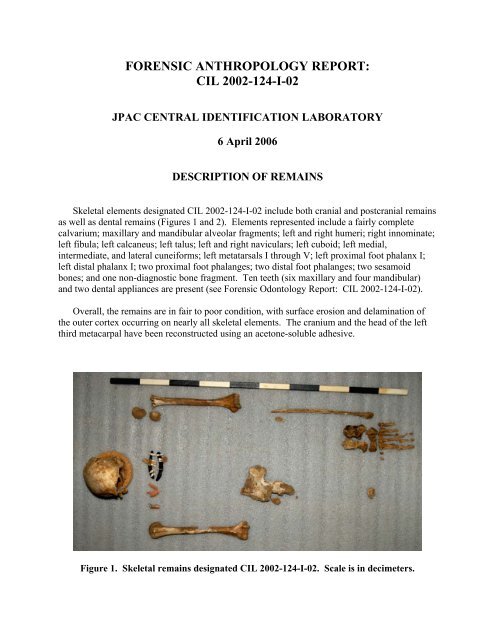
Skeletal elements designated CIL 2002-124-I-02 include both cranial and postcranial remains<br />
as well as dental remains (Figures 1 and 2). Elements represented include a fairly complete<br />
calvarium; maxillary and mandibular alveolar fragments; left and right humeri; right innominate;<br />
left fibula; left calcaneus; left talus; left and right naviculars; left cuboid; left medial,<br />
intermediate, and lateral cuneiforms; left metatarsals I through V; left proximal foot phalanx I;<br />
left distal phalanx I; two proximal foot phalanges; two distal foot phalanges; two sesamoid<br />
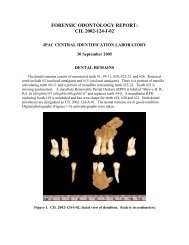
bones; and one non-diagnostic bone fragment. Ten teeth (six maxillary and four mandibular)<br />
and two dental appliances are present (see Forensic Odontology Report: CIL 2002-124-I-02).<br />
Overall, the remains are in fair to poor condition, with surface erosion and delamination of<br />
the outer cortex occurring on nearly all skeletal elements. The cranium and the head of the left<br />
third metacarpal have been reconstructed using an acetone-soluble adhesive.<br />
Figure 1. Skeletal remains designated CIL 2002-124-I-02. Scale is in decimeters.
Forensic Anthropology Report: CIL 2002-124-I-02<br />
Figure 2. Skeletal inventory for CIL 2002-124-I-02; gray denotes skeletal elements that are<br />
present, and blue denotes the presence of non-seriated elements.<br />
Page 2 of 8
Forensic Anthropology Report: CIL 2002-124-I-02<br />
MINIMUM NUMBER OF INDIVIDUALS<br />
One. These remains were segregated from a larger assemblage of skeletal remains with a<br />
minimum number of four individuals. The remains present for analysis in this report are those<br />
that could be associated to this individual via mitochondrial DNA (mtDNA) sequence matches,<br />
odontological association, osteometric comparisons (Byrd and Adams 2003), visual pairmatching,<br />
and taphonomic indicators, as well as through field context (as determined by<br />
consultation with the Recovery Leader/Anthropologist). There is no duplication of elements or<br />
any other evidence of multiple individuals in this set of remains.<br />
SEX<br />
Male. Gross morphological and metric analyses were used to determine sex. Non-metric<br />
cranial characteristics include a small supraorbital ridge, a blunt supraorbital margin, and a large<br />
mastoid process. These traits, with the exception of the first, are more consistent with male<br />
morphology (Bass 1995; Buikstra and Ubelaker 1994; Rogers 2005).<br />
Postcranially, the pelvic region provides the most diagnostic traits for sex determination.<br />
Although most of the key areas for observation in this region are eroded or absent due to<br />
postmortem damage, a few diagnostic traits are present that suggest probable male morphology<br />
(Bass 1995; Buikstra and Ubelaker 1994). The acetabulum is directed laterally, the lunate<br />
surface is broad and deep, and the auricular surface is flat. The greater sciatic notch is deep and<br />
forms an acute angle. Due to the paucity of conventional postcranial traits used for sex<br />
determination, non-conventional traits were also assessed. The distal humerus morphology<br />
demonstrates two features associated with male morphology (asymmetrical trochlea and<br />
triangular olecranon fossa shape) and two features that were indeterminate (slight trochlear<br />
constriction and slightly elevated medial epicondyle) (Ceri et al. 2005; Rogers 1999).<br />
Six postcranial measurements were taken and evaluated against a discriminant model in<br />
FORDISC 2.0 (Ousley and Jantz 1996). The discriminant model has a correct classification rate<br />
of 95.0% when applied to the reference sample of 339 individuals. The remains were classified<br />
by the model as male with a posterior probability of 0.997 and a typicality of 0.580. Overall, the<br />
evidence indicates that this individual’s size and morphology are consistent with males.<br />
AGE<br />
Adult or older juvenile, ≥16 years of age. The estimation of age is based on dental<br />
development and overall appearance of skeletal elements. All secondary centers of epiphyseal<br />
fusion that were available appear to be completely fused. Gross examination of the distal root of<br />
tooth #1 exhibits complete apical closure. This stage of tooth formation occurs on average at<br />
20.2 years, or between 16 and 24.4 years (within two standard deviations) for males (Mincer et<br />
al. 1993).<br />
Page 3 of 8
Forensic Anthropology Report: CIL 2002-124-I-02<br />
RACE<br />
Indeterminate. Gross morphological observations and metric analyses were used to assess<br />
race. Only part of the skull, the cranial vault, was available for analysis. The presence of an<br />
inion hook, absence of wormian bones, and rugged muscle markings are common in caucasoids<br />
(Rhine 1990). Small brow ridges, elliptic external auditory meatus, and complex cranial sutures<br />
are common in mongoloids (Rhine 1990). The lack of more diagnostic cranial elements for the<br />
determination of ancestry and the distribution of observed features between caucasoids and<br />
mongoloids results in an indeterminate assessment.<br />
Six postcranial measurements were taken and evaluated against a discriminant model<br />
including White and Black male populations in FORDISC 2.0 (Ousley and Jantz 1996). The<br />
discriminant model produced a correct classification rate of 77.6% when applied to the reference<br />
sample of 210 individuals. The remains were classified by the model as a White male with a<br />
posterior probability of 0.774 and a typicality of 0.718. Determination of ancestry using<br />
postcranial remains is less reliable than that based on the cranium, and the analysis is further<br />
weakened given the paucity of postcranial remains. Only measurements from the humerus and<br />
calcaneus were available for metric analysis. Thus, an analysis of ancestry for this individual is<br />
indeterminate.<br />
STATURE<br />
65.5 to 71.7 inches. The total length of the right humerus (338 mm) was used to estimate<br />
stature using the White male model of Trotter and Gleser (1952), which was calculated using<br />
FORDISC 2.0 (Ousley and Jantz 1996). The White male equation was selected because it<br />
provides a more robust model due to the larger sample size. This model yields a point estimate<br />
of 68.6 inches with a 95% prediction interval of 65.5 to 71.7 inches.<br />
TRAUMA<br />
Perimortem trauma was identified though fracture angle, shape, edge color, and edge<br />
morphology (Galloway 1999; Ubelaker and Adams 1995). The left distal humerus exhibits a<br />
complete fracture that originates at the trochlea and terminates superior to the medial<br />
supracondylar crest (Figure 3). On the posterior aspect of the bone, the fracture follows the<br />
morphology of the olecranon fossa (Figure 4). On the anterior aspect, the fracture follows the<br />
morphology of the coronoid fossa. The fracture line aligns with the bone grain, which can be<br />
another indicator of perimortem trauma. The fracture edges are sharp and exhibit the same color<br />
patterns as the surrounding cortical bone. Overall morphology suggests that the fracture is a<br />
Type II or high medial condylar fracture (Galloway 1999). This fracture is most common in<br />
falls, but can be the result of a direct blow to the joint surface.<br />
Page 4 of 8
Forensic Anthropology Report: CIL 2002-124-I-02<br />
Figure 3. Fracture of the left distal humerus. View is anterior.<br />
Figure 4. Fracture of the left distal humerus. View is posterior.<br />
Page 5 of 8
Forensic Anthropology Report: CIL 2002-124-1-02<br />
OBSERVATIONS<br />
The overall condition of the remains indicates both surface and subsurface exposure during<br />
the postmortem interval. The remains display a range of color from white to dark brown as a<br />
result of weathering and sediment staining. There is cortical delamination and postmortem<br />
breakage present on nearly all of the skeletal elements. The amount of postmortem breakage is<br />
extensive, especially on the cranium. The anterior aspect of the right innominate, superior to the<br />
arcuate and iliopectinal lines, exhibits a mosaic splitting pattern associated with prolonged<br />
surface exposure (Behrensmeyer 1978). Inferior to the arcuate and iliopectinal lines and on the<br />
posterior aspect of the innominate, the bone color demonstrates evidence of prolonged contact<br />
with soil. Numerous parallel striations indicative of rodent gnawing activity are present on the<br />
broken surface of the ilium (Haglund 1997) (Figure 5).<br />
Two skeletal anomalies were noted during examination. An acetabular fold is present on the<br />
right innominate. The fold is located on the supramedial surface of the acetabular roof. The<br />
etiology of this trait is unknown, but it is believed to be an epigenetic or biomechanical trait<br />
(Mafart 2005). The second anomaly observed involves a rare coalition of the navicular and<br />
cuboid bones (Talkhani and Laing 1999). While a complete bony bridge is not present, both<br />
tarsals have a bony extension, producing a small gap between the articular surfaces. The<br />
articular surfaces of these extensions are rugose and porotic, suggesting that a fibrous<br />
(syndesmosis) or cartilaginous (synchondrosis) union existed. This type of tarsal coalition is<br />
usually asymptomatic; however, in some cases the individual may feel pain or stiffness in the<br />
foot (Talkhani and Laing 1999).<br />
Specimens sampled for mtDNA include teeth (#I, #2, #9, #11, #15, #20, and #22), the right<br />
humeral diaphysis, and the petrous portion of the right temporal bone. For analytical purposes,<br />
each element or fragment in the assemblage has been labeled in pencil with a code that<br />
corresponds to its original field provenience.<br />
CONCLUSIONS<br />
The human skeletal remains designated CIL 2002-124-1-02 consist of one male individual,<br />
greater than or equal to 16 years of age, with a stature between 65.5 and 71.7 inches. There is a<br />
perimortem fracture present on the distal left humerus.<br />
CHRISTIAN M. CROWDER, PhD<br />
Anthropologist<br />
Page 6 of 8
Forensic Anthropology Report: CIL 2002-124-I-02<br />
Figure 5. Shallow grooves on the ilium indicative of rodent gnaw marks.<br />
REFERENCES<br />
Bass, W. M.<br />
1995 Human Osteology: A Laboratory and Field Manual. 4th ed. Special Publication<br />
No. 2 of the Missouri Archaeological Society, Columbia, MO.<br />
Behrensmeyer, A. K.<br />
1978 Taphonomic and ecologic information from bone weathering. Palaeobiology 4:150-<br />
162.<br />
Buikstra, J. E. and D. H. Ubelaker (editors)<br />
1994 Standards for Data Collection from Human Skeletal Remains. Arkansas<br />
Archeological Survey Research Series No. 44, Fayetteville, AR.<br />
Byrd, J. E. and B. J. Adams<br />
2003 Osteometric sorting of commingled human remains. Journal of Forensic Sciences<br />
48:717-724.<br />
Ceri, G. F, H. Schutkowski, and D. A. Weston<br />
2005 The distal humerus—A blind test of the Rogers’ sexing technique using a<br />
documented skeletal collection. Journal of Forensic Sciences 50:1289-1293.<br />
Page 7 of 8
Forensic Anthropology Report: CIL 2002-124-I-02<br />
Galloway, A. (editor)<br />
1999 Broken Bones: Anthropological Analysis of Blunt Force Trauma. Charles C<br />
Thomas, Springfield, IL.<br />
Haglund W. D.<br />
1997 Rodents and human remains. In Forensic Taphonomy: The Postmortem Fate of<br />
Human Remains, edited by W. D. Haglund and M. H. Sorg, pp. 405-414. CRC<br />
Press, Salem, MA.<br />
Mafart, B.<br />
2005 Description, significance and frequency of the acetabular crease of the hip bone.<br />
International Journal of Osteoarchaeology 15:208-215.<br />
Mincer, H. H., E. F. Harris, and H. E. Berryman<br />
1993 A.B.F.O. study of third molar development and its use as an estimator of<br />
chronological age. Journal of Forensic Sciences 38:379-390.<br />
Ousley, S. and R. Jantz<br />
1996 FORDISC 2.0. University of Tennessee, Knoxville, TN.<br />
Rhine, S.<br />
1990 Non-metric skull racing. In Skeletal Attribution of Race: Methods for Forensic<br />
Anthropology, edited by G. W. Gill and S. Rhine, pp. 9-20. Maxwell Museum<br />
Anthropological Papers No. 4, Albuquerque, NM.<br />
Rogers, T. L.<br />
2005 Determining the sex of human remains through cranial morphology. Journal of<br />
Forensic Sciences 50:1-8.<br />
Rogers, T. L.<br />
1999 A visual method for determining sex of skeletal remains using the distal humerus.<br />
Journal of Forensic Sciences 44:57-60.<br />
Talkhani, I. S. and P. Laing<br />
1999 Cuboid-navicular coalition in an adult: A case report. Foot and Ankle Surgery 5:151-<br />
154.<br />
Trotter, M. and G. C. Gleser<br />
1952 Estimation of stature from long bones of American Whites and Negroes. American<br />
Journal of Physical Anthropology 10:463-514.<br />
Ubelaker, D. H. and B. J. Adams<br />
1995 Differentiation of perimortem and postmortem trauma using taphonomic indicators.<br />
Journal of Forensic Sciences 40:509-512.<br />
Page 8 of 8