

Surface modification and antibacterial activity of electrospun ...
Surface modification and antibacterial activity of electrospun ...
Surface modification and antibacterial activity of electrospun ...

Create successful ePaper yourself
Turn your PDF publications into a flip-book with our unique Google optimized e-Paper software.
Journal <strong>of</strong> Membrane Science 320 (2008) 259–267<br />
Contents lists available at ScienceDirect<br />
Journal <strong>of</strong> Membrane Science<br />
journal homepage: www.elsevier.com/locate/memsci<br />
<strong>Surface</strong> <strong>modification</strong> <strong>and</strong> <strong>antibacterial</strong> <strong>activity</strong> <strong>of</strong> <strong>electrospun</strong> polyurethane<br />
fibrous membranes with quaternary ammonium moieties<br />
Chen Yao a,b , Xinsong Li a,∗ , K.G. Neoh b,∗ , Zhilong Shi b , E.T. Kang b<br />
a School <strong>of</strong> Chemistry <strong>and</strong> Chemical Engineering, Southeast University, Sipailou 2, Nanjing 210018, PR China<br />
b Department <strong>of</strong> Chemical <strong>and</strong> Biomolecular Engineering, National University <strong>of</strong> Singapore, Kent Ridge, 119260 Singapore, Singapore<br />
article<br />
info<br />
abstract<br />
Article history:<br />
Received 26 February 2008<br />
Received in revised form 3 April 2008<br />
Accepted 5 April 2008<br />
Available online 15 April 2008<br />
Keywords:<br />
Electrospinning<br />
Antibacterial<br />
<strong>Surface</strong> <strong>modification</strong><br />
Polyurethane<br />
A novel <strong>antibacterial</strong> material was developed by surface <strong>modification</strong> <strong>of</strong> <strong>electrospun</strong> polyurethane (PU)<br />
fibrous membranes, using a process which involved plasma pretreatment, UV-induced graft copolymerization<br />
<strong>of</strong> 4-vinylpyridine (4VP), <strong>and</strong> quaternization <strong>of</strong> the grafted pyridine groups with hexylbromide.<br />
The success <strong>of</strong> <strong>modification</strong> with poly(4-vinyl-N-hexyl pyridinium bromide) groups on these was ascertained<br />
by X-ray photoelectron spectroscopy (XPS). The morphologies <strong>and</strong> mechanical properties were<br />
investigated by scanning electron microscopy (SEM) <strong>and</strong> tensile test, respectively. The results showed<br />
that the morphologies <strong>of</strong> PU fibrous membranes changed slightly during the <strong>modification</strong> process <strong>and</strong><br />
the fiber structures were maintained. The tensile strength <strong>of</strong> PU fibrous membranes decreased after surface<br />
<strong>modification</strong>, with the smallest decrease (90%) [19]. Electrospun cellulose acetate fibers containing<br />
silver nanoparticles showed strong antimicrobial <strong>activity</strong> against<br />
both Gram-negative <strong>and</strong> Gram-positive bacteria [20,21]. Kenawy et<br />
al. modified the poly(vinyl phenol) either by sulphonation or by formation<br />
<strong>of</strong> lithium salt <strong>of</strong> the sulphonated species, <strong>and</strong> investigated<br />
the <strong>antibacterial</strong> activities <strong>of</strong> the modified poly(vinyl phenol) <strong>electrospun</strong><br />
mats [22]. Polyurethane cationomers polymerized from<br />
base PU with chain extenders having a quaternary ammonium<br />
0376-7388/$ – see front matter © 2008 Elsevier B.V. All rights reserved.<br />
doi:10.1016/j.memsci.2008.04.012
260 C. Yao et al. / Journal <strong>of</strong> Membrane Science 320 (2008) 259–267<br />
group were <strong>electrospun</strong> into non-woven nan<strong>of</strong>iber mats for antimicrobial<br />
nan<strong>of</strong>ilter applications [23].<br />
A more straightforward way is to modify the surface <strong>of</strong> polymer<br />
nan<strong>of</strong>ibers without affecting bulk properties <strong>of</strong> the treated<br />
nan<strong>of</strong>ibers. The methods used to impart surface <strong>modification</strong> usually<br />
depend strongly on the nature <strong>of</strong> the fiber-forming polymer<br />
<strong>and</strong> include, but are not limited to, covalent polymer grafting<br />
[24], plasma treatment [25], physisorption (e.g., hydrogen-bonding<br />
interactions) [26], chemisorption [27], <strong>and</strong> chemical derivatization<br />
[28]. Among the various methods, plasma treatment provides a<br />
clean <strong>and</strong> environmentally friendly way for surface <strong>modification</strong><br />
[29]. The free radicals <strong>and</strong> electrons created in the plasma treatment<br />
could be used to modify the polymer nan<strong>of</strong>ibers chemically. Covalent<br />
attachment <strong>of</strong> functional compounds to polymer fiber surfaces<br />
is the preferred approach to introduce functionalities permanently<br />
<strong>and</strong> at reasonably high efficiency.<br />
There are numerous antimicrobials suitable for immobilization<br />
on polymer surfaces. Quaternary ammonium compounds seem<br />
attractive because their target is primarily the microbial membrane<br />
<strong>and</strong> they accumulate in the cell driven by the membrane potential<br />
[30]. To maximize efficiency, quaternary ammonium compound<br />
is used as monomeric link in the polymeric leash <strong>and</strong> poly(4-<br />
vinylpyridine) (PVP) is usually selected as the carrying polymer.<br />
Tiller et al. showed that the surfaces <strong>of</strong> commercial polymers<br />
treated with N-alkylated PVP groups were lethal on contact to both<br />
Gram-positive <strong>and</strong> Gram-negative bacteria, <strong>and</strong> it was also shown<br />
that N-alkyl chain <strong>of</strong> six carbon units in length was the most effective<br />
[31].<br />
The purpose <strong>of</strong> this paper was to develop novel <strong>antibacterial</strong><br />
PU fibrous membranes by electrospinning the polymer followed<br />
by plasma pretreatment, UV-induced graft copolymerization <strong>and</strong><br />
quaternization reaction. Electrospun PU fibrous membranes were<br />
modified with poly(4-vinyl-N-hexyl pyridinium bromide) on the<br />
surfaces to achieve <strong>antibacterial</strong> activities. The modified PU fibrous<br />
membranes were subsequently characterized in terms <strong>of</strong> their<br />
morphologies, surface chemical compositions <strong>and</strong> mechanical<br />
properties. The <strong>antibacterial</strong> activities <strong>of</strong> the fibrous membranes<br />
were assessed against both Gram-positive Staphylococcus aureus (S.<br />
aureus) <strong>and</strong> Gram-negative Escherichia coli (E. coli).<br />
2. Experimental<br />
2.1. Materials<br />
Polyurethane elastomer, Elastollan ® 1180A10, was received from<br />
BASF. 4-Vinylpyridine (4VP) monomer was obtained from Aldrich<br />
Chemical Co. <strong>and</strong> freshly distilled under reduced pressure before<br />
use. Hexylbromide <strong>and</strong> solvents, such as tetrahydr<strong>of</strong>uran (THF),<br />
N,N-dimethyl formamide (DMF), heptane, 2-propanol, were <strong>of</strong><br />
reagent grade <strong>and</strong> used as received from Aldrich Chemical Co. Peptone,<br />
yeast extract, agar <strong>and</strong> beef extract were purchased from<br />
Oxoid. S. aureus (ATCC 25923) <strong>and</strong> E. coli (ATCC DH5) were<br />
obtained from American Type Culture Collection.<br />
2.2. Electrospinning <strong>of</strong> PU<br />
PU was dissolved in a mixed solvent <strong>of</strong> THF <strong>and</strong> DMF (1:1, v/v) to<br />
produce various spinning solutions with PU concentrations ranging<br />
from 5% to 11% (w/v, g/mL). Viscosity, conductivity <strong>and</strong> surface<br />
tension <strong>of</strong> the prepared solutions were measured with a viscometer<br />
(NDJ-9S, ShangPing), a conductivity meter (DDB-303A, Rex) <strong>and</strong><br />
a surface tension meter (BZY-1, HengPing) at 20 ◦ C, respectively. A<br />
syringe pump was used to feed the polymer solution through a 20-<br />
mL plastic syringe fitted with a needle <strong>of</strong> tip diameter <strong>of</strong> 0.6 mm<br />
at a delivery rate <strong>of</strong> 6 mL/h. After high voltage ranging from 10<br />
to 12 kV was applied to the needle, a positive charged jet <strong>of</strong> PU<br />
solution formed from the Taylor cone <strong>and</strong> sprayed to a grounded<br />
drum, approximately 10 cm from the needle tip. With the evaporation<br />
<strong>of</strong> solvent, PU fibers were deposited on the drum to form<br />
fibrous membrane. The as-spun fibrous membrane was then dried<br />
under vacuum <strong>and</strong> annealed at 80 ◦ C for 6 h. All electrospinning<br />
experiments were carried out at 20 ◦ C <strong>and</strong> relative humidity <strong>of</strong> 50%.<br />
2.3. <strong>Surface</strong> <strong>modification</strong> <strong>of</strong> PU fibrous membranes<br />
PU fibrous membranes were subjected to argon plasma pretreatment<br />
in an Anatech SP100 plasma system, equipped with a<br />
cylindrical quartz reactor chamber. The glow discharge was produced<br />
at a plasma power <strong>of</strong> 35 W, an applied oscillator frequency<br />
<strong>of</strong> 40 kHz <strong>and</strong> an argon pressure <strong>of</strong> approximately 80 Pa. After<br />
subjected to glow discharge for 60 s on both surfaces, the fibrous<br />
membrane was exposed to air for 5 min to facilitate formation <strong>of</strong><br />
surface oxide <strong>and</strong> peroxide groups.<br />
The plasma pretreated fibrous membranes were immersed<br />
in 2-propanol solution <strong>of</strong> 20 vol.% 4VP in a Pyrex glass tube.<br />
Degassing <strong>of</strong> the solution was achieved by bubbling nitrogen vigorously<br />
for 30 min <strong>and</strong> sealing the tube with silicon rubber stopper.<br />
The tube was then exposed to UV irradiation in a Riko rotary<br />
photochemical reactor (RH400-10W) for 60 min on each surface.<br />
The graft-copolymerized fibrous membranes were subjected to<br />
washing with copious amounts <strong>of</strong> 2-propanol to remove residual<br />
monomer <strong>and</strong> physically adsorbed homopolymer.<br />
The fibrous membranes with graft-copolymerized 4VP were<br />
then placed in heptane solutions containing 20 vol.% hexylbromide<br />
<strong>and</strong> the reaction mixture was stirred for 48 h at 60 ◦ C. The fibrous<br />
membranes were then thoroughly rinsed with heptane <strong>and</strong> dried<br />
under vacuum.<br />
2.4. <strong>Surface</strong> characterization<br />
The morphology <strong>of</strong> PU fibrous membranes was observed with a<br />
scanning electron microscope (JEOL JSM 5600LV) after gold sputtercoating.<br />
Diameters <strong>of</strong> the <strong>electrospun</strong> fibers were measured directly<br />
from SEM images, with an average value being calculated from at<br />
least 100 measurements.<br />
<strong>Surface</strong> compositions <strong>of</strong> PU fibrous membranes were analyzed<br />
by XPS on an AXIS HSi spectrometer (Kratos Analytical Ltd.) using<br />
the monochromatized Al K X-ray source (1486.6 eV photons) at<br />
a constant dwell time <strong>of</strong> 100 ms <strong>and</strong> a pass energy <strong>of</strong> 40 eV. The<br />
anode voltage was 15 kV, <strong>and</strong> the anode current was 10 mA. The<br />
core-level signals were obtained at a photoelectron take<strong>of</strong>f angle<br />
<strong>of</strong> 90 ◦ (with respect to the membrane surface). To compensate for<br />
surface charging effect, all binding energies (BEs) were referenced<br />
to the C 1s hydrocarbon peak at 284.6 eV. In the peak synthesis,<br />
the line width (full width at half-maximum) <strong>of</strong> the Gaussian<br />
peaks was maintained constant for all components in a particular<br />
spectrum. The peak area ratios for the various elements were corrected<br />
using experimentally determined instrumental sensitivity<br />
factors.<br />
2.5. Mechanical property<br />
Tensile tests <strong>of</strong> PU fibrous membranes were performed using<br />
an Instron universal materials testing machine (Model 5544) with<br />
a 10 N load cell in a constant relative humidity (50%) room at<br />
25 ◦ C. “Dog-bone” shaped samples were cut from the fibrous membranes<br />
(5-mm wide at the narrowest point with a gage length <strong>of</strong><br />
15 mm). The thickness <strong>of</strong> these samples was measured with a digital<br />
micrometer having a precision <strong>of</strong> 1 m. A cross-head speed <strong>of</strong>
C. Yao et al. / Journal <strong>of</strong> Membrane Science 320 (2008) 259–267 261<br />
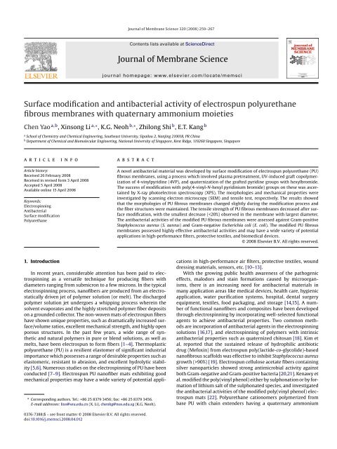
Fig. 1. Electrospinning solution parameters <strong>and</strong> average diameter <strong>of</strong> the as-spun fibers with different PU concentrations.<br />
10 mm/min was used <strong>and</strong> at least five samples were tested for each<br />
type <strong>of</strong> the fibrous membranes.<br />
2.6. Determination <strong>of</strong> <strong>antibacterial</strong> <strong>activity</strong><br />
S. aureus was incubated in 10 mL <strong>of</strong> a 3.1% yeast–dextrose broth<br />
(containing 10 g/L peptone, 8 g/L beef extract, 5 g/L sodium chloride,<br />
5 g/L glucose, <strong>and</strong> 3 g/L yeast extract at a pH 6.8) for 6–8 h at 37 ◦ C<br />
until the exponential growth phase was reached [32].<br />
The bacteria-containing broth was centrifuged at 3000 rpm for<br />
10 min, <strong>and</strong> after removal <strong>of</strong> supernatant, the cells were washed<br />
twice with sterile phosphate-buffered solution (PBS). The bacteria<br />
cells were resuspended to provide a final density <strong>of</strong> 10 6 cells/mL<br />
in PBS (based on st<strong>and</strong>ard calibration with the assumption that<br />
the optical density <strong>of</strong> 1.0 at 540 nm is equivalent to approximately<br />
10 9 cells/mL) [33].<br />
PU fibrous membranes <strong>electrospun</strong> from 10% (w/v) solutions in<br />
THF <strong>and</strong> DMF (1:1, v/v) (either pristine or modified, 8 mm × 8 mm)<br />
were sterilized with ultraviolet irradiation for 30 min, <strong>and</strong> then<br />
immersed in 10 mL <strong>of</strong> the bacterial suspension in an Erlenmeyer<br />
flask <strong>and</strong> shaken at 200 rpm at 37 ◦ C. The viable cell counts <strong>of</strong><br />
bacteria were measured by surface spread plate method. At the<br />
predetermined time, 1 mL <strong>of</strong> bacteria culture was taken from the<br />
flask <strong>and</strong> decimal serial dilutions with PBS were repeated with each<br />
Fig. 2. XPS wide scans <strong>of</strong> (a) pristine PU fibrous membranes, (d) PU membranes grafted with PVP, <strong>and</strong> (g) PU membranes modified with poly(4-vinyl-N-hexylpyridinium<br />
bromide); C 1s core-level spectra <strong>of</strong> (b) pristine PU fibrous membranes, (e) PU membranes grafted with PVP, <strong>and</strong> (h) PU membranes modified with poly(4-vinyl-Nhexylpyridinium<br />
bromide); N 1s core-level spectra <strong>of</strong> (c) pristine PU fibrous membranes, (f) PU membranes grafted with PVP, <strong>and</strong> (i) PU membranes modified with<br />
poly(4-vinyl-N-hexylpyridinium bromide). PU fibrous membranes were <strong>electrospun</strong> from 10% (w/v) PU solutions in THF <strong>and</strong> DMF (1:1, v/v).
262 C. Yao et al. / Journal <strong>of</strong> Membrane Science 320 (2008) 259–267<br />
initial sample. A 0.1-mL drop <strong>of</strong> the diluted sample was then spread<br />
onto solid growth agar plates. After incubation <strong>of</strong> the plates at 37 ◦ C<br />
for 24 h, the number <strong>of</strong> viable cells (colonies) was counted manually,<br />
<strong>and</strong> expressed as mean colony-forming units per milliliter<br />
(CFU/mL) after multiplication with the dilution factor. After 4 h, the<br />
PU fibrous membranes were removed from the bacterial suspension<br />
with sterile forceps <strong>and</strong> gently washed with PBS. The bacteria<br />
retained on membranes were dislodged by mild ultrasonication (for<br />
10 min) in a 100 W ultrasonic bath. Serial 10-fold dilutions were performed<br />
<strong>and</strong> viable counts estimated following the surface spread<br />
plate method. The number <strong>of</strong> colony-forming units on each membrane<br />
surface was computed <strong>and</strong> expressed relative to the apparent<br />
surface area <strong>of</strong> the membrane (CFU/cm 2 ). All experiments were performed<br />
in triplicate <strong>and</strong> the quantitative value was expressed as the<br />
average ± st<strong>and</strong>ard deviation.<br />
The extent <strong>of</strong> bacterial adhesion on PU fibrous membranes<br />
(both pristine <strong>and</strong> modified) were also assessed by examining<br />
these membranes after 4 h immersion in the PBS suspension <strong>of</strong><br />
Fig. 3. SEM images <strong>of</strong> (a), (c) <strong>and</strong> (e) pristine, (b), (d) <strong>and</strong> (f) modified PU fibrous membranes with poly(4-vinyl-N-hexylpyridinium bromide). PU fibrous membranes were<br />
<strong>electrospun</strong> from (a) <strong>and</strong> (b) 6%, (c) <strong>and</strong> (d) 8%, (e) <strong>and</strong> (f) 10% (w/v) solutions in THF <strong>and</strong> DMF (1:1, v/v).
C. Yao et al. / Journal <strong>of</strong> Membrane Science 320 (2008) 259–267 263<br />
10 7 cells/mL S. aureus. Control experiment was carried out with the<br />
filter paper. The membranes were then examined under SEM (JEOL<br />
JSM 5600LV) to assess the adhesion <strong>and</strong> viability <strong>of</strong> the bacteria.<br />
The membrane fixation <strong>and</strong> preparation for SEM were as follows:<br />
the membranes were first washed with PBS <strong>and</strong> 3 vol.% glutaraldehyde<br />
in PBS was added for 5 h <strong>and</strong> stored at 4 ◦ C. The glutaraldehyde<br />
solution was then removed <strong>and</strong> the membranes were washed with<br />
PBS, followed by step dehydration with 25%, 50%, 70%, 95%, <strong>and</strong><br />
100% ethanol for 10 min each. The membranes were dried under<br />
vacuum <strong>and</strong> gold sputter-coated before SEM observation.<br />
For E. coli, the same assay procedures were used as those<br />
described above for S. aureus.<br />
3. Results <strong>and</strong> discussion<br />
3.1. Electrospinning <strong>of</strong> PU<br />
While electrospinning has proven to be a versatile <strong>and</strong> powerful<br />
means <strong>of</strong> fabricating polymer micro/nan<strong>of</strong>ibers, its applicability<br />
to obtain smooth, uniform fibrous structure is not straightforward.<br />
Among various parameters <strong>of</strong> electrospinning process, such<br />
as applied voltage, needle tip-to-receiver distance <strong>and</strong> solution<br />
delivery rate, concentration or corresponding viscosity <strong>of</strong> spinning<br />
solution is one <strong>of</strong> the most effective variables for controlling fiber<br />
morphology <strong>and</strong> diameter. Results obtained from our study showed<br />
that solution concentration was found to be the major factor controlling<br />
the morphology <strong>of</strong> the fibers in the electrospinning <strong>of</strong> PU.<br />
Various PU solutions with concentration in the range <strong>of</strong> 5–11%<br />
(w/v, g/mL) in THF <strong>and</strong> DMF (1:1, v/v) were <strong>electrospun</strong>. A beadon-string<br />
morphology with several big beads was obtained at<br />
PU concentration <strong>of</strong> 5% (w/v). The distribution density <strong>of</strong> beads<br />
decreased when PU concentration increased to 6% (w/v). The shape<br />
<strong>of</strong> the beads became spindle-like. Smooth <strong>and</strong> homogeneous fibers<br />
without beads were produced when PU concentration reached 8%<br />
(w/v). As PU concentration increased from 9% to 11% (w/v), the<br />
<strong>electrospun</strong> fibers became thicker <strong>and</strong> more adhesive at various<br />
bonding sites, which led to a film-like character <strong>and</strong> structural<br />
integrity <strong>of</strong> the fibrous membranes.<br />
The effects <strong>of</strong> electrospinning solution parameters on the average<br />
diameter <strong>of</strong> PU nan<strong>of</strong>ibers were investigated. As shown in<br />
Fig. 1a, viscosity <strong>of</strong> PU solutions increased with increasing PU<br />
concentration. However, both conductivity <strong>and</strong> surface tension <strong>of</strong><br />
PU solutions with different concentrations did not differ much<br />
(Fig. 1b). The viscosity <strong>of</strong> a polymer solution reflects the intermolecular<br />
interactions between polymer chains. Polymer solutions<br />
with higher viscosity usually exhibit longer stress relaxation times,<br />
which may facilitate the formation <strong>of</strong> fibers with large diameters<br />
during electrospinning [34]. When the viscosity <strong>of</strong> PU solutions<br />
increased greatly from 0.186 Pa s up to 0.459 Pa s with increasing<br />
PU concentration from 9% to 11% (w/v), the average fiber diameter<br />
increased dramatically from 820 nm to 1.95 m(Fig. 1a). Therefore,<br />
Fig. 4. (a) Tensile strength, (b) elongation at break <strong>and</strong> (c) Young’s modulus <strong>of</strong> PU fibrous membranes <strong>electrospun</strong> from solutions with different concentrations (both pristine<br />
membranes <strong>and</strong> those modified with poly(4-vinyl-N-hexylpyridinium bromide)).
264 C. Yao et al. / Journal <strong>of</strong> Membrane Science 320 (2008) 259–267<br />
it was considered that the viscosity <strong>of</strong> solution was the major factor<br />
affecting the average diameter <strong>of</strong> <strong>electrospun</strong> PU nan<strong>of</strong>ibers.<br />
3.2. <strong>Surface</strong> <strong>modification</strong> <strong>of</strong> PU fibrous membranes<br />
The success <strong>of</strong> <strong>modification</strong> with poly(4-vinyl-N-hexyl pyridinium<br />
bromide) on <strong>electrospun</strong> PU fibrous membranes can be<br />
ascertained by comparing the XPS spectra <strong>of</strong> the pristine <strong>and</strong> modified<br />
membranes as shown in Fig. 2.<br />
The spectra <strong>of</strong> pristine PU fibrous membranes showed three<br />
main signals corresponding to C 1s (284.6 eV), O 1s (532 eV) <strong>and</strong><br />
N 1s (400 eV) (Fig. 2a). The XPS C 1s core-level spectrum <strong>of</strong> pristine<br />
PU fibrous membrane (Fig. 2b) was resolved into three component<br />
peaks: a hydrocarbon environment (C–H, C–C, 284.6 eV), a carbon<br />
singly bound to oxygen environment (C–O, 286.0 eV), <strong>and</strong> a carbon<br />
in a carbamate environment (–CO–, 289.0 eV) [35]. The corresponding<br />
N 1s spectrum (Fig. 2c) showed an intense peak at the binding<br />
energy (BE) <strong>of</strong> 399.5 eV attributable to the nitrogen (–NH–) in carbamate<br />
<strong>of</strong> PU.<br />
After UV-induced graft copolymerization <strong>of</strong> PVP onto the PU<br />
fibrous membranes, the C 1s core-level spectrum (Fig. 2e) showed<br />
an additional peak at 285.5 eV attributable to C–N species. The<br />
intensity <strong>of</strong> the peak assigned to C–O species became weaker <strong>and</strong><br />
that <strong>of</strong> –CO– species was barely discernible compared with Fig. 2b.<br />
Similarly, the corresponding N 1s spectrum (Fig. 2f) showed an<br />
additional peak at the BE <strong>of</strong> 398.5 eV attributable to imine moiety<br />
(–N ) <strong>of</strong> the pyridine rings [36] <strong>and</strong> a decrease in the intensity <strong>of</strong><br />
–NH– peak component compared with Fig. 2c. The extent <strong>of</strong> surface<br />
grafting <strong>of</strong> PVP can be estimated from the sensitivity factor corrected<br />
ratio <strong>of</strong> the total N 1s peak over the total C 1s peak, expressed<br />
as [N]/[C]. Previous work <strong>of</strong> surface graft copolymerization showed<br />
that graft concentration <strong>of</strong> PVP was affected by the monomer concentration<br />
[37]. In this experiment, 4VP concentration <strong>of</strong> 20% was<br />
chosen for effective surface graft copolymerization. As determined<br />
by XPS, the surface [N]/[C] ratio <strong>of</strong> the 4VP grafted onto PU fibrous<br />
membrane was 0.12, close to the value <strong>of</strong> 0.14, expected for the 4VP<br />
monomeric unit (C 7 H 7 N 1 ). It indicated that the surface was almost<br />
completely covered by 4VP copolymers, which could provide abundant<br />
reactive sites for the subsequent quaternization.<br />
Fig. 2g–i shows the XPS spectra <strong>of</strong> the graft-copolymerized PU<br />
fibrous membrane N-alkylated with hexylbromide. No significant<br />
difference was observed in C 1s core-level spectrum before (Fig. 2e)<br />
<strong>and</strong> after (Fig. 2h) N-alkylation. The corresponding N 1s core-level<br />
spectrum in Fig. 2i showed an additional peak at BE above 400 eV,<br />
attributable to the N + groups <strong>of</strong> the pyridinium ions [38], which<br />
confirmed the derivatization <strong>of</strong> the –N groups by hexylbromide.<br />
On the basis <strong>of</strong> the [N + ]/[N] ratio, the degree <strong>of</strong> alkylation <strong>of</strong> the<br />
pyridine rings on the PU fibrous membrane was around 50–60%.<br />
Fig. 3 shows the typical SEM images <strong>of</strong> PU fibrous membranes<br />
before <strong>and</strong> after surface <strong>modification</strong>. The morphologies <strong>of</strong> the surface<br />
modified PU fibrous membranes <strong>electrospun</strong> from 6%, 8% <strong>and</strong><br />
10% (w/v) solution changed slightly compared with those <strong>of</strong> pristine<br />
PU fibrous membranes. The result indicated that the fiber structures<br />
were maintained during the <strong>modification</strong> process, <strong>and</strong> no<br />
significant change in fiber diameter was observed.<br />
3.3. Mechanical property<br />
PU fibrous membranes both pristine <strong>and</strong> modified with<br />
poly(4-vinyl-N-hexylpyridinium bromide) were tested for their<br />
mechanical integrity in terms <strong>of</strong> tensile strength, elongation at<br />
break <strong>and</strong> Young’s modulus, as indicated in Fig. 4. It can be seen<br />
that the pristine <strong>electrospun</strong> PU fibrous membranes had tensile<br />
strength <strong>of</strong> 3.27–11.8 MPa, elongation at break <strong>of</strong> 159.2–349.7%,<br />
<strong>and</strong> Young’s modulus <strong>of</strong> 1.78–3.73 MPa, depending on the concentration<br />
<strong>of</strong> polymer solution used for electrospinning. PU fibrous<br />
membranes <strong>electrospun</strong> from 7% (w/v) solution had the lowest tensile<br />
strength <strong>of</strong> 3.27 MPa (Fig. 4a). As the concentration increased<br />
to 8% (w/v), the tensile strength increased to 3.88 MPa. The Young’s<br />
modulus <strong>and</strong> elongation at break showed an increasing trend with<br />
concentration as well (Fig. 4b <strong>and</strong> c). This is mainly due to more<br />
interstices between the fibers with smaller diameters, resulting in<br />
a lower number <strong>of</strong> closely adjacent binding in the fibrous membranes.<br />
Since strong “sticky” binding interactions make a major<br />
contribution to the strength <strong>of</strong> the membranes, increasing the fiber<br />
diameters can lead to increases <strong>of</strong> tensile strength [39]. When PU<br />
concentration increased from 9% to 11% (w/v), the tensile strength<br />
<strong>of</strong> the pristine as-spun fibrous membranes increased significantly<br />
from 4.32 to 11.80 MPa, in agreement with the increase in fiber<br />
diameters as shown in Fig. 1a.<br />
On the other h<strong>and</strong>, the tensile strength <strong>and</strong> elongation at break<br />
<strong>of</strong> PU fibrous membranes decreased after surface <strong>modification</strong>,<br />
whereas the Young’s moduli did not show much change. As shown<br />
in Fig. 4a, the tensile strength <strong>of</strong> PU fibrous membranes <strong>electrospun</strong><br />
from 7% (w/v) solution decreased from 3.27 to 1.99 MPa after<br />
<strong>modification</strong>, losing almost 40% <strong>of</strong> tensile strength. As the solution<br />
Fig. 5. Viable cell numbers <strong>of</strong> (a) Staphylococcus aureus, <strong>and</strong> (b) Escherichia coli as a function time in contact with: () control, () pristine <strong>and</strong> () modified PU fibrous<br />
membranes. The cell number was determined by surface spread plate method.
C. Yao et al. / Journal <strong>of</strong> Membrane Science 320 (2008) 259–267 265<br />
concentration increased, smaller decreases in the tensile strength<br />
were observed. With a concentration <strong>of</strong> 11% (w/v) which resulted in<br />
the largest diameter <strong>of</strong> fibers in PU fibrous membranes, the loss <strong>of</strong><br />
tensile strength was approximately 16%. The results indicated that<br />
the surface <strong>modification</strong> may have less adverse effect on the tensile<br />
strength <strong>of</strong> fibrous membranes with larger diameters. In view<br />
<strong>of</strong> these results, the <strong>antibacterial</strong> assays were carried out with surface<br />
modified PU fibrous membranes <strong>electrospun</strong> from 10% (w/v)<br />
solutions in THF <strong>and</strong> DMF (1:1, v/v) in order to achieve a balance<br />
between mechanical properties <strong>and</strong> fibrous structures.<br />
3.4. Antibacterial <strong>activity</strong><br />
Antibacterial efficacy <strong>of</strong> surface modified PU fibrous membranes<br />
with poly(4-vinyl-N-hexylpyridinium bromide) was investigated<br />
by estimating the number <strong>of</strong> viable bacteria cells in the S. aureus<br />
<strong>and</strong> E. coli suspension after being in contact with membranes for<br />
various periods <strong>of</strong> time, <strong>and</strong> the results were shown in Fig. 5. As<br />
expected, no significant loss <strong>of</strong> viable bacteria was detected in the<br />
control experiment (i.e. without the membranes). For pristine PU<br />
fibrous membranes, the viable cell numbers in bacteria suspen-<br />
Fig. 6. SEM images <strong>of</strong> (a) <strong>and</strong> (d) filter paper (control), (b) <strong>and</strong> (e) pristine, <strong>and</strong> (c) <strong>and</strong> (f) modified PU fibrous membranes after immersed in PBS suspension <strong>of</strong> (a)–(c) S.<br />
aureus, or (d)–(f) E. coli at 10 7 cells/mL for 4 h. PU fibrous membranes were <strong>electrospun</strong> from 10% (w/v) solutions in THF <strong>and</strong> DMF (1:1, v/v).
266 C. Yao et al. / Journal <strong>of</strong> Membrane Science 320 (2008) 259–267<br />
sions were similar to the control (Fig. 5a). The surface modified PU<br />
fibrous membranes had a high <strong>antibacterial</strong> efficacy for S. aureus,<br />
reaching 99.9% <strong>and</strong> 99.999% after 1 <strong>and</strong> 4 h contact, respectively.<br />
In comparison, the <strong>antibacterial</strong> efficacy for E. coli was 99.9% after<br />
4 h contact (Fig. 5b), lower than that for S. aureus. Although the<br />
mechanism <strong>of</strong> the <strong>antibacterial</strong> <strong>activity</strong> <strong>of</strong> immobilized quaternary<br />
ammonium groups is not entirely clear. It has been hypothesized<br />
that these immobilized moieties disrupt the integrity <strong>of</strong> the cytoplasmic<br />
membrane to cause cell death, similar to the mechanism<br />
<strong>of</strong> free biocides [40]. The difference in <strong>antibacterial</strong> efficacy is postulated<br />
to be the result <strong>of</strong> the different cell membrane structures<br />
between S. aureus <strong>and</strong> E. coli bacteria. The multilayered cell envelope<br />
structure <strong>of</strong> Gram-negative bacteria may be more resistant to<br />
access by the bactericidal moieties to the inner membrane <strong>of</strong> the<br />
organism [15].<br />
Fig. 6 shows the SEM images <strong>of</strong> different substrates after immersion<br />
in bacteria suspensions. Numerous distinguishable S. aureus<br />
cells can be observed on the filter paper as shown in Fig. 6a. The<br />
number <strong>of</strong> S. aureus cells on the pristine PU fibrous membranes was<br />
significantly less (Fig. 6b). The S. aureus was distributed not only on<br />
the pristine PU fibrous membrane upper surface but also entrapped<br />
in the space between the thin fibers. In contrast, very few sparsely<br />
distributed bacteria cells could be spotted over the entire surface<br />
<strong>of</strong> the modified PU fibrous membrane (Fig. 6c). Similar results were<br />
observed for E. coli on the surface <strong>of</strong> fibrous membranes as shown<br />
in Fig. 6d–f. The results indicated that both filter paper <strong>and</strong> pristine<br />
PU fibrous membranes are good templates for the proliferation <strong>of</strong><br />
bacteria <strong>and</strong> bi<strong>of</strong>ilm formation may occur readily on such surfaces<br />
in contact with bacteria. The presence <strong>of</strong> quaternary ammonium<br />
groups attached to the surface <strong>of</strong> PU fibrous membranes was thus<br />
demonstrated to be very effective in preventing bi<strong>of</strong>ilm formation.<br />
Since the interstitial spaces <strong>of</strong> the pristine PU fibrous membranes<br />
are large enough, it can be expected that some bacteria<br />
would have penetrated into the fibrous membranes during immersion<br />
in the bacteria suspension. Therefore, the bacteria retained<br />
in fibrous membranes were dislodged by mild ultrasonication <strong>and</strong><br />
the number <strong>of</strong> viable cells was counted by surface spread plate<br />
method. For the pristine PU fibrous membranes, the mean number<br />
<strong>of</strong> S. aureus <strong>and</strong> E. coli cells relative to the surface area was<br />
(9.03 ± 0.63) × 10 4 <strong>and</strong> (1.65 ± 0.24) × 10 5 CFU/cm 2 , respectively.<br />
For the modified PU fibrous membranes, the corresponding numbers<br />
were 735 ± 35 <strong>of</strong> S. aureus <strong>and</strong> 2555 ± 315 CFU/cm 2 <strong>of</strong> E. coli<br />
cells. Since very few bacteria cells could be observed on the surface<br />
<strong>of</strong> the modified PU fibrous membrane (Fig. 6c), the bacteria<br />
may be entrapped deep in the membrane either on fibers which<br />
have lower concentration <strong>of</strong> the <strong>antibacterial</strong> N-alkyl pyridinium<br />
units or on dead bacteria deposited on the fibers which provided a<br />
shielding effect from the N-alkyl pyridinium groups.<br />
4. Conclusions<br />
The surface <strong>of</strong> <strong>electrospun</strong> PU fibrous membranes was successfully<br />
modified with poly(4-vinyl-N-hexyl pyridinium bromide)<br />
using a method involving plasma pretreatment, UV-induced<br />
surface graft copolymerization <strong>and</strong> N-alkylation reaction. The morphologies<br />
<strong>of</strong> PU fibrous membranes changed slightly <strong>and</strong> the fiber<br />
structures were maintained after the <strong>modification</strong> process. The<br />
tensile strength <strong>and</strong> elongation at break <strong>of</strong> modified PU fibrous<br />
membranes decreased, whereas the Young’s moduli showed no significant<br />
change. Antibacterial assays showed that the modified PU<br />
fibrous membranes possessed highly effective <strong>antibacterial</strong> activities<br />
against both Gram-positive S. aureus <strong>and</strong> Gram-negative E. coli.<br />
The novel <strong>antibacterial</strong> PU fibrous membranes may have a wide<br />
variety <strong>of</strong> potential applications in high-performance filters, protective<br />
textiles, <strong>and</strong> biomedical devices.<br />
Acknowledgements<br />
Project 50573011 <strong>and</strong> 50673019 supported by the National Natural<br />
Science Foundation <strong>of</strong> China. Grant no.: R 279000202112 from<br />
the National University <strong>of</strong> Singapore, Ministry <strong>of</strong> Education, Singapore.<br />
References<br />
[1] H. Fong, W.D. Liu, C.S. Wang, R.A. Vaia, Generation <strong>of</strong> <strong>electrospun</strong> fibers <strong>of</strong> nylon<br />
6 <strong>and</strong> nylon 6-montmorillonite nanocomposite, Polymer 43 (2002) 775.<br />
[2] X.M. Mo, C.Y. Xu, M. Kotaki, S. Ramakrishna, Electrospun P(LLA-CL) nan<strong>of</strong>iber:<br />
a biomimetic extracellular matrix for smooth muscle cell <strong>and</strong> endothelial cell<br />
proliferation, Biomaterials 25 (2004) 1883.<br />
[3] J.A. Matthews, G.E. Wnek, D.G. Simpson, G.L. Bowlin, Electrospinning <strong>of</strong> collagen<br />
nan<strong>of</strong>ibers, Biomacromolecules 3 (2002) 232.<br />
[4] L. Huang, R.A. McMillan, R.P. Apkarian, B. Pourdeyhimi, V.P. Conticello, E.L.<br />
Chaik<strong>of</strong>, Generation <strong>of</strong> synthetic elastin-mimetic small diameter fibers <strong>and</strong><br />
fiber networks, Macromolecules 33 (2000) 2989.<br />
[5] D.M. Crawford, R.G. Bass, T.W. Haas, Strain effects on thermal transitions<br />
<strong>and</strong> mechanical properties <strong>of</strong> thermoplastic polyurethane elastomers, Thermochim.<br />
Acta 323 (1998) 53.<br />
[6] W. Kuran, M. Sobczak, T. Listos, C. Debek, Z. Florjanczyk, New route to oligocarbonate<br />
diols suitable for the synthesis <strong>of</strong> polyurethane elastomers, Polymer 41<br />
(2000) 8531.<br />
[7] M.M. Demir, I. Yilgor, E. Yilgor, B. Erman, Electrospinning <strong>of</strong> polyurethane fibers,<br />
Polymer 43 (2002) 3303.<br />
[8] A. Pedicini, R.J. Farris, Mechanical behavior <strong>of</strong> <strong>electrospun</strong> polyurethane, Polymer<br />
44 (2003) 6857.<br />
[9] M.S. Khil, D.I. Cha, H.Y. Kim, I.S. Kim, N. Bhattarai, Electrospun nan<strong>of</strong>ibrous<br />
polyurethane membrane as wound dressing, J. Biomed. Mater. Res. Part B: Appl.<br />
Biomater. 67 (2003) 675.<br />
[10] J.H. Hong, E.H. Jeong, H.S. Lee, D.H. Baik, S.W. Seo, J.H. Youk, Electrospinning <strong>of</strong><br />
polyurethane/organically modified montmorillonite nanocomposites, J. Polym.<br />
Sci. Part B: Polym. Phys. 43 (2005) 3171.<br />
[11] S. Kidoaki, I.K. Kwon, T. Matsuda, Structural features <strong>and</strong> mechanical properties<br />
<strong>of</strong> in situ-bonded meshes <strong>of</strong> segmented polyurethane <strong>electrospun</strong> from mixed<br />
solvents, J. Biomed. Mater. Res. Part B: Appl. Biomater. 76 (2006) 219.<br />
[12] T. Matsuda, M. Ihara, H. Inoguchi, I.K. Kwon, K. Takamizawa, S. Kidoaki,<br />
Mechano-active scaffold design <strong>of</strong> small-diameter artificial graft made <strong>of</strong> <strong>electrospun</strong><br />
segmented polyurethane fabrics, J. Biomed. Mater. Res. Part A 73 (2005)<br />
125.<br />
[13] D.I. Cha, H.Y. Kim, K.H. Lee, Y.C. Jung, J.W. Cho, B.C. Chun, Electrospun nonwovens<br />
<strong>of</strong> shape-memory polyurethane block copolymers, J. Appl. Polym. Sci. 96 (2005)<br />
460.<br />
[14] Y.Y. Sun, G. Sun, Novel regenerable N-halamine polymeric biocides. I. Synthesis,<br />
characterization, <strong>and</strong> <strong>antibacterial</strong> <strong>activity</strong> <strong>of</strong> hydantoin-containing polymers,<br />
J. Appl. Polym. Sci. 80 (2001) 2460.<br />
[15] B. Gottenbos, H.C. Mei, F. Klatter, P. Nieuwenhusi, H.J. Busscher, In vitro <strong>and</strong> in<br />
vivo antimicrobial <strong>activity</strong> <strong>of</strong> covalently coupled quaternary ammonium silane<br />
coatings on silicone rubber, Biomaterials 23 (2002) 1417.<br />
[16] A. Melaiye, Z.H. Sun, K. Hindi, A. Milsted, D. Ely, D.H. Reneker, C.A. Tessier,<br />
W.J. Youngs, Silver(I)-imidazole cyclophane gem–diol complexes encapsulated<br />
by <strong>electrospun</strong> eecophilic nan<strong>of</strong>ibers: formation <strong>of</strong> nanosilver particles <strong>and</strong><br />
antimicrobial <strong>activity</strong>, J. Am. Chem. Soc. 127 (2005) 2285.<br />
[17] W.J. Jin, H.J. Jeon, J.H. Kim, J.H. Youk, A study on the preparation <strong>of</strong> poly(vinyl<br />
alcohol) nan<strong>of</strong>ibers containing silver nanoparticles, Synth. Met. 157 (2007) 454.<br />
[18] M. Ignatova, K. Starbova, N. Markova, N. Manolova, I. Rashkov, Electrospun<br />
nano-fibre mats with <strong>antibacterial</strong> properties from quaternised chitosan <strong>and</strong><br />
poly(vinyl alcohol), Carbohydr. Res. 341 (2006) 2098.<br />
[19] K. Kim, Y.K. Luu, C. Chang, D.F. Fang, B.S. Hsiao, B. Chu, M. Hadjiargyrou,<br />
Incorporation <strong>and</strong> controlled release <strong>of</strong> a hydrophilic antibiotic using<br />
poly(lactide-co-glycolide)-based <strong>electrospun</strong> nan<strong>of</strong>ibrous scaffolds, J. Control<br />
Release 98 (2004) 47.<br />
[20] W.K. Son, J.H. Youk, T.S. Lee, W.H. Park, Preparation <strong>of</strong> antimicrobial ultrafine<br />
cellulose acetate fibers with silver nanoparticles, Macromol. Rapid Commun.<br />
25 (2004) 1632.<br />
[21] N.L. Lala, R. Ramaseshan, B.J. Li, S. Sundarrajan, R.S. Barhate, Y.J. Liu, S. Ramakrishna,<br />
Fabrication <strong>of</strong> nan<strong>of</strong>ibers with antimicrobial functionality used as filters:<br />
protection against bacterial contaminants, Biotechnol. Bioeng. 97 (2007) 1357.<br />
[22] E.R. Kenawy, Y.R. Abdel-Fattah, Antimicrobial properties <strong>of</strong> modified <strong>and</strong> <strong>electrospun</strong><br />
poly(vinyl phenol), Macromol. Biosci. 2 (2002) 261.<br />
[23] E.H. Jeong, J. Yang, J.H. Youk, Preparation <strong>of</strong> polyurethane cationomer nan<strong>of</strong>iber<br />
mats for use in antimicrobial nan<strong>of</strong>ilter applications, Mater. Lett. 61 (2007)<br />
3991.<br />
[24] H. Sun, A. Wirsen, A.C. Albertsson, Electron beam-induced graft polymerization<br />
<strong>of</strong> acrylic acid <strong>and</strong> immobilization <strong>of</strong> arginine-glycine-aspartic acid-containing<br />
peptide onto nanopatterned polycaprolactone, Biomacromolecules 5 (2004)<br />
2275.<br />
[25] S. Haiber, X.T. Ai, H. Bubert, M. Heintze, V. Bruser, W. Br<strong>and</strong>l, G. Marginean, Analysis<br />
<strong>of</strong> functional groups on the surface <strong>of</strong> plasma-treated carbon nan<strong>of</strong>ibers,<br />
Anal. Bioanal. Chem. 375 (2003) 875.
C. Yao et al. / Journal <strong>of</strong> Membrane Science 320 (2008) 259–267 267<br />
[26] N.B. Holl<strong>and</strong>, Y.X. Qiu, M. Ruegsegger, R.E. Marchant, Biomimetic engineering<br />
<strong>of</strong> non-adhesive glycocalyx-like surfaces using oligosaccharide surfactant<br />
polymers, Nature 392 (1998) 799.<br />
[27] N.D. Winblade, I.D. Nikolic, A.S. H<strong>of</strong>fman, J.A. Hubbell, Blocking adhesion<br />
to cell <strong>and</strong> tissue surfaces by the chemisorption <strong>of</strong> a poly-l-lysine-graft-<br />
(poly(ethylene glycol); phenylboronic acid) copolymer, Biomacromolecules 1<br />
(2000) 523.<br />
[28] T.K. Lin, B.H. Kuo, S.S. Shyu, S.H. Hsiao, Improvement <strong>of</strong> the adhesion <strong>of</strong> Kevlar<br />
fiber to bismaleimide resin by surface chemical <strong>modification</strong>, J. Adhes. Sci.<br />
Technol. 13 (1999) 545.<br />
[29] R. Wilken, A. Holl<strong>and</strong>er, J. Behnisch, <strong>Surface</strong> radical analysis on plasma-treated<br />
polymers, Surf. Coat. Technol. 116–119 (1999) 991.<br />
[30] L. Kim, A.M. Klibanov, Surpassing nature: rational design <strong>of</strong> sterile-surface<br />
materials, Trends Biotechnol. 23 (2005) 343.<br />
[31] J.C. Tiller, C.J. Liao, L. Kim, A.M. Klibanov, Designing surfaces that kill bacteria<br />
on contact, Proc. Natl. Acad. Sci. U.S.A. 98 (2001) 5981.<br />
[32] D. Cunliffe, C.A. Smart, C. Alex<strong>and</strong>er, E.N. Vulfson, Bacterial adhesion at synthetic<br />
surfaces, Appl. Environ. Microbiol. 65 (1999) 4995.<br />
[33] A.H. Hogt, J. Dankert, J. Feijen, Adhesion <strong>of</strong> coagulase-negative staphylococci to<br />
methacrylate polymers <strong>and</strong> copolymers, J. Biomed. Mater. Res. 20 (1986) 533.<br />
[34] H. Fong, I. Chun, D.H. Reneker, Beaded nan<strong>of</strong>ibers formed during electrospinning,<br />
Polymer 40 (1999) 4585.<br />
[35] M.J. Hearn, B.D. Ratner, D. Briggs, SIMS <strong>and</strong> XPS studies <strong>of</strong> polyurethane surfaces.<br />
1. Preliminary studies, Macromolecules 21 (1988) 2950.<br />
[36] G.H. Yang, E.T. Kang, K.G. Neoh, Y. Zhang, K.L. Tan, <strong>Surface</strong> graft copolymerization<br />
<strong>of</strong> poly(tetrafluoroethylene) films with N-containing vinyl monomers for<br />
the electroless plating <strong>of</strong> copper, Langmuir 17 (2001) 211.<br />
[37] L. Cen, K.G. Neoh, E.T. Kang, <strong>Surface</strong> functionalization technique for conferring<br />
<strong>antibacterial</strong> properties to polymeric <strong>and</strong> cellulosic surfaces, Langmuir 19<br />
(2003) 10295.<br />
[38] K.L. Tan, B.T.G. Tan, E.T. Kang, K.G. Neoh, X-ray photoelectron spectroscopic<br />
studies <strong>of</strong> some polyvinylpyridine–acceptor complexes, J. Mol. Electron. 6<br />
(1990) 5.<br />
[39] C.B. Huang, S.L. Chen, D.H. Reneker, C.L. Lai, H.Q. Hou, High-strength mats<br />
from <strong>electrospun</strong> poly(p-phenylene biphenyltetracarboximide) nan<strong>of</strong>ibers,<br />
Adv. Mater. 18 (2006) 668.<br />
[40] Z.L. Shi, K.G. Neoh, S.P. Zhong, L.Y.L. Yung, E.T. Kang, W. Wang, In vitro <strong>antibacterial</strong><br />
<strong>and</strong> cytotoxicity assay <strong>of</strong> multilayered polyelectrolyte-functionalized<br />
stainless steel, J. Biomed. Mater. Res. Part A 76 (2006) 826.

