Decoherence of matter waves by thermal emission of radiation
Decoherence of matter waves by thermal emission of radiation
Decoherence of matter waves by thermal emission of radiation

You also want an ePaper? Increase the reach of your titles
YUMPU automatically turns print PDFs into web optimized ePapers that Google loves.
..............................................................<br />
<strong>Decoherence</strong> <strong>of</strong> <strong>matter</strong> <strong>waves</strong> <strong>by</strong><br />
<strong>thermal</strong> <strong>emission</strong> <strong>of</strong> <strong>radiation</strong><br />
Lucia Hackermüller, Klaus Hornberger, Björn Brezger*,<br />
Anton Zeilinger & Markus Arndt<br />
1 Institut für Experimentalphysik, Universität Wien, Boltzmanngasse 5,<br />
A-1090 Wien, Austria<br />
* Present address: Fachbereich Physik, Universität Konstanz, D-78457 Konstanz, Germany<br />
.............................................................................................................................................................................<br />
Emergent quantum technologies have led to increasing interest<br />
in decoherence—the processes that limit the appearance <strong>of</strong><br />
quantum effects and turn them into classical phenomena. One<br />
important cause <strong>of</strong> decoherence is the interaction <strong>of</strong> a quantum<br />
system with its environment, which ‘entangles’ the two and<br />
distributes the quantum coherence over so many degrees <strong>of</strong><br />
freedom as to render it unobservable. <strong>Decoherence</strong> theory 1–4<br />
has been complemented <strong>by</strong> experiments using <strong>matter</strong> <strong>waves</strong><br />
coupled to external photons 5–7 or molecules 8 , and <strong>by</strong> investigations<br />
using coherent photon states 9 , trapped ions 10 and<br />
electron interferometers 11,12 . Large molecules are particularly<br />
suitable for the investigation <strong>of</strong> the quantum–classical transition<br />
because they can store much energy in numerous internal<br />
degrees <strong>of</strong> freedom; the internal energy can be converted into<br />
<strong>thermal</strong> <strong>radiation</strong> and thus induce decoherence. Here we report<br />
<strong>matter</strong> wave interferometer experiments in which C 70 molecules<br />
lose their quantum behaviour <strong>by</strong> <strong>thermal</strong> <strong>emission</strong> <strong>of</strong> <strong>radiation</strong>.<br />
We find good quantitative agreement between our experimental<br />
observations and microscopic decoherence theory. <strong>Decoherence</strong><br />
<strong>by</strong> <strong>emission</strong> <strong>of</strong> <strong>thermal</strong> <strong>radiation</strong> is a general mechanism that<br />
should be relevant to all macroscopic bodies.<br />
In this Letter we investigate the decoherence <strong>of</strong> molecular <strong>matter</strong><br />
<strong>waves</strong>. We change the internal temperature <strong>of</strong> the molecules in a<br />
controlled way before they enter a near-field interferometer, and<br />
observe the corresponding reduction <strong>of</strong> the interference contrast.<br />
The idea behind this effort is to demonstrate a most fundamental<br />
decoherence mechanism that we encounter in the macroscopic<br />
world: all large objects, but also molecules <strong>of</strong> sufficient complexity,<br />
are able to store energy and to interact with their environment via<br />
<strong>thermal</strong> <strong>emission</strong> <strong>of</strong> photons. It is generally believed that warm<br />
macroscopic bodies emit far too many photons to allow the<br />
observation <strong>of</strong> de Broglie interferences, whereas individual atoms<br />
letters to nature<br />
or molecules can be sufficiently well isolated to exhibit their<br />
quantum nature. However, there must be a transition region<br />
between these two limiting cases. Interestingly, as we show in this<br />
study, C 70 fullerene molecules have just the right amount <strong>of</strong><br />
complexity to exhibit perfect quantum interference in our experiments<br />
13 at temperatures below 1,000 K, and to gradually lose all<br />
their quantum behaviour when the internal temperature is<br />
increased up to 3,000 K. We can thus trace the quantum-to-classical<br />
transition in a controlled and quantitative way. The complexity <strong>of</strong><br />
large molecules adds a novel quality with respect to previously<br />
performed experiments with atoms 5–7 : the energy in molecules may<br />
be equilibrated in many internal degrees <strong>of</strong> freedom during the free<br />
flight, and a fraction <strong>of</strong> the vibrational energy will eventually be<br />
reconverted into emitted photons. Therefore the internal dynamics<br />
<strong>of</strong> the molecule is also relevant for the quantum behaviour <strong>of</strong> the<br />
centre-<strong>of</strong>-mass state. In contrast to resonance fluorescence, which<br />
was investigated with atoms 5–7 , <strong>thermal</strong> decoherence is omnipresent<br />
in macroscopic systems and it cannot be switched <strong>of</strong>f.<br />
The basic set-up <strong>of</strong> our experiment 14 is sketched in Fig. 1: a beam<br />
<strong>of</strong> C 70 molecules is generated <strong>by</strong> sublimation at about 900 K. The<br />
molecules pass a heating stage where they cross a focused argon ion<br />
laser beam up to 16 times. The fullerenes interact with the laser<br />
approximately every 0.3 mm. The laser heating increases the molecular<br />
temperature <strong>by</strong> 140 K per absorbed photon. We calculate that<br />
they reach up to 5,000 K for very short times, but the re-<strong>emission</strong> <strong>of</strong><br />
<strong>thermal</strong> photons is so efficient that even the hottest molecules are<br />
cooled to below ,3,000 Kwhen they enter the interferometer 7.2 cm<br />
behind the heating stage.<br />
The interferometer consists <strong>of</strong> three identical free-standing gold<br />
gratings with a period <strong>of</strong> d ¼ 991 nm. They are separated <strong>by</strong> the<br />
equal distance <strong>of</strong> L ¼ 38 cm, which is the Talbot length L T ¼ d 2 /l dB<br />
for a typical de Broglie wavelength <strong>of</strong> l dB ¼ 2.6 pm. The first<br />
grating acts as a periodic array <strong>of</strong> narrow slit sources, the second<br />
one as the diffracting element, and the third grating is used as a<br />
scanning detection mask, which modulates the molecular density<br />
pattern produced <strong>by</strong> the Talbot–Lau interference effect 15,16 . The<br />
transmitted molecules are ionized <strong>by</strong> a blue laser beam (wavelength<br />
488 nm, 6.6 mm waist), and their intensity I is recorded as a function<br />
<strong>of</strong> the lateral displacement <strong>of</strong> the third grating. The fringe visibility<br />
V ¼ (I max 2 I min )/(I max þ I min ) characterizes the interferogram<br />
and there<strong>by</strong> the coherence <strong>of</strong> the molecular evolution.<br />
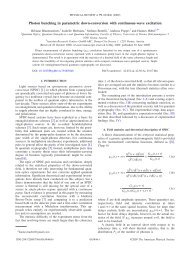
The essence <strong>of</strong> the experiment is to measure the variation <strong>of</strong> the<br />
interference fringe visibility with heating laser power (Fig. 2). Two<br />
observations can be made: first, the interference contrast decreases<br />
Figure 1 Set-up for the observation <strong>of</strong> <strong>thermal</strong> decoherence in a Talbot–Lau molecule<br />
interferometer. A fullerene beam passes from left to right, interacting with a heating stage,<br />
a three-grating (G 1 –G 3 ) <strong>matter</strong>-wave interferometer and an ionizing detection laser beam<br />
in D 2 (wavelength 488 nm, 1/e 2 intensity radius 6.6 mm, 15 W). The gold gratings have a<br />
period <strong>of</strong> 991 nm and slit widths <strong>of</strong> nominally 475 ^ 20 nm. <strong>Decoherence</strong> <strong>of</strong> the fullerene<br />
<strong>matter</strong> <strong>waves</strong> can be induced <strong>by</strong> heating the molecules with multiple laser beams<br />
(514.5 nm, 40 mm waist radius, 0–10 W) before they enter the interferometer. The<br />
resulting molecular temperature can be assessed <strong>by</strong> detecting the heating-dependent<br />
fraction <strong>of</strong> fullerene ions using the electron multiplier D 1 over the heating stage.<br />
NATURE | VOL 427 | 19 FEBRUARY 2004 | www.nature.com/nature 711<br />
© 2004 Nature Publishing Group
letters to nature<br />
Figure 2 Molecule interferograms for C 70 at 190 m s 21 for increasing laser heating<br />
powers, P. The fringe visibility V decreases with increasing heating power P owing to the<br />
rising <strong>emission</strong> probability <strong>of</strong> visible photons: P ¼ 0W(V ¼ 47%), P ¼ 3W(V ¼ 29%),<br />
P ¼ 6W(V ¼ 7%), P ¼ 10.5 W (V ¼ 0%). In contrast to that, the absolute count rate<br />
grows initially with increasing P. This is due to the fact that the <strong>thermal</strong> ionization<br />
probability in detector D 2 increases with the temperature <strong>of</strong> the arriving molecules. At<br />
even higher heating intensities the count rate falls again because <strong>of</strong> ionization and<br />
fragmentation in the heating stage.<br />
monotonically with increasing power, and vanishes at 10 W. This is<br />
the signature <strong>of</strong> decoherence due to the enhanced probability for the<br />
<strong>emission</strong> <strong>of</strong> <strong>thermal</strong> photons that carry ‘which-path’ information.<br />
Second, we notice that the count rate also varies considerably. This is<br />
explained <strong>by</strong> the dependence <strong>of</strong> the ionization efficiency in the<br />
detector D 2 on the internal energy <strong>of</strong> the fullerenes. It proves that<br />
much internal energy remains in the molecules during their flight<br />
through the apparatus.<br />
In order to confirm quantitatively the interpretations <strong>of</strong> both<br />
observations, we model the evolution <strong>of</strong> the distribution <strong>of</strong> the<br />
internal energies on their way through the apparatus. The temperature<br />
dependence <strong>of</strong> the spectral photon <strong>emission</strong> rate (equation (1)<br />
below) then yields the loss <strong>of</strong> fringe visibility as predicted <strong>by</strong><br />
decoherence theory (equation (2) below).<br />
The first photon absorption populates the electronic triplet state<br />
T 1 via the excited singlet S 1 . Given the known C 70 triplet lifetimes<br />
and non-radiative transition rates (see ref. 17 and references<br />
therein), we can assume that all further excitation occurs in the<br />
triplet system and that the absorbed excess energy is rapidly<br />
transferred to the vibrational levels. It is known that fullerenes<br />
may store more than 100 eV for a very short time 17 , and it was<br />
observed that at high temperatures three different cooling mechanisms<br />
start to compete—the <strong>thermal</strong> <strong>emission</strong> <strong>of</strong> photons, electrons<br />
or C 2 dimers 18–22 . These processes are the molecular analogues <strong>of</strong> the<br />
bulk phenomena known as blackbody <strong>radiation</strong>, thermionic <strong>emission</strong><br />
and evaporative cooling. Following the most recent experimental<br />
data 22 , we may safely assume that fragmentation is the least<br />
efficient mechanism. In contrast, <strong>thermal</strong>ly activated ionization is<br />
an important mechanism, which we use both in our fullerene<br />
detector 23 and for molecule thermometry, as discussed below.<br />
Nevertheless, we can safely neglect both delayed ionization and<br />
fragmentation for the discussion <strong>of</strong> the fringe contrast, because<br />
the recoil upon fragmentation and ionization is generally so large<br />
that the affected molecules will miss the narrow detector. We have<br />
also experimentally confirmed that neither C þ 70 ions nor C 68 nor<br />
smaller fragments from the heating region are recorded <strong>by</strong> the<br />
detector D 2 .<br />
However, C þ 70<br />
ions—and potentially ionized fragments—can be<br />
detected immediately above the heating stage <strong>by</strong> the electron<br />
multiplier D 1 (Fig. 1). To get an estimate <strong>of</strong> the molecular temperature<br />
distribution, we record the number <strong>of</strong> ions as a function <strong>of</strong><br />
712<br />
the heating power and <strong>of</strong> the fullerene velocity. By comparing the<br />
data with a model calculation, we can extract the parameters that<br />
govern the molecular heating <strong>of</strong> C 70 . Our model describes the<br />
spatial and velocity-dependent distribution <strong>of</strong> the internal molecular<br />
energy <strong>by</strong> accounting for the stochastic absorption process, the<br />
laser beam characteristics, and the rapid radiative cooling between<br />
the beams as determined <strong>by</strong> equation (1) below. It reproduces the<br />
detected number <strong>of</strong> ions in the heating stage for different laser<br />
powers, different numbers <strong>of</strong> heating beams and all velocities with<br />
the fit parameters for the triplet absorption cross-section,<br />
j(T 1 ) ¼ 2 £ 10 217 cm 2 , and the effective Arrhenius constant for<br />
ionization, A ion ¼ 5 £ 10 9 s 21 . The same calculation also describes<br />
the heating-dependent increase in count rate at the detector D 2<br />
and thus yields independent information on the temperature<br />
distribution in the molecular beam.<br />
The mean temperature in the beam drops rapidly behind the<br />
heating stage through the <strong>emission</strong> <strong>of</strong> <strong>thermal</strong> photons. The <strong>emission</strong><br />
<strong>of</strong> a continuous photon spectrum has already been observed for<br />
fullerenes in other experiments 18,24 . The equation for the <strong>thermal</strong><br />
<strong>radiation</strong> density differs from the macroscopic Planck law for<br />
several reasons. First, the <strong>thermal</strong> wavelengths are much larger<br />
than the size <strong>of</strong> the fullerene, turning it into a coloured emitter.<br />
The mean <strong>emission</strong> probability is proportional to the usual mode<br />
density factor q 2 /p 2 c 2 and the known frequency-dependent<br />
absorption cross-section 25 j abs (q), assuming that it does not<br />
strongly depend on the internal temperature. Second, the particle<br />
is not in <strong>thermal</strong> equilibrium with the <strong>radiation</strong> field. It emits into a<br />
cold environment and stimulated <strong>emission</strong> does not occur. For this<br />
reason, the statistical factor 1=½expðhq=k B TÞ 2 1ÞŠ <strong>of</strong> the Planck<br />
formula now would read expð2hq=k B TÞ: Third, the 204 vibrational<br />
modes <strong>of</strong> C 70 do not constitute an infinite heat bath but have a finite<br />
heat capacity C V . Therefore the <strong>emission</strong> does not take place at a<br />
fixed temperature, although the internal energy is nonetheless<br />
conveniently characterized <strong>by</strong> the micro-canonical temperature<br />
T m . This leads to a further correction in the spectral photon<br />
<strong>emission</strong> rate 26 , which is now fully described <strong>by</strong><br />
© 2004 Nature Publishing Group<br />
Figure 3 Spectral photon <strong>emission</strong> rate R l <strong>of</strong> C 70 molecules, as used for the calculation<br />
<strong>of</strong> <strong>thermal</strong> decoherence. We use the published 25 absorption cross-section for (S 0 ! S 1 )<br />
and a heat capacity <strong>of</strong> C V ¼ 202 k B . The fall-<strong>of</strong>f to short wavelengths is determined <strong>by</strong><br />
the limited internal energy <strong>of</strong> the molecules, while the decrease at long wavelengths is due<br />
to the lack <strong>of</strong> accessible radiative transitions at energies below ,1.5 eV. The figure shows<br />
that in the absence <strong>of</strong> cooling a single molecule at 2,500 K travelling at 190 m s 21 (that is,<br />
with a transit time <strong>of</strong> 4 ms through the interferometer) would emit an integrated number <strong>of</strong><br />
three visible photons. This is sufficient to determine the path <strong>of</strong> the molecule if the<br />
<strong>emission</strong> occurs close to the second grating.<br />
" #<br />
R q ðq; T m Þ¼ q2<br />
p 2 c 2 j absðqÞexp 2 hq 2 k <br />
B hq 2<br />
k B T m 2C V k B T m<br />
ð1Þ<br />
NATURE | VOL 427 | 19 FEBRUARY 2004 | www.nature.com/nature
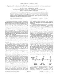
letters to nature<br />
Figure 4 <strong>Decoherence</strong> curves. a, Interference visibility as a function <strong>of</strong> laser heating<br />
power (lower scale). The molecular beam with a mean velocity <strong>of</strong> v m ¼ 190 m s 21<br />
passes a 50 mm central height delimiter comparable to the waist (40 mm) <strong>of</strong> the 16<br />
heating laser beams. We observe a rapid decrease <strong>of</strong> the fringe visibility with increasing<br />
power both in the experiment (circles) and in theory (solid line). The upper axis indicates<br />
the mean temperature <strong>of</strong> the molecules when they enter the interferometer. The<br />
maximum contrast without heating was V 0 ¼ 47%, which is close to the theoretical<br />
value 11 . b, Molecules with v m ¼ 100 m s 21 , selected <strong>by</strong> a 150 mm height delimiter and<br />
heated <strong>by</strong> ten beams <strong>of</strong> the specified incident laser power. The qualitative behaviour is the<br />
same and the quantitative agreement with theory is as good as before. The maximum<br />
contrast for this velocity class was V 0 ¼ 19%. In both experimental arrangements, a<br />
mean number between one and two visible photons is required to reduce the contrast <strong>by</strong> a<br />
factor <strong>of</strong> two.<br />
to the <strong>radiation</strong> wavelength, while the integrals cover all photon<br />
wavelengths l and longitudinal positions vt in the interferometer.<br />
As a result, the visibility is reduced exponentially whenever<br />
photons are emitted whose wavelength is sufficiently small to<br />
resolve the path separation. Our predictions for the loss <strong>of</strong> visibility<br />
are obtained <strong>by</strong> weighting equation (2) with the previously determined<br />
distribution <strong>of</strong> temperature evolutions in the molecular<br />
beam.<br />
In Fig. 4 we compare our decoherence model with the experiments<br />
<strong>by</strong> plotting the interference fringe visibility as a function <strong>of</strong><br />
the laser power. We observe a strong decrease <strong>of</strong> the visibility for<br />
molecules at 190 m s 21 , heated <strong>by</strong> 16 laser beams (Fig. 4a), and for<br />
molecules at 100 m s 21 , heated <strong>by</strong> 10 laser beams (Fig. 4b).<br />
We also observe good agreement between decoherence theory<br />
(solid line) and the experiment (circles). The experiment is reproducible<br />
within the indicated error bars for a given laser alignment,<br />
but small displacements <strong>of</strong> the laser focus will influence the shape<br />
and slope <strong>of</strong> the observed decoherence curve. The difference<br />
between the theoretical and the experimental curve is <strong>of</strong> the order<br />
<strong>of</strong> this variation.<br />
In summary, we have presented conclusive empirical and<br />
numerical evidence for observation <strong>of</strong> the quantum-to-classical<br />
transition <strong>of</strong> a material object caused <strong>by</strong> its own <strong>emission</strong> <strong>of</strong> <strong>thermal</strong><br />
<strong>radiation</strong>. This auto-localization is a fundamental process limiting<br />
the ultimate observability <strong>of</strong> quantum effects in macroscopic<br />
objects. However, for nanometre-sized systems 13,27,28 this mechanism<br />
becomes relevant only at high temperatures, and it is not<br />
expected to be a limitation for interference <strong>of</strong> objects even considerably<br />
larger than the fullerenes, such as proteins. A<br />
Methods<br />
Equation (2) describes the loss <strong>of</strong> <strong>matter</strong> wave coherence due to the <strong>emission</strong> <strong>of</strong> <strong>thermal</strong><br />
photons. It is obtained <strong>by</strong> assuming that the <strong>emission</strong> is isotropic and that the absorbing<br />
walls <strong>of</strong> the apparatus are located in the far-field, where the photon position distribution<br />
reflects its momentum distribution. In this case, a trace over the photon state changes the<br />
fullerene centre-<strong>of</strong>-mass state rˆ according to<br />
ð<br />
r ^! r^<br />
0 ¼ dk pðkÞ<br />
4pk ^U 2 k r^<br />
^U † k<br />
ð3Þ<br />
where the ^U k ¼ expði^rkÞ are momentum translation operators and p(k) is the probability<br />
density for the photon wavenumber k ¼ 2p/l. In the position representation <strong>of</strong> the<br />
density matrix,<br />
In Fig. 3 we plot the wavelength dependence <strong>of</strong> R l ¼ R q jdq=dlj: We<br />
observe that at temperatures below 2,000 K the <strong>emission</strong> rate is<br />
negligible, whereas at higher temperatures the molecules may emit<br />
photons whose wavelengths are comparable to (or even smaller<br />
than) the maximum path separation <strong>of</strong> ,1 mm. They transmit<br />
(partial) which-path information to the environment, leading to a<br />
reduced observability <strong>of</strong> the fullerene wave nature. Around 3,000 K<br />
the molecules have a high probability to emit several visible photons<br />
yielding sufficient which-path information to effect a complete loss<br />
<strong>of</strong> fringe visibility in our interferometer.<br />
A formal description <strong>of</strong> this qualitative picture can be given <strong>by</strong><br />
decoherence theory. It considers the entanglement <strong>of</strong> the molecule<br />
with the emitted photon, and shows how coherences vanish once a<br />
trace over the photon state is performed. For objects with velocity v<br />
and temperature evolution T(t) we obtain a visibility<br />
" ð 2L=v ð 1<br />
V ¼V 0 exp 2 dt dl R l ðl; TðtÞÞ£<br />
0 0<br />
ð2Þ<br />
<br />
1 2 sinc 2p d <br />
L 2 jvt 2 Lj<br />
<br />
l L T<br />
as discussed in the Methods section. V 0 denotes the interference<br />
contrast in the absence <strong>of</strong> photon <strong>emission</strong>. In the exponential,<br />
the sinc function compares the effective molecular path separation<br />
r 0 ðr 1 ; r 2 Þ ;kr 1 jr^<br />
0 jr 2 l ¼ kr 1 jrjr ^ 2 lhðr 1 2 r 2 Þ<br />
we find from equation (3) that the <strong>of</strong>f-diagonal elements are reduced <strong>by</strong> the decoherence<br />
function 29 hðr 1 2 r 2 Þ¼ 1 ð 1<br />
<br />
dl R l ðlÞ sinc 2p jr <br />
1 2 r 2 j<br />
ð5Þ<br />
R tot 0<br />
l<br />
Here p(k) is expressed in terms <strong>of</strong> the spectral <strong>emission</strong> rate R l (see equation (1)) and the<br />
total photon <strong>emission</strong> rate R tot . This sinc-shaped position dependence <strong>of</strong> h is also found in<br />
other experiments with isotropic momentum change 7,8 . It describes the diffraction<br />
limitation <strong>of</strong> a hypothetical microscope used to obtain which-path information on the<br />
molecules.<br />
In the Talbot–Lau geometry 14,16,27 the final molecular fringe pattern w(x) is strictly<br />
periodic in the grating constant d, and can be expanded as a Fourier series, which reads in<br />
the absence <strong>of</strong> decoherence<br />
X<br />
wðxÞ¼ Cl expð2pilx=dÞ<br />
ð6Þ<br />
l<br />
Assuming that a single photon <strong>emission</strong> occurs at the longitudinal position z ¼ vt, a<br />
closed expression for the resulting molecular density pattern can be found. It is obtained<br />
<strong>by</strong> propagating the molecular density matrix in paraxial approximation first to the<br />
position z. We then apply the decohering transformation (equation (4)) followed <strong>by</strong> a<br />
propagation to the final grating. For a set-up with equally spaced and identical<br />
gratings, the new fringe pattern is described <strong>by</strong> a simple modification <strong>of</strong> the Fourier<br />
coefficients<br />
C l ! C l<br />
0<br />
¼ C l hðldðL 2 jvt 2 LjÞ=L T Þ ð7Þ<br />
In order to account for more than one photon <strong>emission</strong>, we make the Markov assumption<br />
that all photon <strong>emission</strong>s are independent <strong>of</strong> each other. Because the modification<br />
NATURE | VOL 427 | 19 FEBRUARY 2004 | www.nature.com/nature 713<br />
© 2004 Nature Publishing Group<br />
ð4Þ
letters to nature<br />
(equation (7)) is independent <strong>of</strong> the molecular density matrix, the change <strong>of</strong> the final<br />
density pattern is governed <strong>by</strong> the differential equation<br />
<br />
<br />
d<br />
dt C L 2 jvt 2 Lj<br />
l ¼ R tot C l h ld 2 C l<br />
ð8Þ<br />
L T<br />
It describes how the final interferogram is blurred as the time interval <strong>of</strong> <strong>emission</strong><br />
increases. Equation (2) follows then immediately after taking into account the<br />
time dependence <strong>of</strong> the <strong>emission</strong> rate due to cooling (equation (1)) and the fact that<br />
for our grating geometry, with a slit width <strong>of</strong> 470 nm and grating constant <strong>of</strong> 990 nm,<br />
only the lowest-order Fourier components contribute to the fringe visibility 30<br />
V ¼ 2jC 1 =C 0 j:<br />
Received 15 September; accepted 8 December 2003; doi:10.1038/nature02276.<br />
1. Joos, E. et al. <strong>Decoherence</strong> and the Appearance <strong>of</strong> a Classical World in Quantum Theory (Springer,<br />
Heidelberg, 2003).<br />
2. Blanchard, P., Giulini, D., Joos, E., Kiefer, C. & Stamatescu, I.-O. (eds) <strong>Decoherence</strong>: Theoretical,<br />
Experimental, and Conceptual Problems (Lecture Notes in Physics 538, Springer, Heidelberg, 2000).<br />
3. Imry, Y. Introduction to Mesoscopic Physics, 2nd edn (Oxford Univ. Press, Oxford, 2000).<br />
4. Zurek, W. H. <strong>Decoherence</strong>, einselection, and the quantum origins <strong>of</strong> the classical. Rev. Mod. Phys. 75,<br />
715–775 (2003).<br />
5. Pfau, T., Spälter, S., Kurtsiefer, Ch., Ekstrom, C. R. & Mlynek, J. Loss <strong>of</strong> spatial coherence <strong>by</strong> a single<br />
spontaneous <strong>emission</strong>. Phys. Rev. Lett. 73, 1223–1226 (1994).<br />
6. Chapman, M. S. et al. Photon scattering from atoms in an atom interferometer: coherence lost and<br />
regained. Phys. Rev. Lett. 75, 3783–3787 (1995).<br />
7. Kokorowski, D. A., Cronin, A. D., Roberts, T. D. & Pritchard, D. E. From single- to multiple-photon<br />
decoherence in an atom interferometer. Phys. Rev. Lett. 86, 2191–2194 (2001).<br />
8. Hornberger, K. et al. Collisional decoherence observed in <strong>matter</strong> wave interferometry. Phys. Rev. Lett.<br />
90, 160401 (2003).<br />
9. Brune, M. et al. Observing the progressive decoherence <strong>of</strong> the “meter” in a quantum measurement.<br />
Phys. Rev. Lett. 77, 4887–4890 (1996).<br />
10. Myatt, C. J. et al. <strong>Decoherence</strong> <strong>of</strong> quantum superpositions through coupling to engineered reservoirs.<br />
Nature 403, 269–273 (2000).<br />
11. Buks, E., Schuster, R., Heiblum, M., Mahalu, D. & Umansky, V. Dephasing in electron interference <strong>by</strong> a<br />
‘which-path’ detector. Nature 391, 871–874 (1998).<br />
12. Ji, Y. et al. An electronic Mach–Zehnder interferometer. Nature 422, 415–418 (2003).<br />
13. Arndt, M. et al. Wave–particle duality <strong>of</strong> C60 molecules. Nature 401, 680–682 (1999).<br />
14. Brezger, B. et al. Matter-wave interferometer for large molecules. Phys. Rev. Lett. 88, 100404<br />
(2002).<br />
15. Patorski, K. in Progress in Optics XXVII (ed. Wolf, E.) 2–108 (Elsevier Science, Amsterdam, 1989).<br />
16. Clauser, J. F. & Li, S. Talbot-von Lau atom interferometry with cold slow potassium. Phys. Rev. A 49,<br />
R2213–R2217 (1994).<br />
17. Dresselhaus, M. S., Dresselhaus, G. & Eklund, P. C. Science <strong>of</strong> Fullerenes and Carbon Nanotubes<br />
(Academic, San Diego, 1996).<br />
18. Mitzner, R. & Campbell, E. E. B. Optical <strong>emission</strong> studies <strong>of</strong> laser desorbed C 60 . J. Chem. Phys. 103,<br />
2445–2453 (1995).<br />
19. Ding, D., Huang, J., Compton, R. N., Klots, C. E. & Haufler, R. E. Cw laser ionization <strong>of</strong> C 60 and C 70 .<br />
Phys. Rev. Lett. 73, 1084–1087 (1994).<br />
20. Hansen, K. & Echt, O. Thermionic <strong>emission</strong> and fragmentation <strong>of</strong> C 60 . Phys. Rev. E 78, 2337–2340<br />
(1997).<br />
21. Kolodney, E., Tsipinyuk, B. & Budrevich, A. The <strong>thermal</strong> energy dependence (10–20 eV) <strong>of</strong> electron<br />
impact induced fragmentation <strong>of</strong> C 60 in molecular beams: experiment and model calculations.<br />
J. Chem. Phys. 102, 9263–9275 (1995).<br />
22. Matt, S. et al. Kinetic energy release distribution and evaporation energies for metastable fullerene<br />
ions. Chem. Phys. Lett. 303, 379–386 (1999).<br />
23. Nairz, O., Arndt, M. & Zeilinger, A. Experimental challenges in fullerene interferometry. J. Mod. Opt.<br />
47, 2811–2821 (2001).<br />
24. Heszler, P., Carlsson, J. O. & Demirev, P. Photon <strong>emission</strong> from gas phase fullerenes excited <strong>by</strong> 193 nm<br />
laser <strong>radiation</strong>. Chem. Phys. 107, 10440–10445 (1997).<br />
25. Coheur, P. F., Carleer, M. & Colin, R. The absorption cross sections <strong>of</strong> C 60 and C 70 in the visible-UV<br />
region. J. Phys. B 29, 4987–4995 (1996).<br />
26. Hansen, K. & Campbell, E. E. B. Thermal <strong>radiation</strong> from small particles. Phys. Rev. E 58, 5477–5482<br />
(1998).<br />
27. Hackermüller, L. et al. Wave nature <strong>of</strong> biomolecules and fluor<strong>of</strong>ullerenes. Phys. Rev. Lett. 91, 090408<br />
(2003).<br />
28. Clauser, J. F. in Experimental Metaphysics (eds Cohen, R. S., Home, M. & Stachel, J.) 1–11 (Kluwer<br />
Academic, Dordrecht, 1997).<br />
29. Joos, E. & Zeh, H. D. The emergence <strong>of</strong> classical properties through interaction with the environment.<br />
Z. Phys. B 59, 223–243 (1985).<br />
30. Brezger, B., Arndt, M. & Zeilinger, A. Concepts for near-field interferometers with large molecules.<br />
J. Opt. B 5, 82–89 (2003).<br />
Acknowledgements We thank S. Uttenthaler for his support in an early stage <strong>of</strong> the experiment.<br />
We acknowledge support <strong>by</strong> the Austrian START programme, the Austrian FWF, the European<br />
TMR and Marie Curie programmes, and the DFG Emmy-Noether programme.<br />
Authors’ contributions L.H. performed most <strong>of</strong> the experiments as a part <strong>of</strong> her Ph.D. thesis. K.H.<br />
developed the decoherence theory, and made the quantitative comparison between experiment<br />
and theory.<br />
Competing interests statement The authors declare that they have no competing financial<br />
interests.<br />
Correspondence and requests for materials should be addressed to M.A.<br />
(markus.arndt@univie.ac.at).<br />
714<br />
..............................................................<br />
High-transition-temperature<br />
superconductivity in the absence<br />
<strong>of</strong> the magnetic-resonance mode<br />
J. Hwang 1 , T. Timusk 1 &G.D.Gu 2<br />
1 Department <strong>of</strong> Physics and Astronomy, McMaster University, Hamilton,<br />
Ontario L8S 4M1, Canada<br />
2 Department <strong>of</strong> Physics, Brookhaven National Laboratory, Upton, New York<br />
11973-5000, USA<br />
.............................................................................................................................................................................<br />
The fundamental mechanism that gives rise to high-transitiontemperature<br />
(high-T c ) superconductivity in the copper oxide<br />
materials has been debated since the discovery <strong>of</strong> the phenomenon.<br />
Recent work has focused on a sharp ‘kink’ in the kinetic<br />
energy spectra <strong>of</strong> the electrons as a possible signature <strong>of</strong> the force<br />
that creates the superconducting state 1–14 . The kink has been<br />
related to a magnetic resonance 13,15–17 and also to phonons 18 .<br />
Here we report that infrared spectra <strong>of</strong> Bi 2 Sr 2 CaCu 2 O 81d<br />
(Bi-2212), shows that this sharp feature can be separated from<br />
a broad background and, interestingly, weakens with doping<br />
before disappearing completely at a critical doping level <strong>of</strong> 0.23<br />
holes per copper atom. Superconductivity is still strong in terms<br />
<strong>of</strong> the transition temperature at this doping (T c < 55 K), so our<br />
results rule out both the magnetic resonance peak and phonons<br />
as the principal cause <strong>of</strong> high-T c superconductivity. The broad<br />
background, on the other hand, is a universal property <strong>of</strong> the<br />
copper–oxygen plane and provides a good candidate signature <strong>of</strong><br />
the ‘glue’ that binds the electrons.<br />
We investigated the Bi-2212 material systematically as a function<br />
<strong>of</strong> doping, including highly overdoped Bi-2212 (see Methods). To<br />
better display the sharp ‘kink’, we do not show the optical conductivity<br />
but focus our attention on a related quantity, the optical<br />
single-particle self-energy, S op (q) (see Methods). In Fig. 1a–d we<br />
plot the imaginary part <strong>of</strong> this quantity, which is simply the<br />
scattering rate <strong>of</strong> the charge carriers. At high frequencies the<br />
scattering rate varies linearly with frequency: this is called the<br />
marginal Fermi-liquid (MFL) behaviour 19 . We note that the overall<br />
scattering rate decreases as the doping increases and that there is a<br />
sharp depression in 1/t(q) below 700 cm 21 at low temperatures,<br />
with an overshoot in the superconducting state. This sharp onset <strong>of</strong><br />
scattering has been in general attributed to the interaction <strong>of</strong> the<br />
charge carriers with a bosonic mode and more recently to the<br />
41-meV neutron resonance 7,13,14 . In the real part <strong>of</strong> the optical selfenergy,<br />
plotted in Fig. 1e–h, we note a sharp peak around 700 cm 21 ,<br />
which tracks the depressions in 1/t(q) in both frequency and<br />
amplitude. One interesting property <strong>of</strong> the peak in the self-energy<br />
is its doping and temperature dependence: the peak gets weaker as<br />
the doping level and the temperature increase. We call the peak the<br />
‘optical resonance mode’. The resonance peak in the self-energy<br />
spectrum can clearly be resolved from the broader background <strong>of</strong><br />
MFL scattering.<br />
The optical self-energy is closely related to the quasiparticle selfenergy<br />
S qp (q), which can be measured in ARPES experiments<br />
although there are important differences in the two quantities 20–22 .<br />
In Fig. 2 we show a comparison <strong>of</strong> the optical self-energy from our<br />
data and the ARPES self-energy at two temperatures: one in the<br />
normal state and the other in the superconducting state. We see that<br />
although the qualitative features are very similar, the magnitudes<br />
and the frequency values <strong>of</strong> the various features differ considerably,<br />
as expected from theory. Part <strong>of</strong> the difference can be attributed to<br />
the uncertainty in locating the unrenormalized baseline in the<br />
ARPES analysis. As noted in ref. 6, a peak in S qp<br />
1 (q) can be clearly<br />
separated from the broad continuum and is closely correlated with<br />
© 2004 Nature Publishing Group<br />
NATURE | VOL 427 | 19 FEBRUARY 2004 | www.nature.com/nature