hTERT-HME1
hTERT-HME1
hTERT-HME1

Create successful ePaper yourself
Turn your PDF publications into a flip-book with our unique Google optimized e-Paper software.
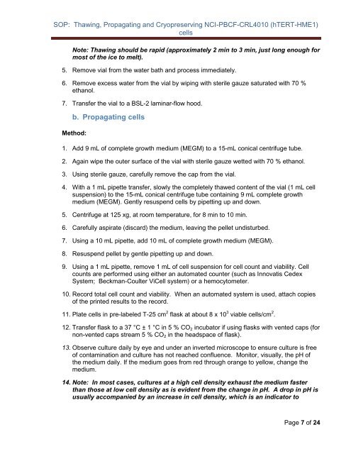
SOP: Thawing, Propagating and Cryopreserving NCI-PBCF-CRL4010 (<strong>hTERT</strong>-<strong>HME1</strong>)<br />
cells<br />
Note: Thawing should be rapid (approximately 2 min to 3 min, just long enough for<br />
most of the ice to melt).<br />
5. Remove vial from the water bath and process immediately.<br />
6. Remove excess water from the vial by wiping with sterile gauze saturated with 70 %<br />
ethanol.<br />
7. Transfer the vial to a BSL-2 laminar-flow hood.<br />
b. Propagating cells<br />
Method:<br />
1. Add 9 mL of complete growth medium (MEGM) to a 15-mL conical centrifuge tube.<br />
2. Again wipe the outer surface of the vial with sterile gauze wetted with 70 % ethanol.<br />
3. Using sterile gauze, carefully remove the cap from the vial.<br />
4. With a 1 mL pipette transfer, slowly the completely thawed content of the vial (1 mL cell<br />
suspension) to the 15-mL conical centrifuge tube containing 9 mL complete growth<br />
medium (MEGM). Gently resuspend cells by pipetting up and down.<br />
5. Centrifuge at 125 xg, at room temperature, for 8 min to 10 min.<br />
6. Carefully aspirate (discard) the medium, leaving the pellet undisturbed.<br />
7. Using a 10 mL pipette, add 10 mL of complete growth medium (MEGM).<br />
8. Resuspend pellet by gentle pipetting up and down.<br />
9. Using a 1 mL pipette, remove 1 mL of cell suspension for cell count and viability. Cell<br />
counts are performed using either an automated counter (such as Innovatis Cedex<br />
System; Beckman-Coulter ViCell system) or a hemocytometer.<br />
10. Record total cell count and viability. When an automated system is used, attach copies<br />
of the printed results to the record.<br />
11. Plate cells in pre-labeled T-25 cm 2 flask at about 8 x 10 3 viable cells/cm 2 .<br />
12. Transfer flask to a 37 °C ± 1 °C in 5 % CO 2 incubator if using flasks with vented caps (for<br />
non-vented caps stream 5 % CO 2 in the headspace of flask).<br />
13. Observe culture daily by eye and under an inverted microscope to ensure culture is free<br />
of contamination and culture has not reached confluence. Monitor, visually, the pH of<br />
the medium daily. If the medium goes from red through orange to yellow, change the<br />
medium.<br />
14. Note: In most cases, cultures at a high cell density exhaust the medium faster<br />
than those at low cell density as is evident from the change in pH. A drop in pH is<br />
usually accompanied by an increase in cell density, which is an indicator to<br />
Page 7 of 24