
Create successful ePaper yourself
Turn your PDF publications into a flip-book with our unique Google optimized e-Paper software.
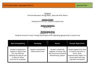
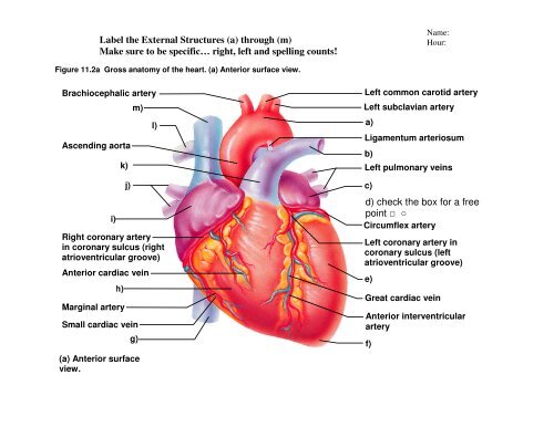
Label the External Structures (a) through (m)<br />
Make sure to be specific… right, left and spelling counts!<br />
Name:<br />
Hour:<br />
Figure 11.2a <strong>Gross</strong> anatomy of the heart. (a) Anterior surface view.<br />
Brachiocephalic artery<br />
Ascending aorta<br />
i)<br />
k)<br />
j)<br />
m)<br />
l)<br />
Right coronary artery<br />
in coronary sulcus (right<br />
atrioventricular groove)<br />
Anterior cardiac vein<br />
h)<br />
Marginal artery<br />
Small cardiac vein<br />
g)<br />
(a) Anterior surface<br />
view.<br />
Left common carotid artery<br />
Left subclavian artery<br />
a)<br />
Ligamentum arteriosum<br />
b)<br />
Left pulmonary veins<br />
c)<br />
d) check the box for a free<br />
point □ ○<br />
Circumflex artery<br />
Left coronary artery in<br />
coronary sulcus (left<br />
atrioventricular groove)<br />
e)<br />
Great cardiac vein<br />
Anterior interventricular<br />
artery<br />
f)
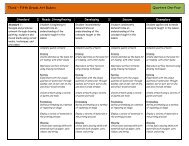
Label the Internal Structures (a) through (r)<br />
Make sure to be specific… right, left and spelling counts!<br />
Figure 11.2c <strong>Gross</strong> anatomy of the heart. (c) Frontal section showing interior chambers and valves.<br />
r)<br />
a)<br />
q)<br />
b)<br />
c)<br />
p)<br />
Right pulmonary<br />
veins<br />
o)<br />
Left pulmonary veins<br />
e)<br />
f)<br />
n)<br />
g)<br />
m)<br />
h)<br />
l)<br />
k)<br />
i)<br />
Myocardium<br />
Visceral pericardium<br />
(c) Frontal section showing interior chambers and valves.