Cosmetic Artecoll: A Long-Lasting Injectable Wrinkle Filler Material ...
Cosmetic Artecoll: A Long-Lasting Injectable Wrinkle Filler Material ...
Cosmetic Artecoll: A Long-Lasting Injectable Wrinkle Filler Material ...
Create successful ePaper yourself
Turn your PDF publications into a flip-book with our unique Google optimized e-Paper software.
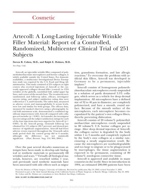
<strong>Cosmetic</strong><br />
<strong>Artecoll</strong>: A <strong>Long</strong>-<strong>Lasting</strong> <strong>Injectable</strong> <strong>Wrinkle</strong><br />
<strong>Filler</strong> <strong>Material</strong>: Report of a Controlled,<br />
Randomized, Multicenter Clinical Trial of 251<br />
Subjects<br />
Steven R. Cohen, M.D., and Ralph E. Holmes, M.D.<br />
San Diego, Calif.<br />
<strong>Artecoll</strong>, an injectable wrinkle filler composed of polymethylmethacrylate<br />
microspheres and bovine collagen, is<br />
widely available outside the United States. For domestic<br />
availability, a multicenter Investigational Device Exemption<br />
study was required by the U.S. Food and Drug Administration.<br />
This study consisted of 251 subjects at eight<br />
centers who received injections of <strong>Artecoll</strong> or the currently<br />
approved collagen dermal filler (control) in 1334<br />
wrinkles of the glabella, nasolabial fold, radial upper lip<br />
lines, and corner-of-the-mouth lines. The treatments were<br />
randomized, and follow-up safety, efficacy, investigator<br />
success rating, and subject satisfaction rating data were<br />
collected at 1, 3, and 6 months. The safety data, measured<br />
as adverse events and immunoglobulin G serum levels,<br />
were low and similar for both groups. The efficacy data,<br />
measured by masked observers using a photographic facial<br />
fold assessment scale, demonstrated a combined significant<br />
improvement with <strong>Artecoll</strong> compared with collagen<br />
at 6 months (p 0.001). At 6 months, the investigator<br />
success ratings and the subject satisfaction ratings for each<br />
of the four injections sites were superior for <strong>Artecoll</strong> (p <br />
0.001). In the <strong>Artecoll</strong> group, 12-month follow-up was<br />
obtained for 111 subjects (86.7 percent) and showed persistence<br />
of significant augmentation. <strong>Artecoll</strong> had fewer<br />
adverse events reported throughout the 12-month safety<br />
study period than the control group did in 6 months,<br />
although the difference was not statistically<br />
significant. (Plast. Reconstr. Surg. 114: 964, 2004.)<br />
Over the past several decades numerous attempts<br />
have been made to develop safe biological<br />
or synthetic materials to permanently fill<br />
wrinkles and scars. 1,2 Virtually all biological materials,<br />
however, are resorbed within 1 year,<br />
and previously used synthetic materials have<br />
been associated with side effects such as migration,<br />
granuloma formation, and late allergic<br />
reactions. 3 To overcome the problems with artificial<br />
skin fillers, <strong>Artecoll</strong> was developed in<br />
Germany to be a permanent, injectable<br />
implant. 4<br />
<strong>Artecoll</strong> consists of homogenous polymethylmethacrylate<br />
microspheres evenly suspended<br />
in a solution of partly denatured 3.5% collagen,<br />
which serves as a vehicle for deep dermal<br />
implantation. All microspheres have a defined<br />
size of 32 to 40 m in diameter, are completely<br />
polymerized, and have a smooth, round surface.<br />
Because of the smooth surface of the<br />
microspheres, each microsphere becomes encapsulated<br />
by the patient’s own collagen fibers,<br />
thereby preventing dislocation.<br />
<strong>Artecoll</strong> consists of 20 volume% polymethylmethacrylate<br />
microspheres evenly suspended<br />
in 80 volume% U.S. bovine collagen per syringe.<br />
After deep dermal injection of <strong>Artecoll</strong>,<br />
the collagen carrier is degraded by the body<br />
within 1 to 3 months and completely replaced<br />
by the body’s own collagen at a similar rate,<br />
ensuring a steady augmentation result. Because<br />
the microspheres are nonbiodegradable<br />
and too large to migrate or to be phagocytosed<br />
by macrophages, the tissue augmentation is<br />
expected to be permanent, consisting of 80<br />
volume% autologous connective tissue.<br />
<strong>Artecoll</strong> is approved and available in many<br />
countries in the world. Since its introduction in<br />
1994, an estimated 200,000 patients have been<br />
From the Division of Plastic Surgery, University of California. Received for publication May 19, 2003; revised November 5, 2003.<br />
Presented in part at the 52nd Annual Meeting of the California Society of Plastic Surgeons, in Rancho Mirage, California, May 25, 2002.<br />
Dr. Cohen and Dr. Holmes are consultants to Artes Medical, San Diego, California.<br />
DOI: 10.1097/01.PRS.0000133169.16467.5F<br />
964
Vol. 114, No. 4 /ARTECOLL INJECTABLE WRINKLE FILLER 965<br />
treated with a reported complication rate of<br />
0.01 percent. 4 On February 28, 2003, the U.S.<br />
Food and Drug Administration’s General and<br />
Plastic Surgery Devices Advisory Panel recommended<br />
that <strong>Artecoll</strong> be approved, with conditions,<br />
for marketing in the United States. <strong>Artecoll</strong><br />
is expected to become the first long-lasting<br />
injectable wrinkle filler to gain Food and Drug<br />
Administration approval since collagen was introduced<br />
in 1981. After approval, <strong>Artecoll</strong> will<br />
be marketed as “Artefill” in the United States,<br />
Canada, and Mexico.<br />
MATERIALS AND METHODS<br />
As an implant material, <strong>Artecoll</strong> is a class III<br />
device requiring Food and Drug Administration<br />
approval via the premarket approval<br />
route. This clinical trial was conducted in accordance<br />
with the Investigational Device Exemption<br />
regulations to obtain safety and efficacy<br />
data for inclusion in the premarket<br />
approval application to the Food and Drug<br />
Administration. The Investigational Device Exemption<br />
application for the <strong>Artecoll</strong> clinical<br />
trial received final approval by the Food and<br />
Drug Administration in August of 1999, and<br />
the trial was completed in September of 2001.<br />
The purpose of this study was to compare the<br />
safety and efficacy of <strong>Artecoll</strong> injections in the<br />
glabellar frown lines, nasolabial folds, radial<br />
upper lip lines, and corner-of-the-mouth (marionette)<br />
lines to the safety and efficacy of collagen<br />
(Zyderm II or Zyplast; Inamed Corporation,<br />
Santa Barbara, Calif.).<br />
The primary objectives of the study were to<br />
compare the cosmetic correction provided by<br />
<strong>Artecoll</strong> at the end of 6 months to that of<br />
Zyderm/Zyplast over the same time period and<br />
to explore the safety of <strong>Artecoll</strong> at 6 and 12<br />
months as an injectable implant for correction<br />
of contour deformities of the dermis of the<br />
face. The secondary objectives of the study<br />
were to characterize the physician’s assessment<br />
of success with respect to how closely the treatment<br />
met the physician’s expectations for correction<br />
and to characterize the subject’s assessment<br />
of satisfaction with respect to the<br />
subject’s personal expectations. Though physicians<br />
were not masked as to the identity of the<br />
treatment, subjects were not told which treatment<br />
they had received until after they had<br />
completed the 6-month evaluation.<br />
The study was performed at eight centers<br />
(four plastic surgery centers and four dermatology<br />
centers) with institutional review board<br />
approval and informed consent from all subjects.<br />
The study was controlled and randomized,<br />
with potential subjects agreeing to be assigned<br />
to either the <strong>Artecoll</strong> or the control<br />
group. The subjects and evaluators were<br />
masked and unaware of which injection material<br />
was received (double-blinded). To be included<br />
in the study, a subject had to fulfill the<br />
following inclusion criteria: age 18 years or<br />
older, realistic expectation of benefits, willing<br />
and able to give informed consent, presenting<br />
for treatment in at least one of the four injection<br />
sites, and willing and able to comply with<br />
follow-up requirements. Exclusion criteria included<br />
pregnancy, treatment with botulinum<br />
toxin type A or collagen or any other wrinkle<br />
augmentation material within 6 months of the<br />
trial, anticipation of cosmetic surgery before<br />
completing the study, chemotherapy or corticosteroid<br />
treatment within 3 months of beginning<br />
the study, ultraviolet light therapy during<br />
the course of the study, anticoagulant therapy,<br />
autoimmune disorder or history thereof, atrophic<br />
skin disease, and extremely thin and/or<br />
flaccid skin. Additional exclusion criteria included<br />
known susceptibility to keloids, known<br />
lidocaine hypersensitivity, history of dietary<br />
beef allergy or undergoing desensitization,<br />
known allergy to collagen, severe allergies (history<br />
of anaphylaxis), cellulitis or infection at<br />
prior implant site, serum immunoglobulin G<br />
levels outside the normal range, and positive<br />
skin test to collagen or two equivocal tests.<br />
Treatment and follow-up consisted of first<br />
screening the interested candidates and enrolling<br />
and randomizing them if they met the<br />
study criteria. A blood sample was then drawn<br />
for serum immunoglobulin G testing, followed<br />
by administration of the collagen skin test appropriate<br />
to the randomization assignment. If<br />
the subject met all criteria, treatment was initiated.<br />
Subjects were permitted to return for as<br />
many as two re-treatments over a maximum<br />
period of 1 month, with no limits on the volume<br />
of <strong>Artecoll</strong> or collagen injected. Follow-up<br />
appointments were then scheduled at 1, 3, 6,<br />
and 12 months, based on the final treatment<br />
date. Safety was assessed by recording all adverse<br />
events and by measuring serum immunoglobulin<br />
G levels at the 1-month visit, as well as<br />
at subsequent visits if it was elevated at the<br />
previous visit. Efficacy was measured by three<br />
masked observers using a Facial Fold Assessment<br />
Scale to rate wrinkles on the subject’s<br />
photographs. Investigator assessment of suc-
966 PLASTIC AND RECONSTRUCTIVE SURGERY, September 15, 2004<br />
cess was recorded at 1, 3, and 6 months using<br />
the following scale: 1 completely successful,<br />
2 very successful, 3 moderately successful,<br />
4 somewhat successful, and 5 not at all<br />
successful. Subject assessment of satisfaction<br />
was recorded at 1-, 3-, and 6-month intervals<br />
using the following scale: 1 very satisfied, 2 <br />
satisfied, 3 somewhat satisfied, 4 dissatisfied,<br />
and 5 very dissatisfied.<br />
Facial Fold Assessment Scale<br />
The Facial Fold Assessment Scale used to<br />
grade wrinkles and furrows is a photographically<br />
based classification of mimetic wrinkles. It<br />
had been previously validated by “live” ratings,<br />
photographic ratings, and profilometric measurement<br />
of wrinkle depth. 5 The scale is an<br />
easy, consistent, and reliable tool for the assessment<br />
of wrinkle depth. <strong>Wrinkle</strong> depth is<br />
graded from 0 (least wrinkle depth) to 5 (most<br />
wrinkle depth). The six-point scale was used in<br />
the present study by three masked observers to<br />
objectively rate wrinkle severity before treatment<br />
and at 1, 3, and 6 months after treatment<br />
with <strong>Artecoll</strong> or collagen. Each rater compared<br />
standardized photographs taken at each study<br />
interval to reference photographs to assign a<br />
grade of 0 to 5 to each of the study wrinkles. 5<br />
Rating was conducted in a randomized manner<br />
and raters were not informed of the treatment<br />
group or evaluation period (pretreatment or<br />
follow-up) for any photograph. Each of the<br />
three raters independently evaluated each<br />
photograph. Efficacy-dependent variables were<br />
expressed as the improvement of Facial Fold<br />
Assessment Scale ratings from baseline, averaged<br />
across the two facial sides in the case of<br />
bilateral treatment. A single overall improvement<br />
score was also computed for each subject<br />
and was also calculated by averaging the improvement<br />
across facial areas.<br />
Injection Technique<br />
Before injection, topical EMLA cream (lidocaine<br />
2.5% and prilocaine 2.5%; AstraZeneca,<br />
Wilmington, Del.) and, for the upper lip, local<br />
anesthesia were employed when indicated by<br />
the investigator. The dermal layer utilized for<br />
<strong>Artecoll</strong> implantation is shown in Figure 1. The<br />
method of implanting <strong>Artecoll</strong> is more technique-sensitive<br />
than that for injecting collagen.<br />
The “tunneling technique” (i.e., moving the<br />
needle in a linear fashion back and forth just<br />
beneath the wrinkle) was utilized. Since the<br />
viscosity of <strong>Artecoll</strong> is three times higher than<br />
FIG. 1. The dermal plane of <strong>Artecoll</strong> implantation.<br />
(Above) The dermal thickness is diminished under a wrinkle.<br />
(Below) <strong>Artecoll</strong> is injected into the deep dermis to “fill” the<br />
wrinkle.<br />
that of Zyplast, a higher constant pressure was<br />
applied throughout the injection procedure. A<br />
27-gauge needle half an inch in length was<br />
utilized. The thickness of the needle and skin<br />
was used to help determine the depth of injection.<br />
The thickness of facial skin varies from 0.2<br />
mm (eyelids) to 0.4 mm (nasolabial folds) to<br />
0.8 mm (glabellar frown lines). 6 The thickness<br />
of the skin in a deep crease is diminished to<br />
about one quarter of its normal thickness. At<br />
the start of the procedure, the needle was<br />
tested by squeezing a small quantity of <strong>Artecoll</strong><br />
out of the tip. <strong>Artecoll</strong> was then implanted<br />
deeply intradermally (e.g., into the reticular<br />
dermis just above the junction between the<br />
dermis and subcutaneous fat). If <strong>Artecoll</strong> was<br />
injected into the papillary dermis, causing a<br />
blanching effect, the injection was stopped and<br />
the needle was placed at a deeper level. At the<br />
end of implantation, the implant was evenly<br />
massaged with the fingernail and slight pressure<br />
was applied to any detected lump. Subjects<br />
were advised that there would be some<br />
swelling for the first 12 to 24 hours and that<br />
areas of light pink coloration along the injection<br />
sites might be present for 2 to 5 days. They<br />
were also advised to minimize mimetic activity<br />
for 1 to 2 days.
Vol. 114, No. 4 /ARTECOLL INJECTABLE WRINKLE FILLER 967<br />
Statistical Analysis<br />
Adverse events were described by counts of<br />
events and counts of subjects experiencing adverse<br />
events. Counts of elevated immunoglobulin<br />
G levels were also provided. Tests for treatment<br />
group differences in number of<br />
treatments and quantity of product were made<br />
using independent t tests. Nonparametric tests<br />
were used for ratings variables. Groups were<br />
compared with Mann-Whitney U tests for improvements<br />
in observer-rated and investigatorrated<br />
Facial Fold Assessment Scale scores and<br />
for investigator success ratings and subject satisfaction<br />
ratings. Within-group tests for improvements<br />
in the <strong>Artecoll</strong> treatment group<br />
were made using Wilcoxon matched-pairs<br />
signed rank data to accommodate the 12-<br />
month observations. Rater reliability for observer<br />
Facial Fold Assessment Scale ratings was<br />
evaluated using intraclass correlation.<br />
RESULTS<br />
There were 251 subjects entered into the<br />
study. One hundred twenty-eight subjects received<br />
<strong>Artecoll</strong> (11 men and 117 women),<br />
while 123 subjects (11 men and 112 women)<br />
received the collagen control (Table I). The<br />
mean age was 53.2 years (range, 28 to 82 years)<br />
for the <strong>Artecoll</strong> subjects and 51.2 years (range,<br />
29 to 78 years) for the control subjects (Table<br />
I). Of these 251 subjects, 247 had at least one<br />
follow-up visit (98.4 percent) and 233 (92.8<br />
percent) had a 6-month follow-up visit. In<br />
the <strong>Artecoll</strong> group, 12-month follow-up was<br />
obtained for 111 subjects (86.7 percent).<br />
Since <strong>Artecoll</strong> treatment was offered to all<br />
subjects in the collagen group at the 6-month<br />
follow-up, no 12-month follow-up could be<br />
obtained for the collagen group. Of the 116<br />
collagen subjects who completed the<br />
6-month follow-up evaluation, 106 (91 percent)<br />
were treated with <strong>Artecoll</strong>.<br />
TABLE I<br />
Subject Data<br />
<strong>Artecoll</strong> Control Total<br />
Sex<br />
Male 11 11 22<br />
Female 117 112 229<br />
Total 128 123 251<br />
Age<br />
Mean 53.2 51.2 52.2<br />
Range 28–82 29–78 28–82<br />
SD 10.3 11.3 10.8<br />
<strong>Artecoll</strong> was injected into the glabellar<br />
frowns of 81 subjects, the nasolabial folds of<br />
108 subjects, the upper lip lines of 69 subjects,<br />
and the mouth corners of 86 subjects; collagen<br />
was injected into the glabellar frowns of 86<br />
subjects, the nasolabial folds of 104 subjects,<br />
the upper lip lines of 59 subjects, and the<br />
mouth corners of 87 subjects (Table II). In<br />
total, 1334 wrinkles were injected: 320 glabellar<br />
frowns, 420 nasolabial folds, 253 lip lines, and<br />
341 mouth corners were treated in the 251<br />
subjects (Table II).<br />
The number of treatments to each of the<br />
facial areas (i.e., glabellar frowns, nasolabial<br />
folds, radial upper lip lines, and corner-of-themouth<br />
lines) was not significantly different (p<br />
0.316 to 0.974) between the <strong>Artecoll</strong> and<br />
control groups (Fig. 2). Almost twice as much<br />
collagen as <strong>Artecoll</strong> was used at each of the<br />
four injection sites (Fig. 3), a statistically significant<br />
difference (p 0.001 in each case).<br />
Results of Primary Objectives<br />
Although adverse reactions were uncommon<br />
in both groups, more redness and swelling and<br />
more lumpiness at the injection site were<br />
noted in the collagen group. There were a total<br />
of 27 adverse events in the <strong>Artecoll</strong> group compared<br />
with 38 in the collagen control group.<br />
These numbers were not statistically significant.<br />
One subject underwent “incidental” removal<br />
and/or drainage in the <strong>Artecoll</strong> group<br />
related to excision of an actinic keratosis in the<br />
vicinity of the previous <strong>Artecoll</strong> injection, and<br />
two subjects in the collagen group required<br />
removal and/or drainage for abscesses (Table<br />
III).<br />
Serum immunoglobulin G levels were elevated<br />
in one subject undergoing <strong>Artecoll</strong> implantation<br />
after 1 month. Levels were elevated<br />
in one subject at 1, 3, and 6 months after<br />
collagen injection (Table IV).<br />
TABLE II<br />
Treatment Data<br />
<strong>Artecoll</strong> Control Total<br />
No. of subjects treated<br />
Glabella 81 86 167<br />
Nasolabial fold 108 104 212<br />
Lip lines 69 59 128<br />
Mouth corners 86 87 173<br />
No. of wrinkles treated<br />
Glabella 155 165 320<br />
Nasolabial fold 214 206 420<br />
Lip lines 137 116 253<br />
Mouth corners 171 170 341
968 PLASTIC AND RECONSTRUCTIVE SURGERY, September 15, 2004<br />
FIG. 2. During the 4 weeks after initial treatment, additional<br />
treatments were permitted. The number of treatments<br />
(mean SE) did not differ significantly between <strong>Artecoll</strong> and<br />
control collagen (p 0.974, 0.316, 0.705, and 0.608, for the<br />
glabellar lines, nasolabial fold, upper lip lines, and mouth<br />
corners, respectively).<br />
FIG. 3. The quantity of <strong>Artecoll</strong> injected (mean SE) was<br />
significantly lower than the amount of control collagen injected<br />
for each facial area (p 0.001 in each case).<br />
Table V summarizes improvement over time<br />
in the masked observers’ Facial Fold Assessment<br />
Scale ratings. Observations 1 month after<br />
injection showed no statistically significant difference<br />
between the two groups for nasolabial<br />
folds, upper lip lines, or mouth corners, while<br />
the control treatment was more effective (p <br />
0.004) than <strong>Artecoll</strong> for glabellar folds. By 3<br />
months, the masked observers’ ratings showed<br />
a statistically significantly greater improvement<br />
in the nasolabial folds (p 0.001) and the<br />
corner-of-the-mouth wrinkles (p 0.001) in<br />
the <strong>Artecoll</strong> group when compared with the<br />
collagen control group. Averaged across facial<br />
areas, the overall result was also significant (p<br />
0.001). At 6 months after injection, the <strong>Artecoll</strong><br />
was statistically better (p 0.001) than<br />
the collagen injection in the nasolabial fold<br />
and overall (p 0.001). Facial Fold Assessment<br />
Scale reliability among the three masked raters<br />
ranged from 0.835 for the glabellar frowns to<br />
0.900 for the corner-of-the-mouth lines.<br />
Results of Secondary Objectives<br />
Table VI summarizes improvement in investigators’<br />
Facial Fold Assessment Scale ratings<br />
over time. Unlike masked observers who rated<br />
from photographs, investigators were not<br />
masked and rated their live experience with<br />
subjects against the reference photographs of<br />
the assessment scale. At 1 month, significantly<br />
greater improvement in glabellar folds was<br />
seen with control collagen than with <strong>Artecoll</strong><br />
(p 0.034), while significantly greater improvement<br />
in mouth corners was seen with<br />
<strong>Artecoll</strong> than with control collagen (p <br />
0.041). By 3 months, all facial areas except for<br />
the glabellar folds (p 0.317) showed significantly<br />
greater improvement with <strong>Artecoll</strong> than<br />
with control collagen (p 0.001 in each case).<br />
The overall average was also significantly<br />
greater for <strong>Artecoll</strong> (p 0.001). By 6 months,<br />
all four facial areas and the overall average<br />
showed significantly greater improvement with<br />
<strong>Artecoll</strong> than with control collagen (p <br />
0.001).<br />
Investigator success ratings over time are<br />
summarized in Figure 4. The ratings for the<br />
two groups were similar at 1 month. However,<br />
by 3 months and 6 months, significantly more<br />
success was noted in the <strong>Artecoll</strong> group than in<br />
the control group (p 0.007 to p 0.001). By<br />
6 months, <strong>Artecoll</strong> ratings were generally in the<br />
very successful range while collagen ratings<br />
were generally in the somewhat successful<br />
range.<br />
A similar presentation for subject ratings of<br />
satisfaction is shown in Figure 5. No significant<br />
differences between treatment groups were<br />
noted at 1 month. By 3 months, the subjects in<br />
the <strong>Artecoll</strong> group reported significantly<br />
greater satisfaction than subjects in the control<br />
group did (p 0.038 to p 0.001). At 6<br />
months, the subjects in the <strong>Artecoll</strong> group continued<br />
to report significantly greater satisfaction<br />
than did the control group subjects (p <br />
0.001 in each case). By 6 months, the means<br />
for the <strong>Artecoll</strong> group were generally in the<br />
satisfied range while the means for the control<br />
group were generally in the dissatisfied range.<br />
12-Month <strong>Artecoll</strong> Efficacy Analysis<br />
Data on improvement at 12 months in Facial<br />
Fold Assessment Scale ratings were available<br />
for the <strong>Artecoll</strong> group only, per protocol, due<br />
to cross-over of collagen subjects to the <strong>Artecoll</strong><br />
treatment at 6 months. Ratings from masked observers<br />
and from investigators were included.<br />
Single-group tests were computed for<br />
masked observer ratings to determine whether<br />
efficacy could be detected 12 months after<br />
treatment. The results showed significant im-
Vol. 114, No. 4 /ARTECOLL INJECTABLE WRINKLE FILLER 969<br />
TABLE III<br />
Adverse Events from <strong>Artecoll</strong> and Control Collagen Injections<br />
Event<br />
Reported<br />
<strong>Artecoll</strong><br />
Removal or<br />
Drainage*<br />
No. of Events<br />
Reported<br />
Control<br />
Removal or<br />
Drainage*<br />
Increased sensitivity 4 1<br />
Sensitization reactions 0 6<br />
Visibility of puncture site 0 2<br />
Granuloma or enlargement of the implant 0 1<br />
Persistent swelling or redness 7 13 (1†)<br />
Abscess 0 3 2<br />
Infection 0 1<br />
Rash, itching more than 48 hours after injection 2 2<br />
Lumpiness at injection site 1 month after injection 8 (1‡) (1†) 1§ 4<br />
Blurred vision (temporary) 1 0<br />
Recurrence of existing herpes labialis 1 0<br />
Flu-like symptoms 1 1†<br />
Other local complications 1 1†<br />
Other systemic complications 1† 0<br />
Severe illness, trauma, death 0 1†<br />
Adverse events 26 1 36 2<br />
Total<br />
Total no. of subjects 21 1 16 2<br />
Total no. of subjects evaluated 128 128 123 123<br />
% of subjects 16.4 0.8 13.0 1.6<br />
* Adverse events with removal or drainage are included in total reported.<br />
† Not related to implant.<br />
‡ Used contrary to protocol lip augmentation.<br />
§ Pathology showed no foreign-body reaction. Diagnosis (seborrheic keratosis) not related to implant.<br />
provement in Facial Fold Assessment ratings<br />
for the each of the four facial areas and the<br />
overall average (p 0.047 to p 0.001).<br />
Similar tests were computed for investigator<br />
Facial Fold Assessment ratings. These showed<br />
significant results in all four of the treatment<br />
areas and overall (p 0.001 in each case).<br />
These ratings for masked observers and investigators<br />
demonstrated effectiveness 12 months<br />
after <strong>Artecoll</strong> treatment.<br />
Investigators’ ratings of success and subjects’<br />
ratings of satisfaction in the <strong>Artecoll</strong> group at<br />
12 months are presented in Figures 4 and 5,<br />
respectively. The success and satisfaction ratings<br />
remained high for the <strong>Artecoll</strong> group at<br />
12 months.<br />
TABLE IV<br />
Abnormal Immunoglobulin G Levels in <strong>Artecoll</strong> and<br />
Control Groups<br />
Follow-Up<br />
Treatment/Level 1 Month 3 Months 6 Months<br />
<strong>Artecoll</strong><br />
Above 1 0 0<br />
Below 0 0 0<br />
Control<br />
Above 1 1 1<br />
Below 6 3 1<br />
DISCUSSION<br />
The results of this study show <strong>Artecoll</strong> to be<br />
a safe and effective soft-tissue filler. Although<br />
<strong>Artecoll</strong> had fewer adverse events reported<br />
throughout the 12-month safety study period<br />
compared with collagen (Zyderm/Zyplast) in a<br />
6-month study period, these results were not<br />
statistically significant. <strong>Artecoll</strong> was more effective<br />
than collagen for correction of nasolabial<br />
folds in masked observers’ ratings at the<br />
6-month effectiveness study period. No statistically<br />
significant difference was noted between<br />
masked observer ratings for <strong>Artecoll</strong> and collagen<br />
in the other injection sites; however, the<br />
quantity of <strong>Artecoll</strong> used was nearly half that of<br />
collagen. Investigators’ success ratings for <strong>Artecoll</strong><br />
were superior to those for collagen at 6<br />
months for each of the four injection sites.<br />
Subjects’ satisfaction ratings for the <strong>Artecoll</strong><br />
group were also higher than for collagen in<br />
each of the injection sites at 6 months after<br />
implantation.<br />
Zyderm was introduced as a dermal filler<br />
into the clinical arena in 1982. 7 It was initially<br />
very well received, but enthusiasm cooled because<br />
of its short duration of action. It is the<br />
general impression of many clinicians that virtually<br />
all biological materials eventually are ab-
970 PLASTIC AND RECONSTRUCTIVE SURGERY, September 15, 2004<br />
TABLE V<br />
Improvement in Masked Observers Ratings Using the Facial Fold Assessment Scale<br />
<strong>Artecoll</strong><br />
Control<br />
No. Mean SD SE No. Mean SD SE<br />
1 Month<br />
Glabellar folds 64 0.17 0.69 0.09 77 0.49 0.68 0.08 0.004<br />
Nasolabial folds 91 0.75 0.76 0.08 91 0.74 0.73 0.08 0.713<br />
Upper lip lines 58 0.31 0.55 0.07 53 0.48 0.60 0.08 0.205<br />
Mouth corners 71 0.46 0.74 0.09 76 0.30 0.65 0.07 0.179<br />
Overall 109 0.53 0.59 0.06 108 0.59 0.55 0.05 0.422<br />
3 Months<br />
Glabellar folds 65 0.25 0.80 0.10 75 0.35 0.60 0.07 0.348<br />
Nasolabial folds 87 0.81 0.81 0.09 88 0.15 0.79 0.08 0.001<br />
Upper lip lines 53 0.18 0.64 0.09 51 0.25 0.52 0.07 0.454<br />
Mouth corners 64 0.45 0.80 0.10 77 0.01 0.66 0.07 0.001<br />
Overall 102 0.53 0.61 0.06 107 0.02 0.48 0.05 0.001<br />
6 Months<br />
Glabellar folds 71 0.34 0.79 0.09 79 0.32 0.68 0.08 0.971<br />
Nasolabial folds 92 0.77 0.87 0.09 91 0.00 0.90 0.09 0.001<br />
Upper lip lines 55 0.08 0.62 0.08 50 0.22 0.48 0.07 0.176<br />
Mouth corners 69 0.26 0.76 0.09 79 0.09 0.74 0.08 0.316<br />
Overall 107 0.50 0.67 0.06 110 0.16 0.57 0.05 0.001<br />
12 Months<br />
Glabellar folds 69 0.41 0.73 0.09<br />
Nasolabial folds 90 0.95 0.95 0.10<br />
Upper lip lines 56 0.24 0.64 0.09<br />
Mouth corners 70 0.17 0.81 0.10<br />
Overall 108 0.55 0.71 0.07<br />
p<br />
sorbed. To provide permanent tissue augmentation,<br />
polymethylmethacrylate, a substance<br />
widely used as a permanent implant, was combined<br />
with a temporary collagen carrier to deliver<br />
smooth, round polymethylmethacrylate<br />
microspheres into the deeper skin layers. 8<br />
Once in position, the bovine collagen carrier is<br />
replaced by autologous connective tissue which<br />
individually encapsulates each of the microspheres,<br />
creating a bulk augmentation that is<br />
TABLE VI<br />
Improvement in Investigator Ratings Using the Facial Fold Assessment Scale<br />
<strong>Artecoll</strong><br />
Control<br />
No. Mean SD SE No. Mean SD SE<br />
1 Month<br />
Glabellar folds 67 1.16 0.79 0.10 79 1.56 1.07 0.12 0.034<br />
Nasolabial folds 91 1.66 0.92 0.10 93 1.59 1.05 0.11 0.405<br />
Upper lip lines 61 1.47 0.74 0.09 54 1.32 0.90 0.12 0.338<br />
Mouth corners 74 1.50 0.97 0.11 76 1.16 0.94 0.11 0.041<br />
Overall 111 1.50 0.68 0.06 111 1.47 0.79 0.07 0.593<br />
3 Months<br />
Glabellar folds 67 1.14 0.90 0.11 74 0.90 1.07 0.12 0.317<br />
Nasolabial folds 88 1.84 0.94 0.10 89 0.51 0.99 0.11 0.001<br />
Upper lip lines 58 1.28 0.69 0.09 51 0.43 0.89 0.12 0.001<br />
Mouth corners 68 1.40 1.25 0.15 75 0.48 0.77 0.09 0.001<br />
Overall 106 1.50 0.83 0.08 108 0.59 0.73 0.07 0.001<br />
6 Months<br />
Glabellar folds 73 1.12 0.95 0.11 82 0.46 1.04 0.12 0.001<br />
Nasolabial folds 96 1.91 1.01 0.10 96 0.01 0.86 0.09 0.001<br />
Upper lip lines 60 1.34 0.95 0.12 54 0.05 0.98 0.13 0.001<br />
Mouth corners 72 1.28 1.41 0.17 80 0.02 0.83 0.09 0.001<br />
Overall 112 1.51 0.95 0.09 115 0.17 0.74 0.07 0.001<br />
12 Months<br />
Glabellar folds 69 1.29 1.01 0.12<br />
Nasolabial folds 91 2.07 1.06 0.11<br />
Upper lip lines 58 1.41 1.02 0.13<br />
Mouth corners 72 1.51 1.23 0.14<br />
Overall 109 1.68 0.94 0.09<br />
p
Vol. 114, No. 4 /ARTECOLL INJECTABLE WRINKLE FILLER 971<br />
FIG. 4. The investigators’ success ratings (mean SE) for <strong>Artecoll</strong> and control<br />
collagen were similar at 1 month (p not significant in each case except for mouth<br />
corners, p 0.011) and significantly higher for <strong>Artecoll</strong> than for control collagen at<br />
both 3 and 6 months (p 0.001 in each case except 3-month glabellar, where p 0.007).<br />
<strong>Artecoll</strong> success ratings remained high at 12 months.<br />
FIG. 5. The subjects’ satisfaction ratings (mean SE) for <strong>Artecoll</strong> and control<br />
collagen were similar at 1 month (p not significant in each case) and significantly<br />
higher for <strong>Artecoll</strong> at 3 and 6 months (p 0.001 in each case except for 3-month<br />
glabellar, where p 0.038). Satisfaction ratings for <strong>Artecoll</strong> remained high at 12<br />
months.<br />
approximately 20 percent synthetic and 80 percent<br />
the patient’s own collagen.<br />
A preliminary product used in humans by<br />
the same inventor, before <strong>Artecoll</strong>, was called<br />
Arteplast. 9 The original suspension consisted<br />
of 30- to 42-m-diameter polymethylmethacrylate<br />
microspheres in gelatin. The first clinical<br />
trials were conducted under the supervision of<br />
the Ethical Commission of Frankfurt University<br />
in 1989. One hundred eighty-seven volunteers<br />
received Arteplast subdermally. In this group,<br />
plus in the 400 subjects who received Arteplast<br />
up until its replacement by <strong>Artecoll</strong> in 1994, a<br />
total of 15 subjects (2.5 percent) developed<br />
foreign-body granulomas from 6 to 18 months<br />
after injection. 4 The majority of these granulomas<br />
were treated with intralesional steroid injection<br />
and rarely with surgical excision. In<br />
1994, a new purification and washing technique<br />
was introduced. 4 The sieving process was<br />
changed from a nylon fabric mesh to a metal<br />
mesh, and a complex washing and ultrasound<br />
procedure was devised that removed virtually<br />
all nanoparticles and electrical surface charges,<br />
which were thought to be the cause of foreignbody<br />
reactions and granuloma formation. Another<br />
change made at the same time was the<br />
use of collagen as a carrier to replace the gelatin<br />
carrier, which is resorbed too quickly and<br />
thereby permits clumping of the particles.<br />
The improved product, named <strong>Artecoll</strong>, was<br />
brought onto the market by Rofil Medical International,<br />
Breda, Holland, in 1994, and it has<br />
since been used in an estimated 200,000 patients<br />
with a reported granulomatous reaction<br />
rate of less than 0.01 percent. 4<br />
In evaluating the literature on safety of permanent<br />
injectable fillers, it is critical for the<br />
clinician to differentiate between Arteplast and<br />
<strong>Artecoll</strong>. Arteplast and <strong>Artecoll</strong> have been con-
972 PLASTIC AND RECONSTRUCTIVE SURGERY, September 15, 2004<br />
fused with each other in the past, making accurate<br />
communication about the safety and<br />
efficacy of <strong>Artecoll</strong> difficult. 10 Electron microscopy<br />
views of the polymethylmethacrylate microspheres<br />
contained in <strong>Artecoll</strong> clearly demonstrate<br />
the absence of microparticles in the<br />
<strong>Artecoll</strong> microspheres (Fig. 6).<br />
Biocompatibility<br />
The chemical inertness and biocompatibility<br />
of polymethylmethacrylate has been well accepted<br />
since Judet 11 introduced the first hip<br />
prosthesis made from polymethylmethacrylate<br />
in 1947. Animal experiments have documented<br />
that an important key to biocompatibility<br />
in the skin is the round shape and<br />
smooth surface and the size of the polymethylmethacrylate<br />
microspheres. 12,13 In comparison,<br />
other synthetic fillers, such as Teflon and silicone<br />
particles, have irregular surfaces and<br />
cause a chronic granulomatous reaction. 14 Microscopically,<br />
the predominant cells seen in<br />
the reaction to Teflon or silicone particles are<br />
FIG. 6. Comparison of scanning electron microscopy images<br />
of polymethylmethacrylate bone cement (above) and<br />
<strong>Artecoll</strong> polymethylmethacrylate 32- to 40-m-diameter microspheres<br />
(below). Note the absence of nanoparticles on the<br />
surface of <strong>Artecoll</strong> microspheres as a result of the washing<br />
procedures.<br />
foreign-body giant cells. In contrast, in the rare<br />
case of foreign-body reaction to <strong>Artecoll</strong>, histologically,<br />
the true granulomas show broad<br />
bands of collagen fibers between microspheres,<br />
which are pushed apart, with rare lymphocytes,<br />
macrophages, and giant cells. 15<br />
These granulomas almost always respond to<br />
intralesional injection with corticosteroid. 4,16<br />
Most materials that are used as biological<br />
fillers to increase the thickness of the dermis in<br />
a wrinkle line are phagocytosed within a few<br />
months. Therefore, a lasting effect can be<br />
achieved only by using either an autogenous<br />
material that becomes vascularized and survives<br />
as a graft or nonresorbable synthetic substances.<br />
There are six million polymethylmethacrylate<br />
microspheres in each 1 cc of<br />
<strong>Artecoll</strong>. Beneath the wrinkle crease, the microspheres<br />
stimulate fibroblasts to encapsulate<br />
each individual microsphere. Collagen is used<br />
as a carrier substance that prevents clumping<br />
during injection and favors tissue ingrowth.<br />
The 20 volume% polymethylmethacrylate microspheres<br />
provide the scaffold for the 80 volume%<br />
autologous connective tissue deposition.<br />
The <strong>Artecoll</strong> serves as a filler that seems to<br />
“splint” the wrinkle crease, preventing further<br />
folding and allowing the dermis to regenerate<br />
in the wrinkle fold.<br />
Treatment Areas<br />
The glabellar lines posed little problem with<br />
injection since the dermis is thick and the underlying<br />
connective tissue provides good support<br />
for the implant (Fig. 7). Slight overcorrection<br />
may be necessary and deeper lines may<br />
require repeated injections. It is difficult to<br />
explain the lack of statistical difference between<br />
collagen and <strong>Artecoll</strong> in the glabellar<br />
frown region using masked observer ratings.<br />
Initial overcorrection was common for collagen<br />
treatment. However, there was a general<br />
reluctance among U.S. clinical trial investigators<br />
to inject as much <strong>Artecoll</strong> as collagen in<br />
each of the four study areas due to its permanent<br />
effect, which may account for the absence<br />
of clear-cut statistical significance, with the exception<br />
of nasolabial folds. Nevertheless, subject<br />
satisfaction ratings and investigator success<br />
ratings were higher for <strong>Artecoll</strong> at the 6-month<br />
point in each of the four study areas.<br />
The results of nasolabial fold augmentation<br />
with <strong>Artecoll</strong> were excellent (Figs. 8 and 9).<br />
Nasolabial creases are best supported by two to<br />
three bands of <strong>Artecoll</strong> implanted parallel and
Vol. 114, No. 4 /ARTECOLL INJECTABLE WRINKLE FILLER 973<br />
FIG. 7. Glabellar lines before (above) and 6 months after<br />
(below) treatment with <strong>Artecoll</strong>.<br />
medial to the fold. During the first several days<br />
after implantation, <strong>Artecoll</strong> can be moved laterally<br />
by facial muscle movement. Care must be<br />
taken not to place the <strong>Artecoll</strong> too superficially.<br />
Otherwise, in patients with thin skin the<br />
implant may appear erythematous for several<br />
weeks and the implant may be visualized as<br />
small white granules. A second session is often<br />
necessary, especially in the inferior aspect of<br />
the nasolabial crease.<br />
Radial lip lines extend upward or downward<br />
from tiny notches in the vermilion-cutaneous<br />
border. In younger patients with nice projection<br />
of the white roll, each wrinkle can be<br />
treated individually. In patients with four or<br />
more vertical lines and in whom the projection<br />
of the white roll is diminished, <strong>Artecoll</strong> can be<br />
injected transversely along the entire white roll<br />
as well as beneath the individual vertical lines<br />
(Fig. 10). There is a natural pocket between<br />
the white roll and the orbicularis oris muscle<br />
that is easily filled centripetally from the corners<br />
of the mouth. Injection into the upper<br />
and lower lips may be painful, and field or<br />
nerve blocks with local anesthesia may be helpful.<br />
<strong>Artecoll</strong> is not intended for injection into<br />
the vermilion of the lip.<br />
<strong>Wrinkle</strong>s at the corners of the mouth and<br />
marionette lines may be difficult to treat, but<br />
they often yield excellent results. First, the<br />
lower white roll itself is treated horizontally<br />
about 1 cm in length from the corner. Next,<br />
five to 10 vertical and horizontal threads of<br />
FIG. 8. Nasolabial fold before (left) and 12 months after (right) treatment with<br />
<strong>Artecoll</strong>.
974 PLASTIC AND RECONSTRUCTIVE SURGERY, September 15, 2004<br />
FIG. 9. Nasolabial folds before (left), 6 months after (center), and 1 year after (right) treatment with <strong>Artecoll</strong>.<br />
FIG. 10. Upper lip lines, marionette lines, and nasolabial folds before (left), 6 months after (center), and 12 months after (right)<br />
treatment with <strong>Artecoll</strong>.<br />
<strong>Artecoll</strong> should be implanted using a crisscrossing<br />
technique (Fig. 11). This supports the<br />
region and slightly lifts the corners of the<br />
mouth. The skin is thin in this area and superficial<br />
injection may lead to telangiectasias. Preferably,<br />
<strong>Artecoll</strong> should be implanted in many<br />
different tunnels in two or more sessions. Injection<br />
of <strong>Artecoll</strong> into the orbicularis oris muscle<br />
is to be avoided as it may result in the<br />
formation of nodules that can be palpated in<br />
the wet mucosa. The marionette lines that extend<br />
vertically from the corners of the mouth<br />
down to the mandibular border can be improved<br />
by linear threading combined with<br />
deep intradermal criss-cross injection of<br />
<strong>Artecoll</strong>.<br />
CONCLUSIONS<br />
The ability to measure the effect of cosmetic<br />
treatments, such as wrinkle fillers, has suffered
Vol. 114, No. 4 /ARTECOLL INJECTABLE WRINKLE FILLER 975<br />
FIG. 11. Extreme nasolabial folds before (left), 1 year after (center), and 4 years after (right) treatment with <strong>Artecoll</strong>.<br />
from the lack of a validated objective rating<br />
scale. The authors hope that the successful<br />
utilization of a photographic facial fold assessment<br />
scale for this Food and Drug Administration<br />
study will encourage development and<br />
adoption of similar scales for cosmetic treatment<br />
evaluations.<br />
This study has demonstrated the safety of<br />
<strong>Artecoll</strong> relative to collagen control, as measured<br />
by relative rates of adverse events. It has<br />
demonstrated the effectiveness of <strong>Artecoll</strong> relative<br />
to collagen control for the treatment of<br />
nasolabial folds, as measured by the objective<br />
rating scale using masked raters. The effectiveness<br />
of <strong>Artecoll</strong> was demonstrated for all areas<br />
treated, using the important outcome measures<br />
of investigator success rating and subject<br />
satisfaction.<br />
Steven R. Cohen, M.D.<br />
8010 Frost Street, Suite 412<br />
San Diego, Calif. 92123<br />
scohen@sdfaces.com<br />
ACKNOWLEDGMENTS<br />
This study was sponsored by Artes Medical, Inc., San Diego,<br />
Calif. It was approved by an institutional review board<br />
and subjects gave informed consent at all eight study centers.<br />
The authors acknowledge the excellent patient care and<br />
study data collection of the other 10 clinical investigators who<br />
took part in this clinical trial: Carl F. Berner, M.D., Seattle,<br />
Wash.; Mariano Busso, M.D., Miami, Fla.; Douglas Hamilton,<br />
M.D., Beverly Hills, Calif.; James J. Romano, M.D., San Francisco,<br />
Calif.; Millard P. Thaler, M.D., and Zeena Ubogy, M.D.,<br />
Mesa, Ariz.; Peter P. Rullan, M.D., and Cole B. Willoughby,<br />
M.D., Chula Vista, Calif.; and Mathew C. Gleason, M.D., and<br />
Thomas R. Vecchione, M.D., San Diego, Calif. The authors<br />
are indebted to Paul Clopton, M.S., Research Service, Veterans<br />
Affairs Medical Center, San Diego, Calif., for his invaluable<br />
statistical work, and PaxMed International, San Diego,<br />
Calif., for its diligent and thorough management of this<br />
study.<br />
REFERENCES<br />
1. Alster, T. S., and West, T. B. Human-derived and new<br />
synthetic injectable materials for soft-tissue augmentation:<br />
Current status and role in cosmetic surgery.<br />
Plast. Reconstr. Surg. 105: 2515, 2000.<br />
2. Kinney, B. M., and Hughes, C. E., III. Soft tissue fillers:<br />
An overview. Aesthetic Surg. J. 21: 469, 2001.<br />
3. Rubin, J. P., and Yaremchuk, M. J. Complications and<br />
toxicities of implantable biomaterials used in facial reconstructive<br />
and aesthetic surgery: A comprehensive review<br />
of the literature. Plast. Reconstr. Surg. 100: 1, 1997.<br />
4. Lemperle, G., Romano, J. J., and Busso, M. Soft tissue<br />
augmentation with <strong>Artecoll</strong>: 10-year history, indications,<br />
technique, and potential side effects. Dermatol.<br />
Surg. 29: 573, 2003.<br />
5. Lemperle, G., Holmes, R. E., Cohen, S. R., and Lemperle,<br />
S. M. A classification of facial wrinkles. Plast. Reconstr.<br />
Surg. 108: 1736, 2001.<br />
6. Tan, C. Y., Statham, B., Marks, R., and Payne, P. A. Skin<br />
thickness measurement by pulses ultrasound: Its reproducibility,<br />
validation, and variability. Br. J. Dermatol.<br />
106: 675, 1982.<br />
7. Knapp, T. R., Kaplan, E. N., and Daniels, J. R. <strong>Injectable</strong><br />
collagen for soft-tissue augmentation. Plast. Reconstr.<br />
Surg. 60: 398, 1977.<br />
8. Lemperle, G., Hazan-Gauthier, N., and Lemperle, M.<br />
PMMA microspheres (<strong>Artecoll</strong>) for skin and soft-tissue<br />
augmentation: Part II. Clinical investigations. Plast.<br />
Reconstr. Surg. 96: 627, 1995.<br />
9. Lemperle, G., Ott, H., Charrier, U., Hecker, J., and Lemperle,<br />
M. PMMA microspheres for intradermal im-
976 PLASTIC AND RECONSTRUCTIVE SURGERY, September 15, 2004<br />
plantation: Part I. Animal research. Ann. Plast. Surg.<br />
26: 57, 1991.<br />
10. McClelland, M., Egbert, B., Hanko, V., Berg, R. A., and<br />
DeLustro, F. Evaluation of <strong>Artecoll</strong> polymethylmethacrylate<br />
implant for soft-tissue augmentation: Biocompatibility<br />
and chemical characterization. Plast. Reconstr. Surg.<br />
100: 1466, 1997.<br />
11. Judet, J. Protheses en resins acrylic. Mem. Acad. Chir. 73:<br />
561, 1947.<br />
12. Morhenn, V. B., Lemperle, G., and Gallo, R. L. Phagocytosis<br />
of different particulate dermal filler substances<br />
by human macrophages and skin cells. Dermatol. Surg.<br />
28: 484, 2002.<br />
13. Lemperle, G., Morhenn, V. B., Pestonjamasp, V., and<br />
Gallo, R. L. Migration studies and histology of injectable<br />
microspheres of different sizes in mice. Plast.<br />
Reconstr. Surg. 113: 1380, 2004.<br />
14. Lemperle, G., Morhenn, V. B., and Charrier, U. Human<br />
histology and persistence of various injectable filler<br />
substances for soft tissue augmentation. Dermatol. Surg.<br />
28: 484, 2002.<br />
15. Requena, C., Izquierdo, M. J., Navarro, M., et al. Adverse<br />
reactions to injectable aesthetic microimplants.<br />
Am. J. Dermatopathol. 23: 197, 2000.<br />
16. Manuskiatti, W., and Fitzpatrick, R. E. Treatment response<br />
of keloidal and hypertrophic sternotomy<br />
scars: Comparison among intralesional corticosteroid,<br />
5-fluorouracil, and 585-nm flashlamp-pumped<br />
pulsed-dye laser treatments. Arch. Dermatol. 138:<br />
1149, 2002.


