Oxygen adsorption on Cu/ZnO (0001) –Zn - Diebold, Tulane
Oxygen adsorption on Cu/ZnO (0001) –Zn - Diebold, Tulane
Oxygen adsorption on Cu/ZnO (0001) –Zn - Diebold, Tulane

You also want an ePaper? Increase the reach of your titles
YUMPU automatically turns print PDFs into web optimized ePapers that Google loves.
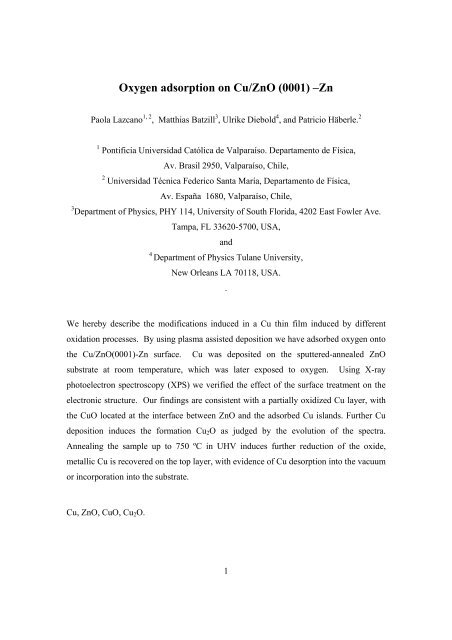
<str<strong>on</strong>g>Oxygen</str<strong>on</strong>g> <str<strong>on</strong>g>adsorpti<strong>on</strong></str<strong>on</strong>g> <strong>on</strong> <strong>Cu</strong>/<strong>ZnO</strong> (<strong>0001</strong>) <strong>–Zn</strong><br />
Paola Lazcano 1, 2 , Matthias Batzill 3 , Ulrike <strong>Diebold</strong> 4 , and Patricio Häberle. 2<br />
1 P<strong>on</strong>tificia Universidad Católica de Valparaíso. Departamento de Física,<br />
Av. Brasil 2950, Valparaíso, Chile,<br />
2 Universidad Técnica Federico Santa María, Departamento de Física,<br />
Av. España 1680, Valparaíso, Chile,<br />
3 Department of Physics, PHY 114, University of South Florida, 4202 East Fowler Ave.<br />
Tampa, FL 33620-5700, USA,<br />
and<br />
4<br />
Department of Physics <strong>Tulane</strong> University,<br />
New Orleans LA 70118, USA.<br />
.<br />
We hereby describe the modificati<strong>on</strong>s induced in a <strong>Cu</strong> thin film induced by different<br />
oxidati<strong>on</strong> processes. By using plasma assisted depositi<strong>on</strong> we have adsorbed oxygen <strong>on</strong>to<br />
the <strong>Cu</strong>/<strong>ZnO</strong>(<strong>0001</strong>)-Zn surface. <strong>Cu</strong> was deposited <strong>on</strong> the sputtered-annealed <strong>ZnO</strong><br />
substrate at room temperature, which was later exposed to oxygen. Using X-ray<br />
photoelectr<strong>on</strong> spectroscopy (XPS) we verified the effect of the surface treatment <strong>on</strong> the<br />
electr<strong>on</strong>ic structure. Our findings are c<strong>on</strong>sistent with a partially oxidized <strong>Cu</strong> layer, with<br />
the <strong>Cu</strong>O located at the interface between <strong>ZnO</strong> and the adsorbed <strong>Cu</strong> islands. Further <strong>Cu</strong><br />
depositi<strong>on</strong> induces the formati<strong>on</strong> <strong>Cu</strong> 2 O as judged by the evoluti<strong>on</strong> of the spectra.<br />
Annealing the sample up to 750 ºC in UHV induces further reducti<strong>on</strong> of the oxide,<br />
metallic <strong>Cu</strong> is recovered <strong>on</strong> the top layer, with evidence of <strong>Cu</strong> desorpti<strong>on</strong> into the vacuum<br />
or incorporati<strong>on</strong> into the substrate.<br />
<strong>Cu</strong>, <strong>ZnO</strong>, <strong>Cu</strong>O, <strong>Cu</strong> 2 O.<br />
1
Introducti<strong>on</strong><br />
The <strong>Cu</strong>/<strong>ZnO</strong> system has been extensively studied primarily because of its importance as a<br />
heterogeneous catalyst for methanol synthesis 1, 2, 3, 4, 5, 6, 7, 8 and the reverse water gas shift<br />
reacti<strong>on</strong> 6, 7, 8, 9 for hydrogen producti<strong>on</strong>. Furthermore, solid state gas sensors utilize the<br />
10, 11, 12<br />
<strong>Cu</strong>O/<strong>ZnO</strong> interface as the active element in humidity sensors and<br />
2<br />
also in the<br />
detecti<strong>on</strong> of harmful gases 12, 13, 14, 15 . In these sensors the fact that copper oxide is a p-<br />
type material, while <strong>ZnO</strong> is mostly an n-type <strong>on</strong>e, is important for their functi<strong>on</strong>ality.<br />
Band bending induced at the p-n juncti<strong>on</strong> reduces the c<strong>on</strong>ductivity of <strong>ZnO</strong>. Up<strong>on</strong> the<br />
reacti<strong>on</strong> of <strong>Cu</strong>O with a test gas, the magnitude of these band bendings are modified <strong>on</strong><br />
the <strong>ZnO</strong> substrate. This effect causes a modificati<strong>on</strong> of the c<strong>on</strong>ductivity, which can then<br />
be easily m<strong>on</strong>itored and used as the “gas resp<strong>on</strong>se” signal.<br />
For an improved understanding of the functi<strong>on</strong>ality of the <strong>Cu</strong>/<strong>ZnO</strong> system in device<br />
applicati<strong>on</strong>s as the <strong>on</strong>e described above, a better characterizati<strong>on</strong> of the <strong>Cu</strong>/<strong>ZnO</strong> interface<br />
is certainly desirable. In particular the dependence of the interface electr<strong>on</strong>ic structure <strong>on</strong><br />
the oxidati<strong>on</strong> state of copper is a relevant piece of informati<strong>on</strong>. To this end we performed<br />
X-ray photoelectr<strong>on</strong> spectroscopy (XPS) studies of <strong>Cu</strong>-deposits <strong>on</strong> a single crystal<br />
<strong>ZnO</strong>(<strong>0001</strong>) substrate under ultra high vacuum c<strong>on</strong>diti<strong>on</strong>s. Different oxidati<strong>on</strong> states of<br />
copper were prepared by using an oxygen- plasma source. The in-situ sample preparati<strong>on</strong><br />
and characterizati<strong>on</strong> procedure enables a detailed look <strong>on</strong> how the interface properties are<br />
altered by the interacti<strong>on</strong> with oxygen.<br />
Previous surface science studies of <strong>Cu</strong> depositi<strong>on</strong> <strong>on</strong> single crystal <strong>ZnO</strong> substrates are<br />
slightly c<strong>on</strong>troversial. One study showed the growth of <strong>Cu</strong> proceeds via a 2D mechanism<br />
up to a critical coverage of 33% of a m<strong>on</strong>olayer and then it c<strong>on</strong>verts into 3D cluster<br />
16, 17<br />
growth. Scanning<br />
tunneling microscopy (STM) studies, performed by members of<br />
our group, dem<strong>on</strong>strated instead the growth proceeds by the formati<strong>on</strong> 3D structures,<br />
already at very low <strong>Cu</strong>-coverage. 18<br />
It has been suggested, this discrepancy arises from<br />
the <strong>Cu</strong>-growth being very sensitive to the substrate preparati<strong>on</strong> and traces of<br />
c<strong>on</strong>taminants at the surface. These differences already hint that small amounts of<br />
adsorbed oxygen may play a role in the morphology of <strong>Cu</strong> clusters. Raim<strong>on</strong>di et al. have<br />
investigated the morphological changes induced by different (oxidizing) reacti<strong>on</strong><br />
c<strong>on</strong>diti<strong>on</strong>s <strong>on</strong> a model catalyst c<strong>on</strong>sisting of <strong>Cu</strong>-particles supported <strong>on</strong> a polycrystalline
<strong>ZnO</strong> film, grown <strong>on</strong> a Si substrate. 21<br />
XPS and AFM studies dem<strong>on</strong>strated that, in the<br />
absence of water, the formati<strong>on</strong> of <strong>Cu</strong> + (<strong>Cu</strong> 2 O) proceeds through the formati<strong>on</strong> of islands<br />
with a large lateral extent, covering most of the <strong>ZnO</strong> substrate. On the other hand, in the<br />
presence of water, the formati<strong>on</strong> of <strong>Cu</strong> 2+ caused an even larger dispersi<strong>on</strong> of the <strong>Cu</strong>islands<br />
size, which resulted in a higher fracti<strong>on</strong> of the <strong>ZnO</strong> surface being covered by <strong>Cu</strong>.<br />
Recently, experimental efforts for understanding the <strong>Cu</strong>/<strong>ZnO</strong> interface have found<br />
support from density functi<strong>on</strong>al theory calculati<strong>on</strong>s.<br />
Meyer and Marx studied <strong>Cu</strong><str<strong>on</strong>g>adsorpti<strong>on</strong></str<strong>on</strong>g><br />
<strong>on</strong> the two polar surfaces. 22<br />
They found that <strong>Cu</strong> atoms adsorb <strong>on</strong> top sites<br />
over oxygen <strong>on</strong> the O-terminated surface, while they preferentially occupy hollow sites<br />
for the Zn-terminated surface. This last result can be rati<strong>on</strong>alized by <strong>Cu</strong> adopting the role<br />
of the next Zn-layer <strong>on</strong> the O-terminated face, while <strong>on</strong> the Zn-terminated surface, the Zn<br />
atoms can be viewed as the first layer of a metal film and <strong>Cu</strong> acts as the c<strong>on</strong>tinuati<strong>on</strong> of<br />
the film, forming <strong>Cu</strong>-Zn b<strong>on</strong>ds. Bromley et al. have also studied <strong>Cu</strong> <strong>on</strong> the <strong>ZnO</strong>(<strong>0001</strong>)-<br />
Zn surface, c<strong>on</strong>centrating primarily <strong>on</strong> the effect of the different oxidati<strong>on</strong> states of<br />
adsorbed <strong>Cu</strong>. 23<br />
Their results show that <strong>Cu</strong> is str<strong>on</strong>gly bound to the surface at Zn-vacancy<br />
sites, forming anchors for subsequent <strong>Cu</strong>-cluster growth. A high abundance of Znvacancies<br />
is expected for the Zn-terminated surface as part of the stabilizati<strong>on</strong> mechanism<br />
24, 25<br />
of the polar surface. For<br />
and, <strong>Cu</strong> 2+<br />
higher oxidati<strong>on</strong> states the binding energy (BE) increases<br />
is the species more str<strong>on</strong>gly bound to the Zn-vacancy sites. The lowest<br />
unoccupied orbital (LUMO) of these <strong>Cu</strong> 2+ i<strong>on</strong>s is, however, below the valence band<br />
maximum of <strong>ZnO</strong> suggesting an electr<strong>on</strong>ic transfer to the <strong>Cu</strong> site, possibly reducing it to<br />
<strong>Cu</strong> + .<br />
3
Experiment<br />
The experiments were carried out under ultrahigh vacuum (UHV) c<strong>on</strong>diti<strong>on</strong>s, in a system<br />
c<strong>on</strong>sisting of i) a preparati<strong>on</strong> chamber, with a base pressure of 6 x 10 -10 mbar, where the<br />
samples were cleaned, metallized and subsequently oxidized, and ii) an analysis chamber<br />
c<strong>on</strong>taining the X-ray photoelectr<strong>on</strong> spectrometer (XPS), operated at a base pressure of<br />
4 x 10 -10 mbar.<br />
The <strong>ZnO</strong> single crystals, mechanically polished and oriented to within 0.5º of the c-axis<br />
of <strong>ZnO</strong>, were obtained from MTI Corporati<strong>on</strong>. The sample’s polarity was c<strong>on</strong>firmed exsitu<br />
by their chemical etching behavior (the O-terminated face of <strong>ZnO</strong> etches more<br />
rapidly in an HCl soluti<strong>on</strong> than the Zn face). They were cleaned, in situ, by cycles of Ar +<br />
i<strong>on</strong> bombardment (1 keV) at room temperature (RT) and annealing up to 700ºC, until no<br />
c<strong>on</strong>taminati<strong>on</strong> could be detected by XPS.<br />
<strong>Cu</strong> (99.999 purity) was vapor-deposited <strong>on</strong>to the clean sample at room temperature using<br />
a Knudsen effusi<strong>on</strong> source (K – cell). The substrates were placed approximately 10 cm<br />
away from the source during <strong>Cu</strong> evaporati<strong>on</strong>. The temperature of the cell was held<br />
c<strong>on</strong>stant at 1100 ºC during <strong>Cu</strong> depositi<strong>on</strong>.<br />
The depositi<strong>on</strong> rate was calibrated by a quartz crystal microbalance before growth.<br />
Depositi<strong>on</strong> rates of 0.008 ! × sec -1 , 0.043 ! × sec -1 and 0.016 ! × sec -1 , were used in all<br />
the experiments for <strong>Cu</strong> films 0.3 ML, 1 ML and 5 ML thick respectively. Sample<br />
oxidati<strong>on</strong> was carried out at room temperature using an oxygen mini plasma source<br />
(OSMiPlas Oxford Scientific), which was operated with an oxygen pressure of 5.5 x 10 -6<br />
mbar and a current of 12 mA. The exposure time was 15 minutes.<br />
The coverage of the deposited metal is given in m<strong>on</strong>olayer (ML) equivalent. 1 ML= 1.7<br />
x 10 15 atoms, corresp<strong>on</strong>ds to the number of <strong>Cu</strong> atoms per unit area in a <strong>Cu</strong> (111) plane.<br />
The typical depositi<strong>on</strong> rate used in this work was 0.42 ML/min. The pressure during <strong>Cu</strong><br />
depositi<strong>on</strong> remained below 6 x 10 -9 mbar, which is <strong>on</strong>ly slightly higher than the normal<br />
operating pressure.<br />
All the XPS spectra reported here were obtained using a X-ray source (VG XR3E2) with<br />
n<strong>on</strong>-chromatized Al K" X-ray radiati<strong>on</strong> (1486.6 eV). A Specs PHOIBOS 100<br />
hemispherical electrostatic energy analyzer with a channeltr<strong>on</strong> energy detector was used,<br />
4
in the medium magnificati<strong>on</strong> mode, which gives an acceptance angle of up to ±7º for a 2<br />
mm spot size. This analyzer has a typical energy resoluti<strong>on</strong> of 15 meV. The energy scale<br />
was calibrated using the 3d 5/2 photoemissi<strong>on</strong> peak from an Ag foil. The detecti<strong>on</strong> was<br />
performed al<strong>on</strong>g the surface normal, except when a different angle is explicitly specified.<br />
The angle between the X-ray source and the analyzer is fixed at 55° for all measurements.<br />
5
Results and Discussi<strong>on</strong><br />
Previous studies by STM of <strong>Cu</strong> growth <strong>on</strong> <strong>ZnO</strong> 17, 18, 19, 20 showed the formati<strong>on</strong> of well<br />
defined clusters. For moderately high <strong>Cu</strong> coverages, <strong>Cu</strong>(111) LEED spots were observed,<br />
indicating a strict crystallographic relati<strong>on</strong>ship of the <strong>Cu</strong> clusters with respect to the <strong>ZnO</strong><br />
substrate. In order to probe the crystalline nature of the <strong>Cu</strong> deposits <strong>on</strong> our sample, we<br />
performed X-ray photoelectr<strong>on</strong> diffracti<strong>on</strong> studies (XPD) al<strong>on</strong>g the 1 100 azimuth of<br />
the <strong>ZnO</strong> substrate. The <strong>Cu</strong> intensity variati<strong>on</strong>s (2 p 3/2 ), versus the polar angle were<br />
m<strong>on</strong>itored after depositing 0.3 ML, 1 ML and 5 ML at room temperature <strong>on</strong> the clean<br />
<strong>ZnO</strong> (<strong>0001</strong>)-Zn terminated surface. The results of these measurements are shown in<br />
Figure 1. For 0.3 ML (trace (i)) the deposited film shows no special features as a<br />
functi<strong>on</strong> angle, bey<strong>on</strong>d the instrumental resoluti<strong>on</strong>. At 1ML (trace (ii)) two weak peaks<br />
appear for 18º and 36º, respectively. Both peaks show a higher intensity for 5ML (trace<br />
(iii)). No variati<strong>on</strong>s in the intensity as a functi<strong>on</strong> of angle are expected for emissi<strong>on</strong> from<br />
an atomically flat surface below 1 ML coverage, if the <strong>Cu</strong> atoms are distributed in a<br />
uniform film or if they form a locally disordered thicker <strong>Cu</strong> film. The fact that we see<br />
two weak peaks at 1ML coverages is an indicati<strong>on</strong> of cluster growth. If the clusters are<br />
formed by <strong>Cu</strong>(111) nanoscale crystals, with the 111 directi<strong>on</strong> al<strong>on</strong>g the surface normal,<br />
and azimuthally oriented in the plane, with <strong>on</strong>e of the high symmetry directi<strong>on</strong>s al<strong>on</strong>g<br />
the 1 100 substrate directi<strong>on</strong>s, the str<strong>on</strong>gest forward focusing peaks are expected at 19º<br />
and -35º (insert in Figure 1). We see both peaks for positive values of the detecti<strong>on</strong><br />
angle; indicating that both possible orientati<strong>on</strong>s of the copper clusters are formed during<br />
growth. For both of these clusters orientati<strong>on</strong>s, the 111 directi<strong>on</strong> is parallel to the<br />
substrate normal, but the ! 112" azimuth is either parallel or antiparallel to the <strong>ZnO</strong><br />
1100 directi<strong>on</strong>.<br />
These results are c<strong>on</strong>sistent with previous measurements for thicker layers (>5 ML), in<br />
which <strong>Cu</strong>(111) LEED spots have been observed in additi<strong>on</strong> to the <strong>ZnO</strong> substrate<br />
maxima. 20<br />
Yoshihara et al. 16 have studied the variati<strong>on</strong> of the <strong>Cu</strong> 2p 3/2 peak intensity as a<br />
functi<strong>on</strong> of polar angle al<strong>on</strong>g the same azimuth at low coverages (0.41 and 0.98 ML).<br />
6
Their results indicate no special angular variati<strong>on</strong> for both coverages. Only after a slight<br />
annealing of the higher coverage film, to 370K, a weak diffracti<strong>on</strong> feature is detected at<br />
about 35º, c<strong>on</strong>sistent with (111) growth.<br />
Figure 2 shows the XPS spectra of Zn 2p (Fig. 2a), <strong>Cu</strong> 2p (Fig. 2b) and O 1s (Fig. 2c) for<br />
different preparati<strong>on</strong> c<strong>on</strong>diti<strong>on</strong>s; traces labeled by (i) in all graphs corresp<strong>on</strong>d to:<br />
<strong>ZnO</strong>(<strong>0001</strong>)-Zn clean surface, (ii): the surface after depositing , at room temperature, 3ML<br />
of <strong>Cu</strong>, (iii): the same sample after oxidati<strong>on</strong> in an oxygen plasma at 10 -6 mbar, (iv): after<br />
an additi<strong>on</strong>al depositi<strong>on</strong> of 3ML of <strong>Cu</strong> <strong>on</strong> the previously-oxidized sample and (v): the<br />
sample annealed to 750°C.<br />
The Zn 2p 1/2 and 2p 3/2 features from the clean <strong>ZnO</strong>(<strong>0001</strong>)-Zn surface (trace (i), Fig. 2a),<br />
obtained after several cleaning cycles, are the two narrow peaks with BEs of 1044.8 eV<br />
and 1021.8 eV and full width at half maximum (FWHM) of 1.88 eV and 1.93 eV,<br />
respectively. From Fig. 2c, trace (i) we can obtain similar informati<strong>on</strong> from the O1s<br />
peak. The corresp<strong>on</strong>ding BE is 530.6 eV and the FWHM is 1.67 eV.<br />
These Zn 2p peaks, with very narrow and symmetrical shapes, show an attenuati<strong>on</strong> of<br />
their intensities after every treatment, induced both by the <strong>Cu</strong> overlayer and the oxygen<br />
exposure (traces (ii), (iii) and (iv)). The peaks are also shifted to lower BEs, for the<br />
treatments in (ii), (iii) and (iv), with respect to their clean surface values (see Table I).<br />
After annealing the oxidized sample to 750ºC, the intensities of the peaks, as well as the<br />
BE and FWHM, are similar to those in (ii) i.e. after the depositi<strong>on</strong> of 3 ML of <strong>Cu</strong>.<br />
After depositing 3ML <strong>Cu</strong>, at room temperature, the trace labeled as (ii), in the Figure 2b,<br />
displays two narrow peaks with FWHM of 1.6 eV and 1.9 eV respectively, and BEs of<br />
932.4 eV and 952.2 eV. These two transiti<strong>on</strong>s are the characteristic 2p 1/2 and 2p 3/2 states<br />
of metallic <strong>Cu</strong>. 26 The substrate-related O 1s peak (Fig. 2c, trace (ii)), is fairly narrow<br />
(1.63 eV) and symmetrical. It also shifts with respect to the oxygen peak in the clean<br />
<strong>ZnO</strong> surface, to a lower BE (530.2 eV). The fact that this system has two comp<strong>on</strong>ents<br />
(<strong>ZnO</strong> & <strong>Cu</strong>), and both the O and Zn features shift their energies by the same amount (-0.4<br />
eV) up<strong>on</strong> <strong>Cu</strong> depositi<strong>on</strong>, without changing significantly the peak shapes, points towards a<br />
band bending mechanism instead of a chemical shift as the reas<strong>on</strong> for these BE<br />
modificati<strong>on</strong>s.<br />
C<strong>on</strong>sistent with our results, Joshirara el at. 16 reported a reducti<strong>on</strong> in the binding energy of<br />
the Zn 2p 3/2 of 0.05 eV for 0.3 ML <strong>Cu</strong> and 0.25 eV for a coverage of 1ML. For the same<br />
7
coverages, Didziulis el at. 36 reported an upward band bending of 0.15 eV and 0.3 eV,<br />
respectively. They do not show results for higher coverages. In Figure 3 we display the<br />
energy shifts in the binding energy due to <strong>Cu</strong> depositi<strong>on</strong> for coverages as large as 10 ML.<br />
The depositi<strong>on</strong> of <strong>Cu</strong> <strong>on</strong> <strong>ZnO</strong>(<strong>0001</strong>) produces <strong>on</strong>ly an upwards band bending. The fact<br />
that the bands need to be bent upwards to align the vacuum levels, agrees with the fact<br />
that the work functi<strong>on</strong> of <strong>ZnO</strong> is smaller than that of <strong>Cu</strong>. From various references, the<br />
different values reported for the work functi<strong>on</strong> are: 3.5 -3.9 eV, 16 3.9 eV 36 for<br />
<strong>ZnO</strong>(<strong>0001</strong>); 4.6-4.9 eV for <strong>Cu</strong>, 36 in agreement with the sign and magnitude we can infer<br />
from our own data for the <strong>Cu</strong> /<strong>ZnO</strong> system.<br />
In the following paragraphs we examine the modificati<strong>on</strong>s of the electr<strong>on</strong>ic structure of<br />
the <strong>Cu</strong> clusters induced by the different sample preparati<strong>on</strong> procedures, as shown by the<br />
spectra in Figure 2b. The effect of oxygen exposure (trace (iii)) induces both a shift and a<br />
broadening, when compared to the previous spectrum (trace (ii)). The <strong>Cu</strong> 2p peaks<br />
present a visible broadening <strong>on</strong> the higher BE side. Since the core-level BE of an atom is<br />
closely related to its chemical envir<strong>on</strong>ment, this additi<strong>on</strong>al intensity could then be<br />
interpreted as a chemical shift associated to some the <strong>Cu</strong> atoms in the overlayer being in a<br />
different chemical state. It is also fair to point out that inhomogeneous surface charging<br />
in the thin layer could also induce a similar broadening, but we are discarding this<br />
possibility since no evidence to this effect have been found of such behavior in previous<br />
studies.<br />
At this point we have assumed this peak (<strong>Cu</strong> 2p 3/2 ) to be formed by two comp<strong>on</strong>ents<br />
which we have fitted to the raw data. One of them at 932.4 eV, corresp<strong>on</strong>ds to metallic<br />
<strong>Cu</strong> and the sec<strong>on</strong>d <strong>on</strong>e at 933.7 eV to <strong>Cu</strong>O 26, 30, 31, 33, 34 , as shown by previous<br />
measurements. Table II shows a summary of different parameters obtained for the <strong>Cu</strong><br />
2p 3/2 features obtained from the traces in Figure 2b. To help in the identificati<strong>on</strong> of the<br />
different spectral features we have also presented in Table III a compilati<strong>on</strong>, from<br />
different sources in the literature, of the BE of copper and oxygen features as measured<br />
by XPS.<br />
In additi<strong>on</strong> to the broadening, a complex satellite peak structure develops a few electr<strong>on</strong><br />
volts above the corresp<strong>on</strong>ding core level positi<strong>on</strong>s. The 942 eV satellite feature is linked<br />
to the 2p 3/2 transiti<strong>on</strong> and has a FWHM of 3 eV (2p 1/2 : 961 eV, FWHM 2.5 eV). These<br />
additi<strong>on</strong>al intensities can be described as shake-ups, 26, 27, 28 as they occur when the<br />
8
outgoing photoelectr<strong>on</strong> simultaneously interacts with a valence electr<strong>on</strong> and excites it<br />
(“shakes it up”) to a higher-energy level. The kinetic energy of the core-derived electr<strong>on</strong><br />
is then reduced slightly, giving rise to a satellite structure a few electr<strong>on</strong> volts above the<br />
main peak in a BE scale. In this system, these str<strong>on</strong>g shake-up satellites peaks are<br />
c<strong>on</strong>sidered an unequivocal signature for the presence of <strong>Cu</strong>O 26, 27, 28, 29, 30, 31, 33, 34 in the<br />
sample.<br />
After a new depositi<strong>on</strong> of 3ML <strong>Cu</strong> (trace (iv)) <strong>on</strong> the previously oxidized sample, we<br />
detect additi<strong>on</strong>al changes in the <strong>Cu</strong> 2p spectra. Both satellite peaks related to <strong>Cu</strong>O (942<br />
eV & 961 eV) disappear, the 2p transiti<strong>on</strong>s become narrower and symmetrical again,<br />
namely the high energy shoulder is str<strong>on</strong>gly attenuated. The new <strong>Cu</strong> 2p 3/2 and 2p 1/2 BEs<br />
are 932.2 eV and 952.0 eV respectively. Additi<strong>on</strong>ally, two small features appear at about<br />
946.3 eV and 964.5 eV, BE. They have been identified as characteristic satellites of <strong>Cu</strong><br />
28, 29, 30, 32, 33, 34<br />
2p in <strong>Cu</strong> 2 O.<br />
Annealing the oxidized sample to different temperatures between 400ºC and 750ºC<br />
reduce gradually the intensity of the satellite peaks associated with <strong>Cu</strong> 2 O. Finally up<strong>on</strong><br />
annealing to 750ºC, this feature is almost imperceptible (trace (v), Fig. 2b) in the data.<br />
The shape of the <strong>Cu</strong> 2p 3/2 feature at 932.2 eV (trace (iv)) remains almost unaltered after<br />
annealing, but the whole peak gets shifted to a higher BE:<br />
temperature goes as high as 750ºC.<br />
932.6 eV, when the<br />
It is tempting to c<strong>on</strong>clude then that we have recovered metallic <strong>Cu</strong> <strong>on</strong> top of the <strong>ZnO</strong><br />
substrate after the reducti<strong>on</strong> process is completed. It is also evident that <strong>Cu</strong> desorbs into<br />
the vacuum, as reflected by the reducti<strong>on</strong> of the corresp<strong>on</strong>ding <strong>Cu</strong> 2p peak area in trace<br />
(v) (Fig. 2b).<br />
Nevertheless, additi<strong>on</strong>al insight is provided by the <strong>Cu</strong> LMM Auger<br />
transiti<strong>on</strong>s as displayed in Figure 4.<br />
The trace labels in Figure 4 are in a <strong>on</strong>e to <strong>on</strong>e<br />
corresp<strong>on</strong>dence with those shown in Figure 2. The salient feature in the spectra after<br />
depositing <strong>Cu</strong> <strong>on</strong> the clean <strong>ZnO</strong> substrate, is the appearance of a peak at a kinetic energy<br />
of 918.5 eV (trace (ii)). <str<strong>on</strong>g>Oxygen</str<strong>on</strong>g> exposure does not change the BE of this feature (trace<br />
(iii)) even though it changes its width. A more detailed analysis show this peak has two<br />
comp<strong>on</strong>ents, <strong>on</strong>e most likely related to <strong>Cu</strong> (918.6 eV KE) and a sec<strong>on</strong>d <strong>on</strong>e derived from<br />
<strong>Cu</strong>O (917.8 KE). Further <strong>Cu</strong> depositi<strong>on</strong> induces additi<strong>on</strong>al changes in the spectrum<br />
(trace (iv)). The LMM transiti<strong>on</strong> becomes now str<strong>on</strong>ger and is also shifted to 916.6 eV<br />
9
KE. As a summary in Table III we display, from different sources in the literature, the<br />
KE of <strong>Cu</strong> LMM as measured by XPS.<br />
Since the 2p-BE of the oxidized <strong>Cu</strong> + and reduced <strong>Cu</strong> have very similar values, it is hard<br />
to distinguish <strong>on</strong>e from the other, just by looking at this res<strong>on</strong>ance. But, the substantial<br />
difference between traces (ii) and (iv) in Figure 4, together with the satellite structure in<br />
Figure 2, seems to c<strong>on</strong>firm that <strong>Cu</strong> depositi<strong>on</strong>, <strong>on</strong> a partially oxidized <strong>Cu</strong>O layer, induces<br />
the formati<strong>on</strong> of <strong>Cu</strong> 2 O. The Auger LMM lines 33, 34 , which show shifts under the<br />
different treatments, allow an easier differentiati<strong>on</strong> between <strong>Cu</strong> and <strong>Cu</strong> 2 O, which would<br />
not be evident just from detecting the changes in the <strong>Cu</strong>-2p core level spectra.<br />
In the Auger spectrum of the “reduced” film (Fig. 4, trace (v)) the clear and sharp <strong>Cu</strong><br />
Auger feature, present in the as-deposited sample, is replaced, in the annealed sample,<br />
(<strong>ZnO</strong>(<strong>0001</strong>)/<strong>Cu</strong>/O/<strong>Cu</strong>-annealed; Fig. 4; trace (v)) by a fairly broad res<strong>on</strong>ance barely<br />
distinguishable from the <strong>ZnO</strong> background. These Auger features, together with the<br />
shape; BE and width of the <strong>Cu</strong>-2p res<strong>on</strong>ance (Fig. 2b), show that the annealing procedure<br />
reduces the <strong>Cu</strong> 2 O <strong>on</strong> the surface to metallic <strong>Cu</strong> with a c<strong>on</strong>siderable desorpti<strong>on</strong> into the<br />
vacuum or possibly cluster coalescence, c<strong>on</strong>sistent with an increased c<strong>on</strong>tributi<strong>on</strong><br />
of <strong>ZnO</strong> in the corresp<strong>on</strong>ding Auger spectrum. From the data available, is not possible to<br />
differentiate between these alternatives.<br />
The changes in the <strong>Cu</strong> peaks are also accompanied by modificati<strong>on</strong>s in the oxygen<br />
spectra, as shown by the corresp<strong>on</strong>ding traces in Figure 2c. After oxygen exposure,<br />
the O 1s peak (trace (iii)) becomes broader and asymmetrical, with a shoulder <strong>on</strong> the high<br />
BE side. This broadening is c<strong>on</strong>sistent with the presence of more than <strong>on</strong>e type of<br />
oxygen in the interfacial regi<strong>on</strong> (<strong>ZnO</strong> and <strong>Cu</strong>O). As a first approximati<strong>on</strong>, we fitted two<br />
comp<strong>on</strong>ents to this peak. One related to the substrate and the other <strong>on</strong>e to <strong>Cu</strong>O.<br />
As a reference, to identify the band bending associated to changes in the surface carrier<br />
c<strong>on</strong>centrati<strong>on</strong>s 37 , we used the Zn-2p BE shift and assume the same value for the O-1s<br />
feature, as expected for photoelectr<strong>on</strong>s emerging from a comm<strong>on</strong> surface. As seen in the<br />
data summarized in Table I, the Zn 2p 3/2 BE decreases by 1.6 eV after O 2 exposure.<br />
C<strong>on</strong>sistent with this shift the <strong>ZnO</strong> comp<strong>on</strong>ent of the O1s peak should be at 529.0 eV.<br />
The sec<strong>on</strong>d comp<strong>on</strong>ent, associated to the broadening, is then determined by the best fit to<br />
the data. In this case the BE related to the presence of <strong>Cu</strong>O <strong>on</strong> the surface is 529.3 eV.<br />
The assignment of the characteristic BEs to different oxidati<strong>on</strong>s states of copper is better<br />
10
understood by comparing our data with previous measurements, as those shown in Table<br />
III. These values, together with the changes displayed by the <strong>Cu</strong> 2p peaks, are in<br />
agreement with the formati<strong>on</strong> of <strong>Cu</strong>O in the adsorbed layer.<br />
After an additi<strong>on</strong>al depositi<strong>on</strong> of 3ML <strong>Cu</strong> <strong>on</strong> the previously-oxidized sample (Figure 2 c,<br />
trace (iv)). The O 1s peak shifts to a slightly higher binding energy and becomes<br />
narrower and more symmetrical. To determine the comp<strong>on</strong>ents of this peak we used the<br />
same procedure described in the previous paragraph. From this procedure, the BE of the<br />
substrate related comp<strong>on</strong>ent is at 529.6 eV, and the oxide (<strong>Cu</strong> 2 O) related feature has a BE<br />
of 530.2 eV.<br />
Finally, in trace (v), after annealing the oxidized sample to 750ºC, the O 1s peak looks<br />
narrower and symmetrical again. The intensity, as well as BE (530.2 eV) and FWHM,<br />
are similar to those in (ii) i.e. after the depositi<strong>on</strong> of a fresh layer of <strong>Cu</strong>. This agreement<br />
with the previously observed Zn and <strong>Cu</strong> features c<strong>on</strong>firms the reducti<strong>on</strong> of <strong>Cu</strong> 2 O to<br />
metallic copper through this annealing procedure.<br />
In Figure 5 we show a series of spectra, with different emissi<strong>on</strong> angles, collected with the<br />
purpose of determining the oxide distributi<strong>on</strong> within the thin layer. The spectra c<strong>on</strong>sider<br />
the <strong>Cu</strong> 2p states, after <strong>Cu</strong> depositi<strong>on</strong> and oxidati<strong>on</strong> (3ML <strong>Cu</strong> + Plasma oxidati<strong>on</strong>), as a<br />
functi<strong>on</strong> of the angle between the analyzer and the surface normal. The intensity<br />
variati<strong>on</strong>s in this type of measurements could be used to determine the compositi<strong>on</strong><br />
distributi<strong>on</strong> in a surface layer and has been used in the past for describing thin passive<br />
films <strong>on</strong> metals and for the detecti<strong>on</strong> of surface segregati<strong>on</strong> in polymers. 27 In our data,<br />
the angular dependence of the XPS spectra manifests itself as a reducti<strong>on</strong> of the relative<br />
intensity of the 2p 3/2 satellite peak, together with a c<strong>on</strong>tinuous narrowing of the 2p <strong>Cu</strong><br />
peaks as the emissi<strong>on</strong> angle is increased. For larger emissi<strong>on</strong> angles the resulting spectral<br />
data is more sensitive to the top layer compositi<strong>on</strong>.<br />
The evident reducti<strong>on</strong> of the relative intensity of the 2p 3/2 satellite peak, together with a<br />
c<strong>on</strong>tinuous narrowing of the <strong>Cu</strong> peaks as the emissi<strong>on</strong> angle is increased, indicates that<br />
the 2p transiti<strong>on</strong>s in the oxidized <strong>Cu</strong> clusters are composed of two distinguishable<br />
comp<strong>on</strong>ents. A high BE shoulder, related to <strong>Cu</strong>O and the main peak corresp<strong>on</strong>ding<br />
mostly to metallic <strong>Cu</strong>. The corresp<strong>on</strong>ding binding energies of the 2p 3/2 comp<strong>on</strong>ents are<br />
932.4 eV and 933.7 eV, respectively. The first of these comp<strong>on</strong>ents is related to metallic<br />
<strong>Cu</strong> and the sec<strong>on</strong>d <strong>on</strong>e to <strong>Cu</strong>O. From this analysis, it is then possible to c<strong>on</strong>clude that<br />
11
<strong>on</strong>ly a fracti<strong>on</strong> of the copper in the clusters is found in a <strong>Cu</strong> 2+ oxidati<strong>on</strong> state and that part<br />
of it retains its metallic character.<br />
The reducti<strong>on</strong> of the intensity of the satellite peak for larger angles is then c<strong>on</strong>sistent with<br />
a gradient in the <strong>Cu</strong>O c<strong>on</strong>centrati<strong>on</strong>, with the higher oxidati<strong>on</strong> states being closer to the<br />
<strong>ZnO</strong> interface. At the same time, the top parts of the clusters are more metallic, since the<br />
spectra for large angles are more c<strong>on</strong>sistent with metallic <strong>Cu</strong>. From this result is then<br />
clear that the stabilized <strong>ZnO</strong> surface does play an active role in the formati<strong>on</strong> of the <strong>Cu</strong><br />
oxide layer at this interface. These partially oxidized clusters show explicit evidence of a<br />
spatial segregati<strong>on</strong> of these two comp<strong>on</strong>ents in the directi<strong>on</strong> of the substrate normal.<br />
Similar gradient distributi<strong>on</strong>s have been reported by Schalow et al. 35 for a Pd/Fe 3 O 4<br />
catalyst. Their results are c<strong>on</strong>sistent with oxygen being reversibly accumulated in a thin<br />
Pd oxide layer, at the metal/oxide interface.<br />
It is not unreas<strong>on</strong>able to think there are traces of <strong>Cu</strong> 2 O in the overlayer after the initial<br />
oxidati<strong>on</strong>, since the free energy is minimized in the <strong>Cu</strong>O-<strong>Cu</strong> reacti<strong>on</strong> which forms <strong>Cu</strong> 2 O.<br />
The high resemblance between the spectra of the clean 2p res<strong>on</strong>ances of <strong>Cu</strong> and <strong>Cu</strong> 2 O<br />
makes it very hard to discard this fact based solely in this informati<strong>on</strong>. Nevertheless the<br />
main difference between them (<strong>Cu</strong> 2 O and <strong>Cu</strong>) is seen in the LMM Auger spectra, as<br />
shown in Figure 4. These spectra (traces ii and iii) are c<strong>on</strong>sistent with the top layer of the<br />
clusters being mostly metallic <strong>Cu</strong>.<br />
The formati<strong>on</strong> of <strong>Cu</strong> 2 O is a temperature dependent process. All our experiments were<br />
carried out with the samples at room temperature, however the different procedures used<br />
to form the oxides, plasma oxidati<strong>on</strong> and <strong>Cu</strong> re-depositi<strong>on</strong>, are not equivalent. An<br />
<str<strong>on</strong>g>Oxygen</str<strong>on</strong>g> plasma source induces a milder temperature effect at the interface when<br />
compared with <strong>Cu</strong> evaporati<strong>on</strong> from a crucible at 1100ºC. Our data indicates this<br />
temperature difference at the interface could be enough to promote <strong>Cu</strong> 2 O formati<strong>on</strong> 28 .<br />
Similar angular dependence measurements were also carried out for the case of <strong>Cu</strong> redepositi<strong>on</strong><br />
<strong>on</strong> the previously oxidized clusters (trace (iv) in Fig. 2). The absence of an<br />
angular dependence of the 2p peaks is then c<strong>on</strong>sistent with a uniform distributi<strong>on</strong> of<br />
<strong>Cu</strong> 2 O throughout the film. C<strong>on</strong>sistent with this measurement, there is almost no variati<strong>on</strong><br />
of the relative intensity of the corresp<strong>on</strong>ding satellite peaks. Thus c<strong>on</strong>firming <strong>Cu</strong> 2 O is<br />
uniformly distributed in the adsorbed film.<br />
12
Summary<br />
We have studied the <strong>Cu</strong>/<strong>ZnO</strong> interface, crystalline structure, its oxidati<strong>on</strong> and the effects<br />
of annealing. The room temperature depositi<strong>on</strong> of <strong>Cu</strong> <strong>on</strong> <strong>ZnO</strong>(<strong>0001</strong>) is known to induce<br />
the formati<strong>on</strong> of islands or cluster of the adsorbed metal. Due to the potential<br />
applicati<strong>on</strong>s of this particular system, it is then reas<strong>on</strong>able to verify the changes induced<br />
in these metallic particles by c<strong>on</strong>trolled temperature treatments under different<br />
atmospheres. We have c<strong>on</strong>centrated our efforts in the determinati<strong>on</strong> of the effects of<br />
oxygen <strong>on</strong> the electr<strong>on</strong>ic structure. The plasma oxidati<strong>on</strong> of the <strong>Cu</strong> clusters, grown at<br />
different coverages, is c<strong>on</strong>sistent with the formati<strong>on</strong> of <strong>Cu</strong>O. Both the angular evoluti<strong>on</strong><br />
of the <strong>Cu</strong>O 2p satellites, together with the changes in shape and energy shifts of the<br />
primary peaks are indicative of the formati<strong>on</strong> of an oxide with a higher c<strong>on</strong>centrati<strong>on</strong> at<br />
the <strong>ZnO</strong>- cluster interface. The rest of the film, closer to the vacuum interface is mostly<br />
metallic <strong>Cu</strong> (Fig. 6(i)). Up<strong>on</strong> further depositi<strong>on</strong> of <strong>Cu</strong> <strong>on</strong>to the oxidized clusters we can<br />
induce the formati<strong>on</strong> of <strong>Cu</strong> 2 O as a dominant comp<strong>on</strong>ent. The <strong>Cu</strong>O features are almost<br />
undetectable after <strong>Cu</strong> re-depositi<strong>on</strong>. The new oxide is better recognized by its Auger<br />
LMM spectra.<br />
By means of the procedure described we can define the initial oxide stoichiometry and<br />
spatial distributi<strong>on</strong> in the adsorbed clusters. Our first treatment (plasma oxidati<strong>on</strong>)<br />
produces an interfacial oxide (<strong>Cu</strong>O) and the sec<strong>on</strong>d <strong>on</strong>e (<strong>Cu</strong>-redepositi<strong>on</strong>) induces the<br />
formati<strong>on</strong> of a uniform oxide in the clusters with <strong>Cu</strong> 2 O stoichiometry (Fig. 6 (ii)).<br />
Metallic Oxides have been shown to have singular catalytic behavior, in particular if the<br />
sizes involved are in the [nm] range, as is the case of a few ML <strong>Cu</strong> in <strong>ZnO</strong>. If we can<br />
modify the catalytic properties by changing the stoichiometry of the oxides, we could<br />
enable new applicati<strong>on</strong>s of these materials. It is also reas<strong>on</strong>able to assume that the actual<br />
chemical behavior of <strong>Cu</strong>/<strong>ZnO</strong> catalysts may be better described under realistic<br />
c<strong>on</strong>diti<strong>on</strong>s, as oxide clusters as the <strong>on</strong>e we have shown to exist rather than pure metal, if<br />
the <strong>Cu</strong> clusters are exposed in the different processes to oxidati<strong>on</strong> c<strong>on</strong>diti<strong>on</strong>s.<br />
13
Acknowledgements<br />
PL thanks the MECESUP program, Chile, for financing her graduate student stipend and<br />
a research visit to <strong>Tulane</strong> University where these experiments were performed. Partial<br />
funding for this research was also provided by PBCT grant ACT027, Chile.<br />
14
References<br />
1. M.S. Spencer, Catal. Lett. 50, 37 (1998).<br />
2. T. Fujitani, J. Nakamura, Appl. Catal. A 191, 111 (2000).<br />
3. K.R. Harikumar and C.N.R. Rao, Appl. Surf. Sci. 125, 245 (1998).<br />
4. G. Meitzner, E. Iglesia, Catal. Today 53, 433 (1999).<br />
5. J. Agrell, M. Bout<strong>on</strong>net, I. Melián-Cabrera, and J.L.G. Fierro, Appl. Catal. A 253,<br />
201 (2003).<br />
6. M.S. Spencer, Top. Catal. 8, 259 (1999).<br />
7. Y. Choi, K. Futagami, T. Fujitani and J. Nakamura, Appl. Catal. A 208, 163<br />
(2001).<br />
8. H. Purnama, T. Ressler, R.E Jentoft, H. Soerijanto, R. Schlögl and R.<br />
Schomäcker, Appl. Catal. A 259, 83 (2004).<br />
9. Shin-Ichiro Fujita, Masahito Usui and Nobutsune Takezawa, Journal of Catalysis<br />
134, 220 (1992).<br />
10. Y. Toyoshima, M. Miyayama, H. Yanagida, and K. Koumoto, Jpn. J. Appl. Phys.<br />
22, 1933 (1983).<br />
11. Ushio Y, Miyayama M, Yanagida H, Sens. Actuators B Chem. B12, 135 (1993).<br />
12. G. Eranna, B.C. Joshi, D. P. Runthala, and R. P. Gupta, Critical Review in Solid<br />
State and Materials Sciences 29, 111 (2004).<br />
13. S–J. Jung and H. Yanagida, Sens. and Actuators B Chem. 37, 55 (1996).<br />
14. Y. Nakamura, H. Zhuang, A. Kishimoto, O. Okada and H. Yanagida, J.<br />
Electrochem. Soc. 145, 632 (1998).<br />
15. Dang Hyok Yo<strong>on</strong>, Ji Haeng Yu and Gye<strong>on</strong>g Man Choi, Sens. And Actuators B<br />
Chem. 46, 15 (1998)<br />
16. J. Yoshihara, J.M. Campbell, and C.T. Campbell, Surf. Sci. 406, 235 (1998).<br />
17. J. Yoshihara, S.C. Parker, C.T. Campbell, Surf. Sci. 439, 153 (1999).<br />
18. L.V. Koplitz, O. Dulub, and U. <strong>Diebold</strong>, J. Phys. Chem. B 107, 10583 (2003).<br />
19. Martin Kroll, Ulrich Köhler, Surf. Sci. 601, 2182 (2007).<br />
20. O. Dulub, Matthias Batzill, and U. <strong>Diebold</strong>, Topics in Catalysis 36, 65 (2005).<br />
21. F. Raim<strong>on</strong>di, J. Wambach, and A. Wokaunn, Phys. Chem. Chem. Phys. 5, 4015<br />
(2003).<br />
22. B. Meyer and D. Marx, Phys. Rev. B 69, 235420 (2004).<br />
15
23. S.T. Bromley, S.A. French, A.A. Sokol, C.R.A Catlow, and P. Sherwood, J. Phys.<br />
Chem. B 107, 7045 (2003).<br />
24. O. Dulub, U. <strong>Diebold</strong>, and G. Kresse, Phys. Rev. Lett. 90, 016102 (2003).<br />
25. G. Kresse, O. Dulub, and U. <strong>Diebold</strong>, Phys. Rev. B 68, 245409 (2003).<br />
26. J.F.Moulder, W.F.Stickle, P.E.Sobol, and K.D.Bomben, Handbook of X-ray<br />
Photoelectr<strong>on</strong> Spectroscopy (Perkin-Elmer Corp., Eden Prairie, Minn, 1992).<br />
27. John F. Watts, John Wolstenholme, An Introducti<strong>on</strong> to surface analysis by XPS<br />
and AES (Wiley, West Sussex, England, 2003).<br />
28. S. Y. Lee, N. Mettlach, N. Nguyen, Y.M. Sun, J.M White, Applied Surface<br />
Science 206, 102 (2003).<br />
29. P. D. Kirsch and J.G. Ekerdt, Journal of Applied Physics 90, 4256 (2001).<br />
30. J. Ghijsen, L.H. Tjeng, J. van Elp, H. Eskes, J. Westerink, G.A. Sawatzkyq, M.<br />
T. Czyzyk, Phys. Rev. B 38, 11322 (1998).<br />
31. R. P. Vasquez, Surface Science Spectra 5, 262 (1998).<br />
32. R. P. Vasquez, Surface Science Spectra 5, 257 (1998).<br />
33. S. Poulst<strong>on</strong>, P.M. Parlett, P. St<strong>on</strong>e and M. Bowker, Surface and Interface Analysis<br />
24, 811 (1996).<br />
34. G. Deroubaix and P. Marcus, Surface and Interface Analysis 18, 39 (1992).<br />
35. T. Schalow, M. Laurin, B. Brandt, S. Schauermann, S. Guim<strong>on</strong>d, H. Kuhlenbeck,<br />
D. Starr, S. Shaikhutdinov, J. Libuda and H. Freund, Angew. Chem. Int. Ed. 44, 2<br />
(2005).<br />
36. Stephen V. Didziulis, Kristine D. Butcher, Susan L. Cohen, and Edward I.<br />
Solom<strong>on</strong>, J. Am. Chem. Soc. 111, 7110-7123 (1989).<br />
37. V.E. Henrich and P.A Cox, The Surface Science of Metal Oxides (Cambridge<br />
University Press, New York 1994).<br />
38. K. Oura, V.G Lifshits, A.A Saranin, A.V. Zotov, M. Katayama, Surface Science:<br />
an Introducti<strong>on</strong> (Springer, Berlin, 2003).<br />
16
Figures & Table Capti<strong>on</strong>s<br />
Figure 1: ARXPS of <strong>Cu</strong> 2p 3/2 after depositing (i) 0.3 ML, (ii) 1 ML and, (iii) 5 ML at<br />
room temperature <strong>on</strong> the clean <strong>ZnO</strong> (<strong>0001</strong>)-Zn surface. Starting at 1 ML coverage the<br />
XPS intensity shows diffracti<strong>on</strong> features c<strong>on</strong>sistent with a crystalline ordering of the <strong>Cu</strong><br />
clusters. The inset shows the angles for the str<strong>on</strong>gest forward focusing peaks expected for<br />
a <strong>Cu</strong> (111) crystal which is oriented azimuthally in the plane al<strong>on</strong>g the 1 100 substrate<br />
directi<strong>on</strong>.<br />
Figure 2: XPS spectra of Zn 2p (a), <strong>Cu</strong> 2p (b) and O 1s (c) for (i) <strong>ZnO</strong>(<strong>0001</strong>)-Zn clean<br />
surface, (ii) after depositing 3ML <strong>Cu</strong>, at room temperature, (iii) the same sample after<br />
oxidati<strong>on</strong> in oxygen plasma at 10 -6 mbar, (iv) after an additi<strong>on</strong>al depositi<strong>on</strong> of 3ML <strong>Cu</strong> <strong>on</strong><br />
the previously-oxidized sample and (v) the sample annealed to 750°C. Clear differences<br />
are evident between <strong>Cu</strong>O and <strong>Cu</strong> 2 O features in the spectra.<br />
Figure 3: Band bending of the Zn 2p 3/2 peak with increasing <strong>Cu</strong> overlayer. We observed<br />
an upwards band bending of the <strong>ZnO</strong> features, c<strong>on</strong>sistent with the <strong>Cu</strong>-Zn O work<br />
functi<strong>on</strong>s difference.<br />
Figure 4: <strong>Cu</strong> + Zn LMM spectra for (i) <strong>ZnO</strong>(<strong>0001</strong>)-Zn clean surface, (ii) after depositing<br />
3ML <strong>Cu</strong>, at room temperature, (iii) the same sample after oxidati<strong>on</strong> in oxygen plasma at<br />
10 -6 mbar, (iv) after an additi<strong>on</strong>al depositi<strong>on</strong> of 3ML <strong>Cu</strong> <strong>on</strong> the previously-oxidized<br />
sample and (v) the sample annealed to 750°C<br />
Figure 5: Series of spectra of the <strong>Cu</strong> 2p states in the oxidized sample (3ML <strong>Cu</strong> + Plasma<br />
oxidati<strong>on</strong>), as a functi<strong>on</strong> of emissi<strong>on</strong> angle. The XPS intensity of the satellite at 942 eV<br />
BE, has it maximum at 0º and decreases gradually as the emissi<strong>on</strong> angle is increased.<br />
This result is c<strong>on</strong>sistent with <strong>Cu</strong> having a higher oxidati<strong>on</strong> state closer to the interface.<br />
17
Figure 6: Schematic illustrati<strong>on</strong> of the different changes induced in the <strong>Cu</strong>/<strong>ZnO</strong> system<br />
by c<strong>on</strong>trolled temperature treatments under different atmospheres. The main compounds<br />
of the clusters are highlighted. The shape and apparent size of the clusters in the<br />
diagrams are arbitrary and not related to experimental results: (i) the <strong>Cu</strong>/<strong>ZnO</strong> system after<br />
an oxidati<strong>on</strong> treatment in oxygen plasma at 10 -6 mbar and, (ii) the oxidized sample after<br />
an additi<strong>on</strong>al depositi<strong>on</strong> of <strong>Cu</strong>.<br />
Table I: BE of Zn 2p 3/2 and O 1s features for: (i) clean surface, (ii) after depositing 3ML<br />
<strong>Cu</strong>, at room temperature, (iii) the same sample after an oxidati<strong>on</strong> treatment in oxygen<br />
plasma at 10 -6 mbar, (iv) after an additi<strong>on</strong>al deposit of <strong>Cu</strong> <strong>on</strong> the previously oxidized<br />
sample and (vi) after the sample was annealed to 750ºC. The labels in parentheses<br />
corresp<strong>on</strong>d to the treatments described in Figure 2 and 4.<br />
Table II: BE for <strong>Cu</strong> 2p 3/2 for: (i) clean surface, (ii) after depositing 3ML <strong>Cu</strong>, at room<br />
temperature, (iiii) the same sample after an oxidati<strong>on</strong> treatment in oxygen plasma at 10 -6<br />
mbar, (iv) after an additi<strong>on</strong>al deposit of <strong>Cu</strong> <strong>on</strong> the previously oxidized sample and (viv)<br />
after the sample was annealed to 750ºC.<br />
Table III: BE of Copper and <str<strong>on</strong>g>Oxygen</str<strong>on</strong>g> features together with KE for the <strong>Cu</strong> LMM<br />
intensity as measured by XPS, from different sources in the literature.<br />
18
Zn 2p 3/2<br />
O 1s<br />
Treatment BE (eV) FWHM (eV) Peak area (cps) BE (eV) FWHM (eV) Peak area (cps)<br />
(i) 1021.8 1.93 532763 530.6 1.67 92963<br />
(ii) 1021.4 1.91 509000 530.2 1.63 84449<br />
(iii) 1020.2 1.87 279284 529.0 529.3 1.55 2.0 49237 31254<br />
(iv) 1020.8 1.78 130962 529.6 530.2 1.30 1.55 23851 36737<br />
(v) 1021.4 1.89 512109 530.2 1.62 86009<br />
TABLE I<br />
<strong>Cu</strong> 2p 3/2<br />
Treatment BE. [eV] FWHM [eV] Peak area [cps]<br />
(ii) 932.4 1.6 236122<br />
(iii) 932.4 933.7 2.3 2.9 89075 59136<br />
(iv) 932.2 1.6 373511<br />
(v) 932.6 1.8 114778<br />
TABLE II<br />
19
BE (eV)<br />
KE (eV)<br />
<strong>Cu</strong> 2p 3/2 O 1s LMM <strong>Cu</strong><br />
Reference<br />
<strong>Cu</strong> <strong>Cu</strong>O <strong>Cu</strong> 2 O <strong>Cu</strong>O <strong>Cu</strong> 2 O <strong>Cu</strong> <strong>Cu</strong>O <strong>Cu</strong> 2 O<br />
31 932.5 933.5 - 529.4 - - 568.6 (*) -<br />
32 932.6 - 932.5 - 530.3 - - 569.7 (*)<br />
33 - 933.8 933.3 530.3 - 918.5 917.1 916.3<br />
34 932.7 933.6 932.5 529.6 530.4 918.6 917.8 916.5<br />
(*) Binding energy.<br />
TABLE III.<br />
20
FIG. 1<br />
21
(a)<br />
(b)<br />
(c)<br />
22
Fig 3<br />
23
FIG 4<br />
24
FIG. 5<br />
25
FIG. 6<br />
26