Vasa previa - SonoWorld
Vasa previa - SonoWorld
Vasa previa - SonoWorld

Create successful ePaper yourself
Turn your PDF publications into a flip-book with our unique Google optimized e-Paper software.
<strong>Vasa</strong> <strong>previa</strong><br />
Yasmine Derbala, MD<br />
Frantisek Grochal, MD<br />
Philippe Jeanty, MD, PhD<br />
All authors from “TheFetus.net” and “Inner Vision Women’s<br />
Ultrasound”, Nashville, TN<br />
Synonyms<br />
<strong>Vasa</strong> praevia.<br />
Pathogenesis<br />
The 2 main causes of vasa <strong>previa</strong> are velamentous insertions<br />
(where the cord inserts directly into the membranes,<br />
leaving unprotected vessels running to the placenta) (25-<br />
62%) and vessels crossing between lobes of the placenta<br />
such as in succenturiate or bilobate placentas (33-<br />
75%) (36, 56). Less commonly, a vessel that courses over<br />
the edge of a marginal placenta or a placenta <strong>previa</strong> may<br />
become a vasa <strong>previa</strong> after extension of the placenta over<br />
better vascularized area (trophotropism) (4) and involution<br />
of the cotyledons that were <strong>previa</strong> (5, 6).<br />
Definition<br />
Fetal vessels crossing or running in close proximity to<br />
the inner cervical os. These vessels course within the<br />
membranes (unsupported by the umbilical cord or placental<br />
tissue) and are at risk of rupture when the supporting<br />
membranes rupture.<br />
Etymology<br />
“<strong>Vasa</strong>” is the plural of “Vas” which comes from Latin<br />
word denoting a vessel or a dish (thus the word “vase”).<br />
“Previa” is a combination of two words: “pre” (or “prae”)<br />
meaning before, and “via” meaning way. “Previa” in<br />
medicine, usually refers to anything obstructing the passage<br />
in childbirth. Literally therefore, vasa <strong>previa</strong> means<br />
“vessels in the way, before the baby”.<br />
History<br />
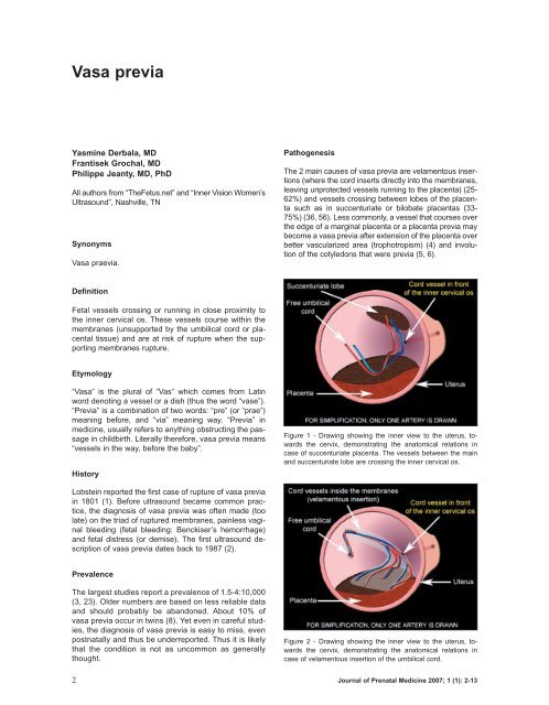
Figure 1 - Drawing showing the inner view to the uterus, towards<br />
the cervix, demonstrating the anatomical relations in<br />
case of succenturiate placenta. The vessels between the main<br />
and succenturiate lobe are crossing the inner cervical os.<br />
Lobstein reported the first case of rupture of vasa <strong>previa</strong><br />
in 1801 (1). Before ultrasound became common practice,<br />
the diagnosis of vasa <strong>previa</strong> was often made (too<br />
late) on the triad of ruptured membranes, painless vaginal<br />
bleeding (fetal bleeding: Benckiser’s hemorrhage)<br />
and fetal distress (or demise). The first ultrasound description<br />
of vasa <strong>previa</strong> dates back to 1987 (2).<br />
Prevalence<br />
The largest studies report a prevalence of 1.5-4:10,000<br />
(3, 23). Older numbers are based on less reliable data<br />
and should probably be abandoned. About 10% of<br />
vasa <strong>previa</strong> occur in twins (8). Yet even in careful studies,<br />
the diagnosis of vasa <strong>previa</strong> is easy to miss, even<br />
postnatally and thus be underreported. Thus it is likely<br />
that the condition is not as uncommon as generally<br />
thought.<br />
Figure 2 - Drawing showing the inner view to the uterus, towards<br />
the cervix, demonstrating the anatomical relations in<br />
case of velamentous insertion of the umbilical cord.<br />
2 Journal of Prenatal Medicine 2007; 1 (1): 2-13
<strong>Vasa</strong> <strong>previa</strong><br />
Sonographic findings<br />
Figure 3 - Drawing showing the inner view to the uterus, towards<br />
the cervix, demonstrating the anatomical relations in<br />
case of marginal placenta with vessels running at the edge of<br />
placenta and crossing the inner cervical os. By trophotropism,<br />
the marginal edge of the placenta regresses, leaving the vessel<br />
in front of the inner cervical os.<br />
Risk factors<br />
Conditions associated with vessels that run close to the<br />
cervix, such as low-lying placenta (7, 8), placenta <strong>previa</strong><br />
(9), multiple pregnancies (10), and of course multi-lobate<br />
placentas and velamentous insertion [1% of singleton<br />
pregnancy (38), 10% in multifetal pregnancies (11-<br />
13)]. About 2% of velamentous insertions are associated<br />
with a vasa <strong>previa</strong> (14-16).<br />
Placenta membranacea (22) is also a risk factor. It is<br />
less clear why, but in-vitro fertilization increases the risk<br />
of vasa <strong>previa</strong> (17-20), (about 1:300 pregnancies) (21).<br />
Many of these conditions present with vaginal bleeding<br />
which should be considered a possible alert symptom<br />
for vasa <strong>previa</strong>.<br />
Although vasa <strong>previa</strong> can be recognized in grey-scale<br />
as linear structures in front of the inner os (22, 23), the<br />
diagnosis is considerably simpler by putting a flash of<br />
color Doppler (color or power) (24, 25), over the cervix.<br />
Arterial flow but also venous flow can be recognized. Although<br />
some have obtained the diagnosis by perineal<br />
scan (26), a transvaginal image is clearly superior to an<br />
abdominal scan. Some have also advocated the use of<br />
3D (27, 59). Our impression is that 3D does not contribute<br />
much either in the diagnosis nor the mapping of<br />
the vessels since this is quite straightforward from 2D<br />
alone. Since 3D is not universally available, its unavailability<br />
should not be construed as a reason to not seek<br />
vasa <strong>previa</strong>. Nevertheless, 3D allows review of the volume<br />
if an unexpected finding is found at delivery. Another<br />
recent idea is to attempt to diagnose the cord insertion<br />
in the first trimester during the nuchal lucency<br />
screening, at a time when the fetus is less likely to obscure<br />
the cord insertion (28).<br />
Other diagnostic procedures<br />
Alternative methods of diagnosis such as digital palpation<br />
of a vasa <strong>previa</strong>, amnioscopy, Apt, Ogita (29) or<br />
similar (30) tests (fetal blood detection), and palpation<br />
have mostly a historical significance. MRI has been suggested<br />
too (31, 32). All these methods require a greater<br />
expertise then color Doppler thus cannot compare in<br />
speed and availability.<br />
Implications for targeted examinations<br />
In all pregnancies, we recommend sonographic examination<br />
for the placental cord insertion.<br />
In cases where the cord insertion is central and there<br />
is no succenturiate lobe, the likelihood of a vasa <strong>previa</strong><br />
is negligible. Only those cases where the placenta is<br />
low-lying should be examined more carefully. In prac-<br />
Figure 4 - Pathological specimen shows the fetal side of bilobate<br />
placenta with velamentous insertion of the umbilical cord<br />
between the placental lobes. (Courtesy Francois Manson and<br />
TheFetus.net).<br />
Figure 5 - Pathological specimen shows the maternal side of<br />
the bilobate placenta. (Courtesy Francois Manson and TheFetus.net).<br />
Journal of Prenatal Medicine 2007; 1 (1): 2-13 3
Y. Derbala et al.<br />
Figure 6 - Second trimester vaginal 2D sonography shows a<br />
sagittal section through the cervix. In this gray scale mode no<br />
vessels are visible crossing the inner cervical os.<br />
Figure 8 - Second trimester vaginal 2D sonography shows a<br />
sagittal section through the cervix with the marginal placenta<br />
<strong>previa</strong> localized at the dorsal wall of the uterus.<br />
Figure 7 - The same scan as in image 5 using color Doppler<br />
shows a vasa <strong>previa</strong> crossing the inner cervical os.<br />
Figure 9 - The same scan as in image 3 using color Doppler<br />
showing a vessel crossing the inner cervical os (vasa <strong>previa</strong>).<br />
tice, a short sweep with color Doppler over the internal<br />
os will usually detect abnormal vessels over the cervix.<br />
If anything is seen in color, greater attention needs to<br />
be paid to the region. Transvaginal (TV) sonography<br />
with color Doppler is ideal, not only because the proximity<br />
of the transducer to the os and the vessels but also<br />
vessels that are in a coronal plane of the patient are<br />
easier to recognize on transvaginal exam than on abdominal<br />
sonography. However, due to the extra time<br />
required and the invasiveness, this is only justified<br />
when there is a sufficient presumption on the abdominal<br />
scan or risk factors (low–lying placenta, multi-lobed<br />
placenta, multiple pregnancies, in-vitro fertilization,<br />
unidentified cord insertion, or abnormal flow over the<br />
cervix) or when there is an additional reason to do a TV<br />
scan.<br />
The following is a proposed diagnostic algorithm for the<br />
second-trimester detection of vasa <strong>previa</strong> (Fig. 12).<br />
During the second trimester examination (or later examination<br />
if the previous information is missing), observation<br />
of the placental cord insertion and the lower margin<br />
of the placenta shows that they are both clearly far from<br />
the inner os. In those cases there is essentially no risk<br />
of vasa <strong>previa</strong> and no further assessment for vasa <strong>previa</strong><br />
is required.<br />
Or, during the exam a succenturiate or multilobate placenta,<br />
velamentous insertion, a multifetal pregnancy, a<br />
low placenta, or an in-vitro fertilization is found or exists.<br />
Then an abdominal scan of cervix with color Doppler is<br />
suggested. If it is clearly normal then we go back to the<br />
“No risk” category.<br />
Or if the exam is not obviously normal, then a transvaginal<br />
color Doppler should be performed. If it is normal,<br />
we go back to the “no or low risk” category. If the<br />
transvaginal color Doppler is “Suspicious or abnormal”,<br />
then manage the patient as having a vasa <strong>previa</strong>. If<br />
during the initial abdominal exam there is any additional<br />
reason for performing a transvaginal examination,<br />
perform one and if it is strictly normal, the matter can<br />
be dropped, otherwise manage the patient as having a<br />
vasa <strong>previa</strong>.<br />
This should cover most clinical situation but exceptions<br />
4 Journal of Prenatal Medicine 2007; 1 (1): 2-13
<strong>Vasa</strong> <strong>previa</strong><br />
Figure 10 - Proposed diagnostic algorithm for the second-trimester detection of the vasa <strong>previa</strong>.<br />
Figure 11 - A second trimester vaginal 2D ultrasonographic<br />
scan shows sagittal section through the cervix with amniotic fluid<br />
above.<br />
are bound to happen and should be judged as they<br />
arise.<br />
Although some studies have claimed that adding a<br />
transvaginal ultrasound to an abdominal ultrasound only<br />
adds about a minute of examination time (33-35), this<br />
does not include the time to explain the procedure to the<br />
patient, obtain verbal consent, as well as patient preparation.<br />
Several studies, have shown that when specifically<br />
sought, velamentous insertions and thus vasa <strong>previa</strong><br />
can be reliably recognized (36-39), and that further, in<br />
prenatally detected vasa <strong>previa</strong>, the newborn survival<br />
rate ranged from 97-100% in the study group. Yet, other<br />
studies have demonstrated that velamentous insertions<br />
are regularly missed (40, 41).<br />
Even in skilled centers specifically attempting to identify<br />
vasa <strong>previa</strong>, some cases are likely to be missed<br />
(42). In one study 1 or possibly 2 out of 11 (or 12) cases<br />
was missed, and false positive ranged from 10-16%<br />
(36, 37). Even when specifically sought, a predisposing<br />
factor such as velamentous insertion which some<br />
authors report to recognize with 100% (39) accuracy, is<br />
only recognized by others in 62% (43), with higher result<br />
in anterior placenta (92%) and worst result in fundal<br />
(40%) or posterior (50%) placenta. In less skilled<br />
environment, the diagnosis can be missed even in the<br />
presence of risk factors (44). Some of these studies<br />
are getting a little old and results are improving.<br />
When a vasa <strong>previa</strong> is identified, serial scans, decreased<br />
maternal activity and close attention to early<br />
signs of labor or bleeding should be recommended<br />
(36).<br />
The bottom line is that although it is unlikely that all<br />
vasa <strong>previa</strong> will be recognized, awareness of the risk<br />
Journal of Prenatal Medicine 2007; 1 (1): 2-13 5
Y. Derbala et al.<br />
factors and adoption of a protocol, such as the one<br />
suggested below, to specifically seek vasa <strong>previa</strong> plus<br />
careful examination should substantially decrease the<br />
number of unsuspected cases at delivery and baring<br />
technical problems of maternal obesity or scarring a<br />
majority (90-95%) should be recognized.<br />
Differential diagnosis<br />
“Linear structures” in front of the inner os in grey-scale<br />
may also represent marginal placental sinus,<br />
chorioamniotic separation and simple folds of the<br />
membranes. The differential diagnosis of those is easily<br />
established by color Doppler. Pulsed Doppler will<br />
demonstrate a fetal umbilical or venous waveform if it<br />
is a vasa <strong>previa</strong>. Sometimes marginal placental sinus<br />
may present with flow, but it will be a maternal heart<br />
frequency.<br />
Pitfalls and artifacts<br />
Although the diagnosis of vasa <strong>previa</strong> appears straight<br />
forward, the diagnosis of cord insertion by the abdominal<br />
approach is not always feasible in obese patients,<br />
those with scars or even simply difficult fetal presentations.<br />
In case where the inner os is not seen on abdominal<br />
scans, a transvaginal examination would be recommended.<br />
Even on transvaginal examination there are possible pitfalls<br />
such as motion artifacts. Motion artifacts can occasionally<br />
give the impression on transvaginal color<br />
Doppler of <strong>previa</strong> flow simply due to sloshing of amniotic<br />
fluid resulting from fetal motion. This artifact can be recognized<br />
by its irregular nature and lack of reproducibility.<br />
Another pitfall is to confuse a funic presentation for a<br />
vasa <strong>previa</strong>. These are differentiated by the shifting in<br />
position of the cord, easily done by gently tapping with<br />
the transducer over the region.<br />
Figure 12 - The same second trimester vaginal sonography as<br />
in figure 2 using color Doppler showing a flushing artefact,<br />
caused by the movement of the amniotic fluid during fetal movement,<br />
imitating vasa <strong>previa</strong>.<br />
Figure 14 - The same scan as in figure 15 using the color<br />
Doppler clearly show that the suspicious structure is without<br />
Doppler signal and thus is not a vessel.<br />
Figure 13 - A second trimester vaginal 2D ultrasonographic<br />
scan shows sagittal section through the cervix with suspicious<br />
vessels crossing inner cervical os (arrow).<br />
Figure 15 - Second trimester vaginal Doppler image shows a<br />
high frequency fetal hart rate at the level of vasa <strong>previa</strong>. This<br />
helps to distinguish vasa <strong>previa</strong> from maternal cervical vessels.<br />
6 Journal of Prenatal Medicine 2007; 1 (1): 2-13
<strong>Vasa</strong> <strong>previa</strong><br />
Table I - Review of studies attempting to identify vasa <strong>previa</strong>.<br />
Author No. of Objectives G.A (wks) Methods of Umbilical cord Pathogenesis Associated Conclusions<br />
cases of the Study at diagnosis Investigation insertion of vasa <strong>previa</strong> complications<br />
to identify identification<br />
Nomiyama 587 Identify umbilical Mid- trimester Color Doppler Specificity Velamentous cord Premature rupture 99.8% (586/587)<br />
et al. (1998) (555 cord insertion scan during routine 99.8% (580/581) insertion: of membrane of all cord insertions<br />
singletons, sonography as identified<br />
16 sets Velamentous cord 18-20 wks (TVS*) & (TAS) 1 not seen turned – positive predictive<br />
of twin) insertion out normal – value 83% (5/6)<br />
<strong>Vasa</strong> <strong>previa</strong> – negative predictive<br />
– value 100% (580/<br />
– 580)<br />
Lee et al. 93,874 Prenatal ultra-sound Average 26 Abdominal and Record shows Velamentous: 10 Antepartum Identify<br />
(2000) diagnosis, and weeks transvaginal all cases were bleeding in 6/18 asymptomatic<br />
clinical out-comes with Doppler identified Bilobed placenta: 3 (some with fetal patients before<br />
of vasa <strong>previa</strong> Earliest 16 ultrasound intraventricular delivery<br />
weeks Succenturiate hemorrhage)<br />
placenta: 2 Delivery at<br />
Fetal heart 35-36 wks after<br />
Marginal: 2 rate abnormalities maturity<br />
amniocentesis<br />
Intra-uterine fetal<br />
death & preterm<br />
delivery<br />
Low Apgar scores<br />
Catanzarite 33,208 Specificity of Most at Abdominal and Velamentous: 2 Scimitar syndrome 10 of 11 cases<br />
et al. (2001) sonographic 20-24 wks transvaginal identified (91%)<br />
diagnosis of (67%) approach Multilobar placenta: 8 Ventriculomegaly 1 marginal placenta<br />
vasa <strong>previa</strong><br />
and pregnancy + color or power Delivery at 32-37<br />
Doppler weeks<br />
Sepulveda<br />
832 Identifying 2nd and 3rd Color Doppler 825/832 (99%) Velamentous: 7/8 Infant with transient 825/832 (99%)<br />
et al. (2003) velamentous trimester ultrasound tachypnea cord insertion<br />
insertion of the + (2D) + (3D) Eccentric: 1/8 detected<br />
cord in routine (at least 16 Cephalocele<br />
obstetric ultrasound weeks)<br />
Trisomy 21<br />
Mean = 23 wk<br />
Respiratory distress<br />
syndrome<br />
Journal of Prenatal Medicine 2007; 1 (1): 2-13 7
Y. Derbala et al.<br />
Table I - Review of studies attempting to identify vasa <strong>previa</strong>.<br />
Author No. of Objectives G.A (wks) Methods of Umbilical cord Pathogenesis Associated Conclusions<br />
cases of the Study at diagnosis Investigation insertion of vasa <strong>previa</strong> complications<br />
to identify identification<br />
Hasegawa 340 Detection of cord 9-11 weeks Gray-scale 318/340 (93.5%) 4/283 + 10/35 = SGA = 4/35 Cord insertion in<br />
et al. (2006) insertion site transvaginal 283 normal cord velamentous cord ower 1/3 of the<br />
(in the lower third sonography insertion insertion PROM = 9/35 luterus in 1st<br />
of uterus) during the trimester lead to<br />
late first trimester 35 cord insertion 4/283 + 5/35 = Emergency frequent<br />
in lower 1/3 of the accessory C.S. = 2/35 developmental<br />
uterus placenta abnormalities of<br />
placenta & cord.<br />
Useful for the<br />
identification of<br />
high-risk<br />
pregnancies<br />
Hasegawa 3446 Umbilical cord 18-20 weeks Gray-scale 3367/ 3421 Velamentous 25/40 Fetal heart rate Pregnancy with<br />
et al. (2006) insertion ultrasonography and 10/3367 abnormalities velamentous cord<br />
and color-flow 40 suspected insertion should be<br />
imaging velamentous Marginal 28/39 Low Apgar high risk<br />
cord insertion and 10/3367 scores pregnancy<br />
39 suspected<br />
Marginal cord<br />
insertion<br />
G.A. = gestational age; SGA = small for gestational age; PROM = premature rupture of membranes; CS = cesarean section; TAS = trans-abdominal sonography; TVS = trans-vaginal sonography.<br />
8 Journal of Prenatal Medicine 2007; 1 (1): 2-13
<strong>Vasa</strong> <strong>previa</strong><br />
Table II - Review of reported cases 1990-2006.<br />
Reference No. of Vaginal G.A. (wks) TAS TVS Pathology Number of Time and mode Associated anomalies<br />
cases bleeding at diagnosis + + vessels of delivery or neonatal complications<br />
as initial CDU CDU<br />
presentation<br />
Harding et al. 1 Yes 24 wk 4d 1 Succenturiate One large vessel<br />
(1990) lobe<br />
Nelson et al. 1 No 26 wk 1 Velamentous Four major vessels<br />
(1990) Cord insertion<br />
Hsieh et al. 1 No 30 wk 1 Succenturiate Fetal vessels<br />
(1991) Lobe<br />
Arts et al. 1 Yes 39 wk 1* Succenturiate Several vessels 39 wk, Elective C.S Normal<br />
(1993) Lobe +<br />
Velamentous<br />
Cord insertion<br />
Meyer et al. 1 No 27 wk 1 1 Marginal Large vein and<br />
(1993) Cord insertion small artery<br />
Hata et al. 1 Yes 30 wk 1 Velamentous Network of vessels<br />
(1994) Cord insertion<br />
Fleming et al. 1 No 1 1 Bilobed Several vessels<br />
(1996) Placenta<br />
Sauerbrei and 1 No 36 wk 1 1 Velamentous Several vessels 36 wk, Elective C.S. Normal<br />
Davies (1998) Cord insertion<br />
1 Yes 28 wk 1 1 Placenta 4 Vessels 33 wk, Elective C.S. Normal<br />
Membranacea<br />
Devesa et al. 1 No 20 wk 1 1 Velamentous cord Large vessel 37 wk, Elective C.S. Normal<br />
(1996) insertion<br />
Fung et al. 1 Yes 35 wk 1 Bilobed placenta Network of vessels 36 wk, Elective C.S. Normal<br />
(1998)<br />
1 Yes (heavy) Not diagnosed 1 Velamentous cord Network of vessels 39 wk, Elective C.S. Resuscitation + blood<br />
insertion Transfusion + convulsion<br />
1 No Not diagnosed Network of vessels 39 wk, Emergency C.S.<br />
Velamentous cord Normal<br />
insertion 37 wk, Elective C.S.<br />
Journal of Prenatal Medicine 2007; 1 (1): 2-13 9
Y. Derbala et al.<br />
Table II - Review of reported cases 1990-2006.<br />
Reference No. of Vaginal G.A. (wks) TAS TVS Pathology Number of Time and mode Associated anomalies<br />
cases bleeding at diagnosis + + vessels of delivery or neonatal complications<br />
as initial CDU CDU<br />
presentation<br />
Oyelese et al. 1 Yes 38 wk 1 Velamentous cord Network of vessels 38 wk, Normal Stillborn ex sanguinated<br />
(1998) insertion Vaginal Delivery fetus<br />
1 No (clear 23 wk 1 1 Network of vessels<br />
fluid only) Succenturiate lobe 24 wk, Emergency C.S. Died at 7 days of life<br />
1 34 wk 1 2 Vessels (A & V) (chorio-amionitis) sepsis and prematurity<br />
Yes Velamentous cord<br />
insertion 36 wk, Elective C.S. Normal<br />
Lee et al. 1 No 21 wk 1d 1(3D) Bilobed Large vessel 33 wk, Elective C.S. Normal<br />
(2000) Multiplanar Placenta +<br />
1 No 34 wk 3d volume (3D) Velamentous cord Large vessel 35 wk, Elective C.S. Normal<br />
Flightpath<br />
Velamentous cord<br />
Canterino 1 No 19 wk 1(3D) 1 Velamentous cord Network of vessels 35 wk, Elective C.S. Respiratory distress<br />
et al. (2004) insertion Syndrome<br />
Stafford 1 Yes 28wk 3 Bilobed Network of vessels 30 wk 2d, elective C.S. Normal<br />
et al (2004) Placenta (low-lying )<br />
Oyelese 1 No 30 wk 1 1 + 3D Bilobed placenta Large vessel 35 wk, elective C.S. Normal<br />
et al. (2004)<br />
1 Yes 24 wk 1 (3D) 1 + 3D Velamentous cord Network of vessels 34 wk, elective C.S. Normal<br />
insertion<br />
Hsieh 1 Yes 27 wk 1 (3D) Velamentous cord Network of vessels 34 wk, elective C.S. Normal<br />
et al. (2006) insertion<br />
1 Yes 35 wk 1 (3D) Network of vessels 35 wk 2d, elective C.S. Normal<br />
Succenturiate lobe<br />
Ushakov 1 15 wk 1 Velamentous cord Fetal vessels (in all Elective C.S. was done<br />
et al. (2006) insertion 4 cases) as a to all the cases<br />
1 22 wk 1 network of vessels<br />
(All 4 cases)<br />
1 26 wk 1<br />
1 30 wk 1<br />
G.A. = gestational age; SGA = small for gestational age; PROM = premature rupture of membranes; CS = cesarean section; TAS = trans-abdominal sonography; TVS = trans-vaginal sonography; CDU = Color-<br />
Doppler ultrasonography.<br />
10 Journal of Prenatal Medicine 2007; 1 (1): 2-13
<strong>Vasa</strong> <strong>previa</strong><br />
Finally, a vessel seen during a first trimester transvaginal<br />
scan should not be assumed to represent a vasa<br />
<strong>previa</strong>. Too often the vessel will be of maternal origin<br />
and be confused because of lateral resolution issues.<br />
Pulse Doppler will demonstrate a maternal pulse. The<br />
diagnosis of vasa <strong>previa</strong> is thus best made in the 2nd to<br />
3rd trimester. Should a suspicious vessel be found in the<br />
first trimester, a repeat scan in the second trimester is<br />
suggested.<br />
Review of the literature is provided in Tables I and II.<br />
Since vasa <strong>previa</strong> have been considered difficult to diagnose,<br />
have not specifically been sought and are not<br />
common, there are unfortunately no large prospective<br />
studies of the condition, and the evidence about the<br />
benefit of antenatal diagnosis relies on many small series<br />
or case report.<br />
Associated anomalies<br />
The various reported associated anomalies are probably<br />
coincidental and include cephalocele (38), Scimitar<br />
syndrome (36) and Trisomy 21 (38). A few others can be<br />
related to compression or damage of the vessels by the<br />
presenting parts and includes heart rate anomalies (43),<br />
small for gestational age, and intra-ventricular hemorrhage<br />
in a twin or even intra-uterine fetal death (23).<br />
Prognosis<br />
The major complication from vasa <strong>previa</strong> is the rupture<br />
of the vessels carrying fetal blood. This occurs at or near<br />
delivery if the condition is undetected. These results in a<br />
perinatal mortality of 56% (56) in undiagnosed cases,<br />
and 3% in those diagnosed prenatally (56). The median<br />
Apgar score (1 and 5 min) is 8 and 9 when detected prenatally<br />
versus only 1 and 4 for survivors of undetected<br />
cases (56). Further, transfusion is required in 58% of<br />
newborn without prenatal diagnosis, versus only 3% of<br />
those diagnosed prenatally (56). A less well quantified<br />
complication is the compression of the vasa <strong>previa</strong> by<br />
the presenting part resulting in decreased flow to the fetus<br />
and possibly hypoxia (57). Postnatal complications<br />
are related to either prematurity (due to early C-section<br />
with no confirmation of lung maturity) and include hyaline<br />
membrane disease, bronchopulmonary dysplasia,<br />
transient tachypnea, respiratory distress syndrome, or<br />
to partial exsanguination and complications related to<br />
anemia, hypovolemic shock (23) or complications of<br />
transfusions (8).<br />
Recurrence risk<br />
No reported increased risk.<br />
Management<br />
The outcome is markedly improved (97% survival versus<br />
44%) when a prenatal diagnosis is followed by elective<br />
C-section is performed at 35 weeks or earlier if<br />
signs of labor or membrane rupture occurs (56). Some<br />
have advocate hospitalization from 30-32 weeks with<br />
corticosteroids to assist in promoting lung maturity when<br />
the cervix is not demonstrated to be long and closed<br />
(58). When time permits, an amniocentesis to assess<br />
lung maturity is justified (59).<br />
Advocacy<br />
In the UK – UKVP raising awareness (http://www.vasapraevia.co.uk)<br />
has been very active in raising awareness<br />
on the issue (and their originators Daren & Natalie<br />
Samat deserve a lot of credit for their tireless work). The<br />
authors express their gratitude for their work and of the<br />
work of the International <strong>Vasa</strong> Previa Foundation<br />
(http://www.IVPF.org). Further, Dr. Oyelese has had the<br />
great kindness to review this manuscript and his many<br />
corrections are greatly appreciated.<br />
Conclusions<br />
Although no large-scale prospective studies are there to<br />
support these conclusions, personal experiences, case<br />
reports and smaller studies all concur to demonstrate a<br />
marked improvement in outcome when a vasa <strong>previa</strong> is<br />
detected prenatally. The obvious conclusion, until<br />
proven otherwise, is that a substantial improvement in<br />
outcome will depend only on prenatal detection. This implies<br />
a greater awareness of the condition and an effort<br />
at detecting it. The purpose of this manuscript is to help<br />
alert those who do prenatal examination that vasa <strong>previa</strong><br />
are not difficult to recognize when sought and that<br />
they are common enough to be worth seeking.<br />
References<br />
11. Lobstein J. Archives de L’art des Accouchements 1801;<br />
Strasbourg; p. 320.<br />
12. Gianopoulos J, Carver T, Tomich PG, Karlman R, Gadwood<br />
K. Diagnosis of vasa <strong>previa</strong> with ultrasonography.<br />
Obstet Gynecol 1987 Mar;69(3 Pt 2):488-91.<br />
13. Oyelese KO, Turner M, Lees C, Campbell S. <strong>Vasa</strong> <strong>previa</strong>:<br />
an avoidable obstetric tragedy. Obstet Gynecol Surv 1999<br />
Feb;54(2):138-45.<br />
14. Strassmann P. Placenta <strong>previa</strong>. Arch Gynecol 1902;<br />
67:112.<br />
15. Oyelese Y, Chavez MR, Yeo L, Giannina G, Kontopoulos<br />
EV, Smulian JC, Scorza WE. Three-dimensional sonographic<br />
diagnosis of vasa <strong>previa</strong>. Ultrasound Obstet Gynecol<br />
2004 Aug;24(2):211-5.<br />
16. Francois K, Mayer S, Harris C, Perlow JH. Association of<br />
vasa <strong>previa</strong> at delivery with a history of second-trimester<br />
placenta <strong>previa</strong>. J Reprod Med 2003 Oct;48(10):771-4.<br />
17. Lee W, Kirk JS, Comstock CH, Romero R. <strong>Vasa</strong> <strong>previa</strong>:<br />
prenatal detection by three-dimensional ultrasonography.<br />
Ultrasound Obstet Gynecol 2000 Sep;16(4):384-7.<br />
18. Fung TY, Lau TK. Poor perinatal outcome associated with<br />
vasa <strong>previa</strong>: is it preventable? A report of three cases and<br />
review of the literature. Ultrasound Obstet Gynecol 1998<br />
Dec;12(6):430-3.<br />
19. Francois K, Mayer S, Harris C, Perlow JH. Association of<br />
vasa <strong>previa</strong> at delivery with a history of second-trimester<br />
placenta <strong>previa</strong>. J Reprod Med 2003 Oct;48(10):771-4.<br />
Journal of Prenatal Medicine 2007; 1 (1): 2-13 11
Y. Derbala et al.<br />
10. Raga F, Ballester MJ, Osborne NG, Bonilla-Musoles F.<br />
Role of color flow Doppler ultrasonography in diagnosing<br />
velamentous insertion of the umbilical cord and vasa <strong>previa</strong>.<br />
A report of two cases. J Reprod Med 1995 Nov;40<br />
(11):804-8.<br />
11. Bernirschke K, Kaufmann P. Pathology of the human placenta.<br />
New York, NY Springer-Verlag 2000:353-359.<br />
12. Israel R. <strong>Vasa</strong> <strong>previa</strong> in binovular twins. Report of a case.<br />
Obstet Gynecol 1961 Jun;17:691-4.<br />
13. Whitehouse DB, Kohler HG. <strong>Vasa</strong> praevia in twin pregnancy:<br />
report of two cases. J Obstet Gynaecol Br Emp 1960<br />
Apr;67:281-3.<br />
14. Vago T, Caspi E. Antepartum bleeding due to injury of velamentous<br />
placental vessels. Obstet Gynecol 1962 Nov;<br />
20:671-4.<br />
15. Quek SP, Tan KL. <strong>Vasa</strong> <strong>previa</strong>. Aust N Z J Obstet Gynecol<br />
1972;12:206-9.<br />
16. Toivonen S, Heinonen S, Anttila M, Kosma VM, Saarikoski<br />
S. Reproductive risk factors, Doppler findings, and outcome<br />
of affected births in placental abruption: a population-based<br />
analysis. Am J Perinatol 2002 Nov;19(8):451-<br />
60.<br />
17. Spellacy WN. <strong>Vasa</strong> <strong>previa</strong>, multiple pregnancies, and in<br />
vitro fertilization clarification. Fertil Steril 2003 May;79<br />
(5):1254-5.<br />
18. Schachter M, Tovbin Y, Arieli S, Friedler S, Ron-El R, Sherman<br />
D. In vitro fertilization is a risk factor for vasa <strong>previa</strong>.<br />
Fertil Steril 2002 Sep;78(3):642-3.<br />
19. Oyelese Y, Spong C, Fernandez MA, McLaren RA. Second<br />
trimester low-lying placenta and in-vitro fertilization?<br />
Exclude vasa <strong>previa</strong>. J Matern Fetal Med. 2000 Nov-Dec;<br />
9(6):370-2.<br />
20. Oyelese KO, Schwarzler P, Coates S, Sanusi FA, Hamid<br />
R, Campbell S. A strategy for reducing the mortality rate<br />
from vasa <strong>previa</strong> using transvaginal sonography with color<br />
Doppler. Ultrasound Obstet Gynecol 1998 Dec;12(6):434-<br />
8.<br />
21. Schachter M, Tovbin Y, Arieli S, Friedler S, Ron-El R, Sherman<br />
D. In vitro fertilization is a risk factor for vasa <strong>previa</strong>.<br />
Fertil Steril 2002 Sep;78(3):642-3.<br />
22. Sauerbrei EE, Davies GL. Diagnosis of vasa <strong>previa</strong> with<br />
endovaginal color Doppler and power Doppler sonography:<br />
report of two cases. J Ultrasound Med 1998 Jun;17<br />
(6):393-8.<br />
23. Lee W, Lee VL, Kirk JS, Sloan CT, Smith RS, Comstock<br />
CH. <strong>Vasa</strong> <strong>previa</strong>: prenatal diagnosis, natural evolution, and<br />
clinical outcome. Obstet Gynecol 2000 Apr;95(4):572-6.<br />
24. Clerici G, Burnelli L, Lauro V, Pilu GL, Di Renzo GC. Prenatal<br />
diagnosis of vasa <strong>previa</strong> presenting as amniotic<br />
band. ’A not so innocent amniotic band’. Ultrasound Obstet<br />
Gynecol 1996 Jan;7(1):61-3.<br />
25. Devesa R, Munoz A, Torrents M, Carrera JM. Prenatal diagnosis<br />
of vasa <strong>previa</strong> with transvaginal color Doppler ultrasound.<br />
Ultrasound Obstet Gynecol 1996 Aug;8(2):139-<br />
41.<br />
26. Hertzberg BS, Kliewer MA. <strong>Vasa</strong> <strong>previa</strong>: prenatal diagnosis<br />
by transperineal sonography with Doppler evaluation. J<br />
Clin Ultrasound 1998 Oct;26(8):405-8.<br />
27. Araujo Junior E, Filho HA, Pires CR, Zanforlin Filho SM,<br />
Moron AF. Prenatal diagnosis of vasa <strong>previa</strong> through color<br />
Doppler and three-dimensional power Doppler ultrasonography.<br />
A case report. Clin Exp Obstet Gynecol 2006;33<br />
(2):122-4.<br />
28. Sepulveda W. Velamentous insertion of the umbilical cord:<br />
a first-trimester sonographic screening study. J Ultrasound<br />
Med 2006 Aug;25(8):963-8.<br />
29. Odunsi K, Bullough CH, Henzel J, Polanska A. Evaluation<br />
of chemical tests for fetal bleeding from vasa <strong>previa</strong>. Int J<br />
Gynaecol Obstet 1996 Dec;55(3):207-12.<br />
30. Lindqvist PG, Gren P. An easy-to-use method for detecting<br />
fetal hemoglobin-A test to identify bleeding from vasa <strong>previa</strong>.<br />
Eur J Obstet Gynecol Reprod Biol 2006:22.<br />
31. Oyelese Y, Jha RC, Moxley MD, Collea JV, Queenan JT.<br />
Magnetic resonance imaging of vasa praevia. BJOG 2003<br />
Dec;110(12):1127-8.<br />
32. Nimmo MJ, Kinsella D, Andrews HS. MRI in pregnancy:<br />
the diagnosis of vasa <strong>previa</strong> by magnetic resonance imaging.<br />
Bristol Med Chir J 1988 May;103(2):12.<br />
33. Lauria MR, Smith RS, Treadwell MC, Comstock CH, Kirk<br />
JS, Lee W, Bottoms SF. The use of second-trimester transvaginal<br />
sonography to predict placenta <strong>previa</strong>. Ultrasound<br />
Obstet Gynecol 1996 Nov;8(5):337-40.<br />
34. Farine D, Peisner DB, Timor-Tritsch IE. Placenta <strong>previa</strong>--is<br />
the traditional diagnostic approach satisfactory? J Clin Ultrasound<br />
1990 May;18(4):328-30.<br />
35. Leerentveld RA, Gilberts EC, Arnold MJ, Wladimiroff JW.<br />
Accuracy and safety of transvaginal sonographic placental<br />
localization. Obstet Gynecol 1990 Nov;76(5 Pt 1):759-<br />
62.<br />
36. Catanzarite V, Maida C, Thomas W, Mendoza A, Stanco L,<br />
Piacquadio KM. Prenatal sonographic diagnosis of vasa<br />
<strong>previa</strong>: ultrasound findings and obstetric outcome in ten<br />
cases. Ultrasound Obstet Gynecol 2001 Aug;18(2):109-<br />
15.<br />
37. Lee W, Lee VL, Kirk JS, Sloan CT, Smith RS, Comstock<br />
CH. <strong>Vasa</strong> <strong>previa</strong>: prenatal diagnosis, natural evolution, and<br />
clinical outcome. Obstet Gynecol 2000 Apr;95(4):572-6.<br />
38. Sepulveda W, Rojas I, Robert JA, Schnapp C, Alcalde JL.<br />
Prenatal detection of velamentous insertion of the umbilical<br />
cord: a prospective color Doppler ultrasound study. Ultrasound<br />
Obstet Gynecol 2003 Jun;21(6):564-9.<br />
39. Nomiyama M, Toyota Y, Kawano H. Antenatal diagnosis of<br />
velamentous umbilical cord insertion and vasa <strong>previa</strong> with<br />
color Doppler imaging. Ultrasound Obstet Gynecol 1998<br />
Dec;12(6):426-9.<br />
40. Heinonen S, Ryynanen M, Kirkinen P, Saarikoski S. Perinatal<br />
diagnostic evaluation of velamentous umbilical cord<br />
insertion: clinical, Doppler, and ultrasonic findings. Obstet<br />
Gynecol 1996 Jan;87(1):112-7.<br />
41. Eddleman KA, Lockwood CJ, Berkowitz GS, Lapinski RH,<br />
Berkowitz RL. Clinical significance and sonographic diagnosis<br />
of velamentous umbilical cord insertion.Am J Perinatol.<br />
1992 Mar;9(2):123-6.<br />
42. Robert JA, Sepulveda W. Fetal exsanguination from ruptured<br />
vasa <strong>previa</strong>: still a catastrophic event in modern obstetrics.<br />
J Obstet Gynaecol 2003 Sep;23(5):574.<br />
43. Hasegawa J, Matsuoka R, Ichizuka K, Sekizawa A, Farina<br />
A, Okai T. Velamentous cord insertion into the lower third<br />
of the uterus is associated with intrapartum fetal heart rate<br />
abnormalities. Ultrasound Obstet Gynecol 2006 Apr;27<br />
(4):425-9<br />
44. Lijoi AF, Brady J. <strong>Vasa</strong> <strong>previa</strong> diagnosis and management.<br />
J Am Board Fam Pract. 2003 Nov-Dec;16(6):543-8.<br />
45. Hasegawa J, Matsouka R, Ichizuka K, Otsuki K, Sekizawa<br />
A, Farina A, Okai T. Cord insertion into the lower third of<br />
the uterus in the first trimester is associated with placental<br />
and umbilical cord abnormalities. Ultrasound Obstet Gynecol<br />
2006;28:183-186.<br />
46. Hasegawa J, Matsuoka R, Ichizuka K, Sekizawa A, Farina<br />
A, Okai T. Velamentous cord insertion into the lower third<br />
of the uterus is associated with intrapartum fetal heart rate<br />
abnormalities. Ultrasound Obstet Gynecol 2006;27:425-<br />
429.<br />
47. Nelson LH, Malone PJ, King M. Diagnosis of vasa <strong>previa</strong><br />
12 Journal of Prenatal Medicine 2007; 1 (1): 2-13
<strong>Vasa</strong> <strong>previa</strong><br />
with transvaginal and color flow Doppler ultrasound. Obstet<br />
Gynecol 1990;76:506.<br />
48. Hseih F-J, Chen H-F, Ko T-M, et al. Antenatal diagnosis of<br />
vasa <strong>previa</strong> by color flow mapping. J Ultrasound Med<br />
1991;10:397.<br />
49. Arts H, van Eyck. Antenatal diagnosis of vasa <strong>previa</strong> by<br />
transvaginal color Doppler sonography. Ultrasound Obstet<br />
Gynecol 1993;3:276.<br />
50. Meyer WJ, Blumenthal L, Cadkin A, et al. <strong>Vasa</strong> <strong>previa</strong>: Prenatal<br />
diagnosis with transvaginal color Doppler flow imaging.<br />
Am J Obstet Gynecol 1993;169:1627.<br />
51. Hata K, Hata T, Fujiwaki R, et al. An accurate antenatal diagnosis<br />
of vasa <strong>previa</strong> with transvaginal color Dopplerultrasonography.<br />
Am J Obstet Gynecol 1994;171:265.<br />
52. Fleming AD, Johnson C, Targy M, et al. Diagnosis of vasa<br />
<strong>previa</strong> with ultrasound and color flow Doppler: A case report.<br />
Nebraska Med J 1996;81:191.<br />
53. Stafford IP, Neumann E, Jarrell H. Abnormal Placental<br />
Structure and <strong>Vasa</strong> Previa. J Ultrasound Med 2004;23:<br />
1521-2.<br />
54. Hsieh CTC, Wu JL, Wu HH, Yeh GP. Prenatal diagnosis of<br />
vasa <strong>previa</strong> with three-dimensional ultrasonography: report<br />
of two cases. Ultrasound in Obstetrics & Gynecology<br />
2006;28:512-14.<br />
55. Ushakov F, Herman A, Tovbin Y, Sherman D, Maymon R.<br />
<strong>Vasa</strong> <strong>previa</strong>: early antenatal diagnosis. Ultrasound in Obstetrics<br />
& Gynecology 2006;28:412.<br />
56. Oyelese Y, Catanzarite V, Prefumo F, Lashley S,<br />
Schachter M, Tovbin Y, Goldstein V, Smulian JC. <strong>Vasa</strong> <strong>previa</strong>:<br />
the impact of prenatal diagnosis on outcomes. Obstet<br />
Gynecol 2004 May;103(5 Pt 1):937-42.<br />
57. Harding JA, Lewis DF, Major CA, Crade M, Patel J, Nageotte<br />
MP. Color flow Doppler--a useful instrument in the<br />
diagnosis of vasa <strong>previa</strong>. Am J Obstet Gynecol 1990 Nov;<br />
163(5 Pt 1):1566-8.<br />
58. Oyelese Y, Smulian JC. Placenta <strong>previa</strong>, placenta accreta,<br />
and vasa <strong>previa</strong>. Obstet Gynecol 2006 Apr;107(4):927-41.<br />
59. Canterino JC, Mondestin-Sorrentino M, Muench MV, Feld<br />
S, Baum JD, Fernandez CO. <strong>Vasa</strong> <strong>previa</strong>: prenatal diagnosis<br />
and evaluation with 3-dimensional sonography and<br />
power angiography. J Ultrasound Med. 2005 May;24(5):<br />
721-4.<br />
Journal of Prenatal Medicine 2007; 1 (1): 2-13 13