The History of Sclerosing Foams
The History of Sclerosing Foams
The History of Sclerosing Foams

You also want an ePaper? Increase the reach of your titles
YUMPU automatically turns print PDFs into web optimized ePapers that Google loves.
<strong>The</strong> <strong>History</strong> <strong>of</strong> <strong>Sclerosing</strong> <strong>Foams</strong><br />
JAN-CHRISTOPH G. R. WOLLMANN, MD<br />
Bad Kreuznach, Germany<br />
BACKGROUND. <strong>The</strong> use <strong>of</strong> foamed sclerosants in phlebology is<br />
undergoing a renaissance. <strong>The</strong> use <strong>of</strong> foam sclerotherapy was<br />
relaunched only a few years ago. Despite this, the early<br />
developments, pioneer findings, and improvements, especially<br />
in foaming techniques, are not widely recognized.<br />
OBJECTIVE. <strong>The</strong> objective <strong>of</strong> this study was to give an overview<br />
from the very beginnings <strong>of</strong> foam sclerotherapy until the most<br />
recent and progressive techniques, as described by Tessari or the<br />
double syringe system technique.<br />
RESULTS. <strong>The</strong> publications found after a thorough research for<br />
literature about foam sclerotherapy allow us to examine what<br />
has been invented between Orbach’s work in 1944 and now<br />
RESEARCH AND TRAVEL EXPENSES WERE PROVIDED BY KREUSSLER.<br />
THE USE <strong>of</strong> foamed sclerosants in phlebology is not<br />
new. Nevertheless, the worldwide use <strong>of</strong> this treatment<br />
was ‘‘relaunched’’ only a few years ago. <strong>The</strong> major<br />
national and international journals reflected the<br />
interest for this treatment option and for sclerotherapy<br />
in general, indicated by the increasing numbers <strong>of</strong><br />
lectures or publications on this subject. <strong>The</strong>y usually<br />
start with a reference to the ‘‘basic invention’’: the<br />
work <strong>of</strong> Egmont James Orbach in 1944. 1 Another<br />
frequently cited publication is the one by Juan Cabrera<br />
Garrido, who presented the results <strong>of</strong> his development<br />
in Spain in 1995. 2 A thorough research on literature<br />
about foam sclerotherapy revealed a series <strong>of</strong> highly<br />
interesting articles. <strong>The</strong>y allow us to look at what has<br />
been invented between 1944 and 1995, and—surprisingly—even<br />
before 1944. Basically, the literature<br />
shows that remarkable work was carried out in the<br />
field <strong>of</strong> noncommercial foam sclerotherapy and that<br />
sclerosing foams have been used by numerous doctors<br />
continuously for the past six decades, especially for the<br />
treatment <strong>of</strong> varicose veins <strong>of</strong> the lower limbs.<br />
Results<br />
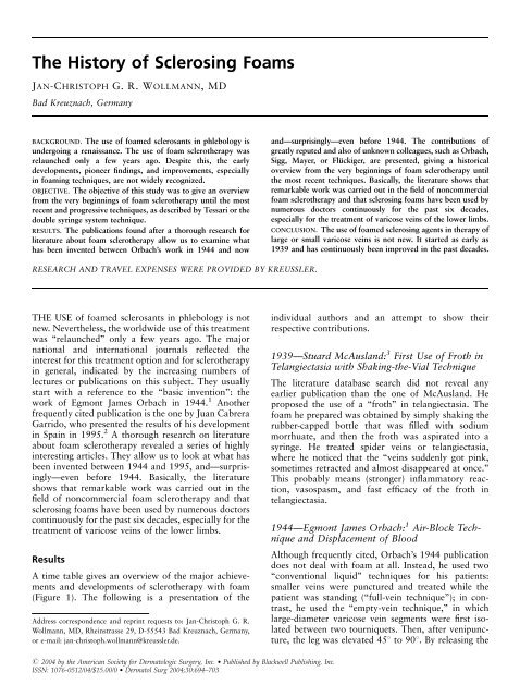
A time table gives an overview <strong>of</strong> the major achievements<br />
and developments <strong>of</strong> sclerotherapy with foam<br />
(Figure 1). <strong>The</strong> following is a presentation <strong>of</strong> the<br />
Address correspondence and reprint requests to: Jan-Christoph G. R.<br />
Wollmann, MD, Rheinstrasse 29, D-55543 Bad Kreuznach, Germany,<br />
or e-mail: jan-christoph.wollmann@kreussler.de.<br />
r 2004 by the American Society for Dermatologic Surgery, Inc. Published by Blackwell Publishing, Inc.<br />
ISSN: 1076-0512/04/$15.00/0 Dermatol Surg 2004;30:694–703<br />
and—surprisingly—even before 1944. <strong>The</strong> contributions <strong>of</strong><br />
greatly reputed and also <strong>of</strong> unknown colleagues, such as Orbach,<br />
Sigg, Mayer, or Flückiger, are presented, giving a historical<br />
overview from the very beginnings <strong>of</strong> foam sclerotherapy until<br />
the most recent techniques. Basically, the literature shows that<br />
remarkable work was carried out in the field <strong>of</strong> noncommercial<br />
foam sclerotherapy and that sclerosing foams have been used by<br />
numerous doctors continuously for the past six decades,<br />
especially for the treatment <strong>of</strong> varicose veins <strong>of</strong> the lower limbs.<br />
CONCLUSION. <strong>The</strong> use <strong>of</strong> foamed sclerosing agents in therapy <strong>of</strong><br />
large or small varicose veins is not new. It started as early as<br />
1939 and has continuously been improved in the past decades.<br />
individual authors and an attempt to show their<br />
respective contributions.<br />
1939—Stuard McAusland: 3 First Use <strong>of</strong> Froth in<br />
Telangiectasia with Shaking-the-Vial Technique<br />
<strong>The</strong> literature database search did not reveal any<br />
earlier publication than the one <strong>of</strong> McAusland. He<br />
proposed the use <strong>of</strong> a ‘‘froth’’ in telangiectasia. <strong>The</strong><br />
foam he prepared was obtained by simply shaking the<br />
rubber-capped bottle that was filled with sodium<br />
morrhuate, and then the froth was aspirated into a<br />
syringe. He treated spider veins or telangiectasia,<br />
where he noticed that the ‘‘veins suddenly got pink,<br />
sometimes retracted and almost disappeared at once.’’<br />
This probably means (stronger) inflammatory reaction,<br />
vasospasm, and fast efficacy <strong>of</strong> the froth in<br />
telangiectasia.<br />
1944—Egmont James Orbach: 1 Air-Block Technique<br />
and Displacement <strong>of</strong> Blood<br />
Although frequently cited, Orbach’s 1944 publication<br />
does not deal with foam at all. Instead, he used two<br />
‘‘conventional liquid’’ techniques for his patients:<br />
smaller veins were punctured and treated while the<br />
patient was standing (‘‘full-vein technique’’); in contrast,<br />
he used the ‘‘empty-vein technique,’’ in which<br />
large-diameter varicose vein segments were first isolated<br />
between two tourniquets. <strong>The</strong>n, after venipuncture,<br />
the leg was elevated 451 to 901. By releasing the
Dermatol Surg 30:5:May 2004 WOLLMANN: THE HISTORY OF SCLEROSING FOAMS 695<br />
Figure 1. Overview about the contributions to foam sclerotherapy.<br />
proximal tourniquet, blood could flow into the central<br />
or more proximal direction, while the distal tourniquet<br />
reduced or eliminated the supply <strong>of</strong> new blood from<br />
distally. This technique caused less dilution <strong>of</strong> the<br />
liquid sclerosing agent.<br />
Nevertheless, he sometimes noticed therapeutic<br />
failures, so he had the ‘‘famous idea’’: to intensify the<br />
contact between the sclerosant and the endothelium,<br />
the diameter <strong>of</strong> the vein should not only be reduced as<br />
far as possible prior to injection, but the vein should be<br />
rather free from any blood. <strong>The</strong>refore, he injected a<br />
small amount <strong>of</strong> air into the venous segment to be<br />
treated to completely displace the remaining blood (see<br />
Figure 2).<br />
Experimentally, he had shown that 1 cm 2 <strong>of</strong> air<br />
injected into a 6-mm infusion tube pending vertically<br />
and filled with water did not separate into several<br />
ascending bubbles but remained in the area <strong>of</strong><br />
injection as one large bubble, if the injection was not<br />
given too slowly. He then injected a colored solution<br />
into this air bubble, which, over a period <strong>of</strong> 3 to 5 s,<br />
did not get mixed with water but remained undiluted<br />
in the ‘‘air pocket.’’<br />
Clinically, he used the ‘‘air-block technique’’ only<br />
for smaller and medium-sized varicose veins; He<br />
recommended the conventional technique, without<br />
air injection, for larger veins. Unfortunately, Orbach’s<br />
article does not reveal the reasons for this recommendation.<br />
It remains unclear why the method that he<br />
considered to be more effective should not be suited<br />
for large varicose veins which usually were more<br />
difficult to treat. In 1970, Stemmer 4 et al. showed in an<br />
experiment that the air-block technique was only<br />
reliable with vessel diameters <strong>of</strong> up to 4 mm and was<br />
never successful with diameters <strong>of</strong> more than 8 mm.<br />
Sclerosis was rather impeded with such diameters: <strong>The</strong><br />
air bubble floating on the blood column protected the<br />
vessel from contact with the sclerosant at the upper<br />
Figure 2. <strong>The</strong> air-block technique: a small amount <strong>of</strong> air was injected<br />
before (top) the injection <strong>of</strong> sclerosant (bottom) to avoid dilution <strong>of</strong> the<br />
liquid.<br />
Figure 3. <strong>The</strong> air-block technique in large vessels: A floating air<br />
bubble (top) was able to prevent contact between the endothelium on<br />
the upper vessel wall and the sclerosant.<br />
circumference (see Figure 3), thus being effective only<br />
partially. <strong>The</strong> original air-block technique is basically<br />
no longer used today, but its development to a ‘‘foam<br />
block’’ (air block with large-bubbled foams) is still<br />
used today by some phlebologists under various names<br />
for smaller vessels. Small advantages in efficacy<br />
(increase by 20%) are believed to be outweighed by<br />
the disadvantages (plus 100% side effects). 5 <strong>The</strong><br />
maximum amount <strong>of</strong> air injected was 3 mL, a limit<br />
that became an orientation for most physicians using<br />
the air-block technique.<br />
1944—Robert Rowden Foote: 6 Foam in Feeder<br />
Veins with Shaking-the-Syringe Technique<br />
In the same year as Orbach’s publication, a book by<br />
Robert Rowden Foote was published in London. In<br />
addition to an overview <strong>of</strong> his ‘‘empty-vein technique,’’<br />
he wrote about the treatment <strong>of</strong> spider veins: ‘‘<strong>The</strong><br />
feeder vein should be dealt with first, whenever<br />
possible. <strong>The</strong> best injection fluid is the soapy froth<br />
obtained by shaking up 1 cc <strong>of</strong> ethamoline (ethanolamine<br />
oleate) in a 2-cc syringe. [It should be<br />
mentioned that the ratio <strong>of</strong> 1 plus 1 is, strictly<br />
speaking, not foam but an air/liquid dispersion.<br />
Usually a mixture is defined as foam if the gas portion<br />
is higher than 0.52. 7 A characteristic order <strong>of</strong><br />
magnitude <strong>of</strong> gas/fluid systems is the gas proportion<br />
j. Depending on the gas proportion, distinction is<br />
made between gas dispersion, spherical foam (wet<br />
foam), and polyhedral foam (dry foam). In gas<br />
dispersions, there are individual air bubbles in liquid;<br />
the gas proportion j is less than 0.52. In the spherical<br />
foam, the number <strong>of</strong> individual bubbles is higher—<br />
there is still a rather high amount <strong>of</strong> liquid; the gas
696 WOLLMANN: THE HISTORY OF SCLEROSING FOAMS Dermatol Surg 30:5:May 2004<br />
proportion is 0.524j40.74. In the polyhedral foam,<br />
in contrast, the amount <strong>of</strong> liquid between the bubbles<br />
is so small that the individual bubbles get closer and<br />
closer to each other, thus forming polyhedrons. <strong>The</strong><br />
gas proportion j is greater than 0.74. In a foaming<br />
fluid (e.g., beer), all types generally occur, the<br />
polyhedral foam being above the spherical foam and<br />
the gas dispersion and liquid below.] Once the needle<br />
is in the vein, the bubbles can be made to course along<br />
the minute veins and can be followed visually in their<br />
transit.’’ Nowadays, ethanolamine oleate is no longer<br />
available in most countries. <strong>The</strong> ‘‘agitation technique’’<br />
was refined in the following years and is no longer in<br />
use. <strong>The</strong> air:sclerosant ratio <strong>of</strong> 1 plus 1 described by<br />
Foote suggests that the dispersion was very fluid so<br />
that it could not be used for displacing the blood in<br />
larger diameter veins.<br />
1949—Karl Sigg: 8 Foam-Block Technique and<br />
Viscosity <strong>of</strong> Foam<br />
In 1949, Karl Sigg picked up the air-block technique<br />
described 5 years before: Also for other varices than<br />
spider veins, i.e., larger veins, he used the new<br />
technique and reported more than 4000 treatments<br />
performed ‘‘without problems.’’ Sigg describes the<br />
rationale for the use <strong>of</strong> the air-block technique in a<br />
similar manner as Orbach and Foote: ‘‘<strong>The</strong> purpose is<br />
to prevent the dilution <strong>of</strong> the solutions for injection in<br />
the vein by the blood and to ensure that the sclerosant<br />
gets in contact with the intima <strong>of</strong> the vein in a rather<br />
concentrated formy. Even with this small-scale use,<br />
the air bubble chased the blood before the injection<br />
fluid arrived and thus cleared the way for a closer<br />
contact <strong>of</strong> the sclerosant with the venous intima.’’<br />
Later, Sigg combined Orbach’s air-block technique<br />
and Foote’s foam application and thus introduced a<br />
new aspect into the therapeutic options known to that<br />
date: ‘‘Orbach’s method becomes even more beneficial<br />
if foam is injected instead <strong>of</strong> airy. This foam is less<br />
rapidly washed away in the varicose vein than it<br />
happens with the pure injection <strong>of</strong> air.’’ Thus Sigg<br />
introduced for the first time the idea (even if not<br />
pronounced) <strong>of</strong> increased viscosity <strong>of</strong> foam. With the<br />
use <strong>of</strong> foam, he improved the air-block technique<br />
(introducing a foam block), but without omitting the<br />
fluid sclerosant (see Figure 4). Sigg described his own<br />
procedure for the manufacture <strong>of</strong> foam; the glass<br />
syringe filled with fluid sclerosant was held with the<br />
opening pointing downward. Approximately 1 mL <strong>of</strong><br />
air was aspirated through it, pearls developed in the<br />
solution, and thus more or less large bubbles were<br />
generated in the syringe.<br />
Figure 4. <strong>The</strong> foam-block technique: First, foam was injected to<br />
maintain the displacing effect <strong>of</strong> the air block for a longer time (top),<br />
and then normal liquid sclerosant was injected (bottom).<br />
1950—Egmont James Orbach: 9,10 Vasospasm<br />
after Foam Sclerotherapy<br />
A later publication by Orbach reveals the first attempt<br />
to compare the efficacy <strong>of</strong> foam (administered as air<br />
block and foam) with the efficacy <strong>of</strong> fluid sclerosants.<br />
<strong>The</strong> end point was the length <strong>of</strong> the sclerothrombus<br />
generated by the injection <strong>of</strong> the respective substance.<br />
<strong>The</strong> efficacy <strong>of</strong> the foam generated by agitation <strong>of</strong> the<br />
syringe or drug vial was increased approximately 3.5to<br />
4-fold compared with the same amount <strong>of</strong> ‘‘conventional’’<br />
fluid. <strong>The</strong> observation <strong>of</strong> a ‘‘marked<br />
vasospasm’’ after injection <strong>of</strong> foam is nowadays<br />
considered to be an important immediate end point<br />
for the assessment <strong>of</strong> the efficacy <strong>of</strong> modern sclerotherapy.<br />
11–13 This (reversible) vasospasm, which is<br />
<strong>of</strong>ten clearly detectable in duplex-guided sclerotherapy,<br />
can be considered to be a sign <strong>of</strong> initial vascular<br />
damage after sclerotherapy. After administration <strong>of</strong> a<br />
viscous foam, vasospasm is more common and more<br />
pronounced than after sclerotherapy with conventional<br />
fluid. 42 An important factor in this respect is<br />
that a given volume <strong>of</strong> a blood-displacing foam can<br />
spread over a much longer venous segment after a<br />
vasospasm occurs and can even act at a certain<br />
distance from the site <strong>of</strong> administration. (For example,<br />
1 mL <strong>of</strong> a viscous foam completely fills a vein <strong>of</strong> 8 mm<br />
in diameter over a length <strong>of</strong> approximately 20 mm. A<br />
reduction <strong>of</strong> the vessel diameter owing to spasm to<br />
2 mm distributes the same foam volume to a length <strong>of</strong><br />
almost 32 cm. In vivo, a spasm <strong>of</strong> a mean length <strong>of</strong><br />
28 cm could be provoked with vessels <strong>of</strong> 4 to 8 mm in<br />
diameter and a foam application <strong>of</strong> 2 to 2.5 mL <strong>of</strong><br />
double-syringe-system foam.) As long as vasospasm<br />
exists, venipuncture at the same site is aggravated. <strong>The</strong><br />
use <strong>of</strong> small venous cannulae or catheters may help<br />
dealing with this.<br />
Because foams in current use are very different from<br />
those used by Orbach, it is impossible to draw<br />
conclusions about the general efficacy <strong>of</strong> foam from<br />
this experiment. Nevertheless, according to all findings<br />
and based on theoretical considerations, the efficacy <strong>of</strong><br />
foam is always higher than that <strong>of</strong> the same amount<br />
and concentration <strong>of</strong> fluid. <strong>The</strong> level <strong>of</strong> increased<br />
efficacy cannot be predicted in the individual case<br />
without sufficient standardization. <strong>The</strong> increased
Dermatol Surg 30:5:May 2004 WOLLMANN: THE HISTORY OF SCLEROSING FOAMS 697<br />
strength <strong>of</strong> foams needs to be respected if thinking<br />
about sclerosing smaller vessels with foam.<br />
1953—Arve Ree: 14 First Use <strong>of</strong> ‘‘Pure Foam’’<br />
<strong>The</strong> Norwegian doctor Ree introduced a new era in<br />
1953: He was the first to inject ‘‘pure’’ foam, not in<br />
addition to air and common fluid sclerosant but<br />
instead <strong>of</strong> the sclerosant and, in particular, without<br />
the previous injection <strong>of</strong> air. With his technique<br />
(agitation <strong>of</strong> a detergent solution in the vial and<br />
subsequent aspiration <strong>of</strong> the bubbles into the syringe)<br />
he first treated a series <strong>of</strong> 50 patients very successfully.<br />
He injected 2 to 7 mL <strong>of</strong> foam, depending on the vessel<br />
diameter, corresponding to an amount <strong>of</strong> air <strong>of</strong> up to<br />
6.6 mL. <strong>The</strong> bubble sizes seem to have been in the<br />
millimeter range with that technique. Owing to the<br />
higher kinetic energy (agitation <strong>of</strong> a vial and subsequent<br />
passage through a bottleneck with turbulent<br />
flow), it may be considered that a foam with smaller<br />
bubbles than with the previous techniques could be<br />
generated.<br />
1956—Peter Flückiger: 15 Retrograde Injection,<br />
Aspiration Technique, and Leg Elevation<br />
Flückiger recommended the technique known as<br />
‘‘retrograde sclerotherapy,’’ a technique characterized<br />
by leg elevation and sclerosant injection into saphenous<br />
veins from a proximal injection site in the distal<br />
direction. Thus, the sclerosant could reach all insufficient<br />
collaterals and the saphenous vein by one<br />
injection. He obtained better results by using sclerosing<br />
foam than with fluid sclerosants: ‘‘Basically, the<br />
procedure consists in injecting y Varsyl foam (ethanolamine<br />
oleate) into an appropriate vein. Due to the<br />
buoyancy, the foam has only a low tendency to move<br />
in central direction with the blood flowy. After<br />
removal <strong>of</strong> the needle, the location <strong>of</strong> the ‘‘foam seal’’<br />
can be determined by palpationy. <strong>The</strong> foam usually<br />
spreads in the surrounding <strong>of</strong> the injection site in distal<br />
and proximal direction. After injection, the intravascular<br />
foam depot y is displaced by manual massage<br />
against the periphery.’’ Flückiger additionally mentioned<br />
major aspects <strong>of</strong> modern foam sclerotherapy in<br />
his publication: He saw that the foam he used<br />
remained in the region <strong>of</strong> the injection site as a<br />
‘‘depot,’’ noticed that it was able to displace the blood<br />
in the proximal direction and was not washed away<br />
from the blood, and noticed that the foam could be<br />
directed to other regions by manual manipulation.<br />
Moreover, he postulated and discussed further treatment-relevant<br />
foam properties for the first time:<br />
Increased efficacy by increase <strong>of</strong> the effective surface<br />
<strong>of</strong> foam compared to fluid sclerosants;<br />
A stronger sclerosing effect with smaller amounts <strong>of</strong><br />
sclerosants;<br />
Postulation <strong>of</strong> a minimized bubble size; and<br />
Postulation <strong>of</strong> homogeneity <strong>of</strong> foam.<br />
It is obvious that a given amount <strong>of</strong> sclerosant in the<br />
form <strong>of</strong> foam has a much larger surface than it has in<br />
an unchanged, fluid state: the smaller the bubbles <strong>of</strong><br />
the foam are, the greater the surface. Since damage to<br />
the intima y is based on the contact <strong>of</strong> the intima<br />
with the sclerosant, the use <strong>of</strong> foam allows an<br />
extended sclerosation with a small amount <strong>of</strong> sclerosant.<br />
<strong>The</strong> preparation <strong>of</strong> a homogenous, fine-bubbled<br />
foam is an important condition for the success <strong>of</strong> the<br />
procedure.<br />
(<strong>The</strong> homogeneity <strong>of</strong> foam, i.e., a foam whose<br />
bubbles are as homogenous as possible, is an<br />
important prerequisite for its flow behavior (viscosity)<br />
and its stability. Ideally, a foam has only bubbles <strong>of</strong> the<br />
same diameter, which is, in reality, barely or never<br />
feasible. Foam is subjected to aging, which involves<br />
mainly two aspects: In contrast, the fluid that is<br />
between the gas bubbles in a young foam drains owing<br />
to the force <strong>of</strong> gravity, so that the upper bubbles slowly<br />
break down because <strong>of</strong> the ‘‘fluid deficiency.’’ In<br />
contrast, bubbles disappear because a gas diffusion<br />
takes place between the foam bubbles. <strong>The</strong> internal gas<br />
pressure is higher in small bubbles owing to the higher<br />
surface tension, so that gas moves from those bubbles<br />
into larger bubbles. Smaller bubbles <strong>of</strong> a foam get even<br />
smaller and large ones even larger—until they break<br />
down. In a homogenous foam, all bubbles would have<br />
the same inner gas pressure. A gas diffusion from one<br />
bubble to the adjacent bubble would not take place<br />
and the foam would remain stable until just the drain<br />
process would contribute to aging.)<br />
Flückiger considered the various known foam<br />
manufacturing procedures, and suggested his own<br />
new technique: ‘‘In order to obtain a homogenous finebubbled<br />
foam, the procedure consisting in the agitation<br />
<strong>of</strong> a syringe filled with air and sclerosant<br />
[Orbach’s technique 1950] is completely inappropriate.<br />
<strong>The</strong> drawing <strong>of</strong> air through a sclerosant in the<br />
syringe [Sigg’s technique 1949] does not yield a small<br />
fine-bubbled foam either. Moreover, none <strong>of</strong> these<br />
methods is able to convert the entire amount <strong>of</strong><br />
sclerosant into foam. <strong>The</strong>re is always a certain amount<br />
<strong>of</strong> remaining fluid, which cannot be used for retrograde<br />
sclerotherapy with the elevated leg. A perfect,<br />
fine-bubbled foam is obtained by the simultaneous<br />
aspiration <strong>of</strong> sclerosant y and air through a thin<br />
injection needley. <strong>The</strong> thin injection needle (the<br />
narrower the lumen, the finer the foam) y is
698 WOLLMANN: THE HISTORY OF SCLEROSING FOAMS Dermatol Surg 30:5:May 2004<br />
Figure 5. <strong>The</strong> aspiration technique: <strong>The</strong> tip <strong>of</strong> the needle was placed<br />
at the liquid–air interface, thus allowing coaspiration <strong>of</strong> air and<br />
sclerosant that would generate foam in one run.<br />
introduced into the ampoule in such a way that<br />
approximately two thirds <strong>of</strong> the opening <strong>of</strong> the bevel<br />
are imbibed in the fluid (see Figure 5). When drawing<br />
back the plunger, the syringe fills with Varsyl foam<br />
under a sizzling noise, the foam remaining stable for<br />
several minutes.’’<br />
1957: Heinz Mayer and Hans Brücke: 16 First<br />
Micr<strong>of</strong>oam Device (Double-Piston Syringe)<br />
A milestone regarding the improvement and standardization<br />
<strong>of</strong> sclerosing foam was the publication by two<br />
surgeons Mayer and Brücke: they described the first<br />
device that had been developed specifically for the<br />
preparation <strong>of</strong> viscous sclerosing foams, a syringe with<br />
a double plunger (see Figure 6). (A double-plunger<br />
syringe following the principle <strong>of</strong> Mayer-Brücke was<br />
‘‘reinvented’’ several times in the following decades. A<br />
very homogenous, extremely fine microbubble foam<br />
can be produced using a simple reproduction <strong>of</strong> that<br />
syringe—unfortunately, the original is no longer<br />
available. More than 40 years ago, Mayer and Brücke<br />
worked with foams that are now suggested to be<br />
classified as ‘‘micr<strong>of</strong>oam.’’ 17 )<br />
‘‘Before the main plunger, there is a second, thin<br />
plunger with numerous holes, whose piston leads to a<br />
central bore <strong>of</strong> the main plunger and protrudes it.<br />
When the main plunger is fixed, the plunger provided<br />
with holes can rapidly be moved forward and backward<br />
and the Phlebocid (ethanolamin oleate) can be<br />
mixed with the air contained in the syringe. A finebubbled,<br />
viscous foam developsy. We have tested<br />
Figure 6. <strong>The</strong> Mayer-Brücke device: <strong>The</strong> inner plunger with numerous<br />
tiny holes can rapidly be moved forward and backward to mix<br />
sclerosant and air contained in the syringe. <strong>The</strong> outer plunger was used<br />
for injection <strong>of</strong> the viscous and fine-bubbled foam.<br />
most sclerosants <strong>of</strong> similar composition and selected<br />
Phlebocid, which provides the stiffest foam upon<br />
agitation with a homogenous distribution <strong>of</strong> air<br />
bubblesy. We consider the greatest advantage <strong>of</strong> the<br />
use <strong>of</strong> foam y to be the homogenous distribution <strong>of</strong><br />
the sclerosanty.’’ Clinically, they stated after administration<br />
<strong>of</strong> foam: ‘‘<strong>The</strong> fibrous tissue <strong>of</strong> the venous<br />
lumen is so complete that in general, recanalizationydoes<br />
not occur. We never observed recurrent<br />
varicose veins after using Phlebocid foam. We never<br />
observed complications after foam filling such as air<br />
embolisms or skin necroses.’’<br />
1962—Peter Flückiger: 18 Turbulent Flow<br />
In 1962, Flückiger described another technique for the<br />
preparation <strong>of</strong> foam: pumping <strong>of</strong> air and sclerosant<br />
forward and backward between a drug vial and the<br />
attached syringe. Later, this technique was modernized<br />
and improved by Alessandro Frullini 19,20 by adding an<br />
adapter between the syringe and the bottle seal.<br />
Furthermore, Flückiger changed some points since<br />
his first publication in 1956: he maintained leg<br />
elevation not only during sclerotherapy but also for<br />
some minutes thereafter, to allow the foam to degrade.<br />
He warned therapists to be cautious while ‘‘stroking’’<br />
the foam seal in a peripheral direction to prevent any<br />
unintended movement <strong>of</strong> foam into the deep venous<br />
system. Today, it is recommended that you wait some<br />
minutes after foam sclerotherapy <strong>of</strong> large veins before<br />
applying compression. 42<br />
1963—Peter Lunkenheimer: 21 First Use <strong>of</strong><br />
Polidocanol Foam<br />
Even before polidocanol received its first marketing<br />
authorization, a German physician had the chance to<br />
use the novel sclerosing solution for research purposes<br />
for the first time. Kreussler’s archives revealed a letter<br />
from that phlebologist, which contains a written ‘‘case<br />
report’’ about the first patient treatment: ‘‘<strong>The</strong> first<br />
patient was treated with 2 mL Aethoxysklerol s<br />
foamy.’’
Dermatol Surg 30:5:May 2004 WOLLMANN: THE HISTORY OF SCLEROSING FOAMS 699<br />
1969—Walter Gillesberger: 22 Low-Pressure<br />
Technique<br />
Gillesberger’s technique was based on the generation<br />
<strong>of</strong> a negative pressure in a (glass) syringe, so that air<br />
could enter through the capillary gap between the<br />
syringe piston and the plunger and thus generate<br />
bubbles (see Figure 7). Gillesberger’s syringe was not<br />
closed hermetically like the Monfreux syringe tip later<br />
on, but the negative pressure was strong enough to<br />
create a foam and was not compensated by the inflow<br />
<strong>of</strong> the sclerosant from the vial. <strong>The</strong> technique by<br />
Gillesberger or its refinement by Monfreux, who<br />
generated foam with an ‘‘absolute’’ negative pressure,<br />
is very simple. What is problematic is that there is no<br />
standardized ratio between air and sclerosant. <strong>The</strong>refore,<br />
the foams produced in that manner are different,<br />
depending on the syringe type, needle, generated<br />
suction, and the resulting gas content; they may be<br />
more fluid one time and more viscous another time.<br />
This leads to different behavior in the vessel (degree<br />
and duration <strong>of</strong> the displacement <strong>of</strong> blood) and thus<br />
probably to an unpredictable efficacy in vivo. (<strong>The</strong><br />
ability <strong>of</strong> foam to displace blood depends on its quality<br />
or characteristics: foams obtained with low sclerosant<br />
concentrations, with small amounts <strong>of</strong> air, and/or with<br />
rather large bubbles have a more or less fluid behavior;<br />
i.e., they are more easily washed away by liquids<br />
(blood) and have no or just a small and short-term<br />
displacing effect. Viscous foams (higher concentration,<br />
higher gas content, and small bubbles), in contrast,<br />
basically have a displacing effect in larger vessels. 41 If<br />
sclerosing foam is to be used for small diameter veins,<br />
precaution is recommended owing to the enhancement<br />
<strong>of</strong> the efficacy. A viscous foam would probably have<br />
too a strong effect in small veins; moreover, the foam<br />
breaks down as a result <strong>of</strong> the passage through the<br />
required fine needles. 42<br />
Figure 7. <strong>The</strong> low-pressure technique: Pulling down the piston<br />
allowed air to enter the syringe through the tiny gap between the<br />
syringe piston and the plunger.<br />
1984—Gerald Hauer: 23 Twin-Syringe Technique<br />
Hauer patented a twin-syringe set for foam preparation:<br />
Two parallel syringes, one filled with air, the<br />
other one filled with sclerosing solution, are simultaneously<br />
emptied into a ‘‘mixing chamber’’ by pressure,<br />
which leads to the formation <strong>of</strong> bubbles. <strong>The</strong> ratio <strong>of</strong> 1<br />
plus 1 (sclerosing agent and air) suggests a dispersion<br />
that should have the effect <strong>of</strong> a foam block.<br />
1986—Michael Grigg: 24 Double Syringes and<br />
Connecting Tubes<br />
In 1986, Grigg demonstrated a new foam production<br />
procedure: the principle was the generation <strong>of</strong> a<br />
turbulent flow between two syringes, connected via a<br />
plastic infusion tube, so that fluid and air could be<br />
pumped forward and backward. Belcaro later improved<br />
this technique by the addition <strong>of</strong> a further<br />
strongly foaming detergent solution in small amounts<br />
(0.1–0.2 mL). (‘‘Improved’’ in this context means<br />
prolongation <strong>of</strong> half-life. Owing to the commonly<br />
relevant amount <strong>of</strong> silicone in disposables (syringes<br />
and infusion tubes, etc.), the half-life <strong>of</strong> the foam was<br />
evidently limited because silicone, which destroys the<br />
surfactant arrangement <strong>of</strong> the foam lamellae, is a very<br />
strong foam breaker. <strong>The</strong>refore, anything that contributes<br />
to the maintenance <strong>of</strong> foam bubbles supports<br />
the foam stability: this is, in addition to an increase <strong>of</strong><br />
the surfactant concentration, in particular the minimization<br />
<strong>of</strong> the amount <strong>of</strong> silicone.) According to him,<br />
the foam half-life was approximately 4 min. This socalled<br />
‘‘Irvine technique’’ (named after the laboratory<br />
in which the technique was demonstrated) can be<br />
considered to be a precursor <strong>of</strong> Tessari’s technique and<br />
the double-syringe system (DSS). Belcaro and coworkers<br />
24 first performed investigations on the safety <strong>of</strong><br />
the foam generated according to that technique: 5 to<br />
10 mL <strong>of</strong> the foam produced with that technique did<br />
not cause any change <strong>of</strong> the pulmonary ventilation<br />
scintigraphy or perfusion scintigraphy in 12 patients.<br />
1995—Juan Cabrera Garrido: 2 Rotating Brush<br />
Technique<br />
In 1995, Cabrera Garrido published data from the<br />
clinical use <strong>of</strong> ‘‘micr<strong>of</strong>oam.’’ He had treated venous<br />
malformations and saphenous veins including collaterals<br />
with high volumes <strong>of</strong> foam. His objective was to<br />
fill the complete venous lumen by injecting high doses<br />
<strong>of</strong> foam at one time. A new aspect <strong>of</strong> the production<br />
was the use <strong>of</strong> a high-speed rotating brush (a modified<br />
dental bur), so that foam was agitated—like cream<br />
with a food mixer—and the facultative addition <strong>of</strong><br />
CO2 as a carrier gas. In experiments, CO2—if
700 WOLLMANN: THE HISTORY OF SCLEROSING FOAMS Dermatol Surg 30:5:May 2004<br />
Figure 8. Foam prepared with 3% polidocanol solution according to<br />
the double-syringe system (DSS). Sterile air or CO2 was used as gas.<br />
<strong>The</strong> degradation <strong>of</strong> foam is faster if CO2 is used instead <strong>of</strong> air and even<br />
more pronounced if the ratio is changed from 1:5 to 1:10.<br />
employed as the only gas—led to foams with quickly<br />
degrading bubbles (see Figure 8). Regarding the highdose<br />
treatment, more recent publications by Cabrera<br />
Garridos mentioned that the foam could flow from the<br />
greater saphenous vein into the deep venous system via<br />
the saphen<strong>of</strong>emoral junction or other connections,<br />
where it may provoke thromboses. 25 <strong>The</strong> number <strong>of</strong><br />
deep venous thromboses in a first study on this foam<br />
(6% thromboses when administering 20 mL <strong>of</strong><br />
Varisolve foam and more) 26 does not lend support to<br />
the high-dose foam therapy and requires special safety<br />
measures.<br />
1997—Alain Monfreux: 27 Low-Pressure<br />
Technique and ‘‘Méthode MUS’’<br />
Monfreux’s technique, published in 1997 under the<br />
name <strong>of</strong> ‘‘Méthode MUS,’’ was picked up primarily by<br />
French phlebologists. <strong>The</strong> principle was based on<br />
Gillesberger’s technique: generation <strong>of</strong> a negative<br />
pressure in the glass syringe filled with sclerosing<br />
solution, thus inlet <strong>of</strong> air through the fine gap between<br />
syringe barrel and plunger with corresponding pearling<br />
<strong>of</strong> the solution and transformation into a foam.<br />
Unlike Gillesberger, Monfreux generated an ‘‘absolute’’<br />
negative pressure by placing a cap on the syringe.<br />
Although all concentrations <strong>of</strong> sclerosants can be used<br />
with this method, the efficacy cannot be predicted in<br />
each individual case: a defined ratio <strong>of</strong> gas and<br />
sclerosant or a defined bubble size cannot be determined,<br />
because many variables (e.g., the width <strong>of</strong> the<br />
capillary gap between the syringe barrel and plunger,<br />
force, and duration <strong>of</strong> traction at the plunger and thus<br />
the order <strong>of</strong> magnitude <strong>of</strong> the effective shearing force<br />
which is responsible for the pearling <strong>of</strong> the solution)<br />
depend on the brand and on the user.<br />
1998—Symon Sadoun/Jean-Patric Benigni: 5,28<br />
Advanced Low-Pressure Technique<br />
Both authors improved Monfreux’s technique by<br />
making it usable for plastic syringes: <strong>The</strong> principle—<br />
generation <strong>of</strong> a negative pressure in a syringe closed by<br />
a Luer stopper—remained similar: pulling down the<br />
piston to generate subatmospheric pressure was<br />
followed by quickly releasing the piston several times.<br />
As with Monfreux and his predecessors, a defined<br />
gas:sclerosant ratio could not be stated.<br />
1998—Miguel Santos Gaston: 29 Advanced<br />
Low-Pressure Technique<br />
Gaston basically adopted the Monfreux technique, but<br />
added some more steps to the preparation procedure:<br />
once the foam was prepared according to Monfreux,<br />
he emptied the foam into a glass container and then<br />
aspirated the foam again. This was repeated several<br />
times, making—on the one hand—the foam more fine,<br />
but—on the other hand—more dry.<br />
1999—Javier Garcia Mingo: 30,31 High-Pressure<br />
Technique and ‘‘Foam Medical System’’<br />
Mingo was the first to describe a sterilizable, reusable<br />
device for the preparation <strong>of</strong> foam, which generates<br />
foam by the introduction <strong>of</strong> various gases from a<br />
pressure-gas cylinder and subsequent passage <strong>of</strong> the<br />
mixture through a fine nozzle, the ‘‘foam medical<br />
system.’’ <strong>The</strong> handling and cleaning <strong>of</strong> the device,<br />
which appears a little complicated, has prevented its<br />
wide use so far, although Mingo’s clinical results are<br />
considered very promising.<br />
2000—Lorenzo Tessari: 32,33 Double Syringes,<br />
Three-Way Tap, and the ‘‘Tourbillon Technique’’<br />
Tessari’s Tourbillon technique is at present, together<br />
with the similar DSS technique, the most commonly<br />
used technique. It corresponds basically to Irvine’s<br />
technique 24 but has clear advantages over the latter.<br />
Tessari uses two syringes as well (various sizes being<br />
described), but Tessari’s technique does not require<br />
‘‘foaming aids’’: <strong>The</strong> two syringes are connected by a<br />
three-way stopcock, and the sclerosing solution and air<br />
are drawn back and forth by pump movements. Owing<br />
to the absence <strong>of</strong> a connecting tube, a lot <strong>of</strong> disturbing<br />
silicone is no longer present. A more detailed<br />
description <strong>of</strong> the pumping procedure (at least 10<br />
times) to improve the standardization <strong>of</strong> the foam was<br />
given in 2001. 34<br />
<strong>The</strong> three-way stopcock has an additional ‘‘technical<br />
finesse’’: it is possible to vary the size <strong>of</strong> the passage
Dermatol Surg 30:5:May 2004 WOLLMANN: THE HISTORY OF SCLEROSING FOAMS 701<br />
by turning the cock—a narrow passage generates high<br />
turbulences and generates smaller blubbles than a wide<br />
passage. This procedures uses 2 to 2.5 mL <strong>of</strong> air and<br />
0.5 mL <strong>of</strong> sclerosing solution (all concentrations,<br />
mainly 1 and 3% for large and very large vessels).<br />
Irrespective <strong>of</strong> the concentration, the gas proportion j<br />
is approximately 0.7 to 0.83; the gas bubbles are very<br />
fine, and the half-life depends on the concentration and<br />
on the syringe. <strong>The</strong> foam prepared according to the<br />
Tessari method has successfully been tested in clinical<br />
trials. 35,36<br />
2000—Alessandro Frullini: 17–19 Advanced<br />
Turbulent Flow<br />
In 2000, Frullini improved the technique developed by<br />
Flückinger in 1962 and made it usable for disposable<br />
syringes by adding an adapter 18 and in 2001 by<br />
optionally using sterile air. 17,19<br />
2001—Gilles Gachet: 37 Aspiration Technique<br />
<strong>The</strong> aspiration technique published by Gachet in 2001<br />
is, regarding foam preparation, very similar with the<br />
description <strong>of</strong> Flückiger’s method from 1956.<br />
2001—Double-Syringe System (DSS), 11,12,38–40<br />
Two-Way Connector, and Pressure<br />
When searching for a suitable technique for the<br />
preparation <strong>of</strong> foam, it was noticed that the methods<br />
available to date all had at least one disadvantage that<br />
aggravated experiments with sclerosing foam or even<br />
made it impossible from the scientific point <strong>of</strong> view:<br />
the foams were not reproducible, because important<br />
foam characteristics (e.g., the gas proportion, the<br />
sclerosant concentration, the bubble size, or others)<br />
were stated incompletely or not at all, because the<br />
required materials were not described in detail, or<br />
because the very preparation was not defined clearly.<br />
<strong>The</strong> foams prepared for laboratory experiments and<br />
preclinical experiments all had a different consistence<br />
and an inhomogeneous flow behavior, if minor<br />
changes were made. Various types, manufacturers,<br />
and sizes <strong>of</strong> syringes; changes to the needle diameters<br />
and lengths; different temperatures during preparation;<br />
and other factors led to completely diverging<br />
results. <strong>The</strong>refore, a preparation variant was searched<br />
for that was simple, fast, sterile and especially<br />
reproducible. In a laboratory experimental series, the<br />
various variables that may affect the stability <strong>of</strong> the<br />
foam were changed systematically and the best<br />
possible combination <strong>of</strong> syringe type, gas:fluid ratio,<br />
sclerosant concentration, and manufacturing instructions<br />
was looked for.<br />
<strong>The</strong> most stable and fine-bubbled foam was obtained<br />
according to the following instructions: the required<br />
materials were a 10-mL Omnifix syringe, a 10-mL<br />
Inject syringe (each with a Luer-Lock connection), one<br />
Combidyn adapter (to connect the syringes), and a 0.2mm<br />
filter (for sterilization <strong>of</strong> air) (see Figure 9).<br />
Exactly 8 mL air is drawn into the Inject syringe via<br />
the sterile filter; afterwards, after removal <strong>of</strong> the filter,<br />
exactly one ampoule (2 mL) <strong>of</strong> polidocanol 3%<br />
(Aethoxysklerol). <strong>The</strong> two syringes are connected to<br />
the adapter. First some pumping movements (5 )are<br />
performed against resistance (by thumb pressure onto<br />
the other syringe piston) until the two components are<br />
well mixed. Afterward, the foam is pumped again<br />
quickly forth and back seven times between the two<br />
syringes without resistance like in the Tessari technique,<br />
until a homogenous foam has formed (see Figure 10).<br />
<strong>The</strong> double-syringe-system foam has a fixed sclerosant:air<br />
ratio <strong>of</strong> 1:5 ( 5 1 plus 4), a half-life <strong>of</strong><br />
approximately 150 s, with an initial mean bubble size<br />
<strong>of</strong> 70 mm. Divergent syringes, concentrations, scler-<br />
Figure 9. Components used in the double-syringe technique.<br />
Figure 10. Double-syringe-system (DSS) foam.
702 WOLLMANN: THE HISTORY OF SCLEROSING FOAMS Dermatol Surg 30:5:May 2004<br />
osant:air ratios, or pump procedures make the foam<br />
less stable and less viscous.<br />
<strong>The</strong> first prospective, randomized multicenter study<br />
compared foam sclerotherapy with double-syringe<br />
system with conventional fluid sclerotherapy in 88<br />
patients with greater saphenous vein insufficiency. A<br />
single injection (the protocol did not include further<br />
injections) <strong>of</strong> 2 to 2.5 mL <strong>of</strong> double-syringe-system<br />
foam or 3% liquid polidocanol showed a 2-year<br />
occlusion rate <strong>of</strong> 84% in the foam group versus 40%<br />
in the fluid group. Vasospasm was clearly more<br />
frequent and more pronounced in the group treated<br />
with foam (mean length 28 cm) than in the control<br />
group, the rate <strong>of</strong> side effects being identical. 11,12<br />
Conclusion and Discussion<br />
<strong>The</strong> use <strong>of</strong> foamed sclerosing agents in therapy <strong>of</strong> large<br />
or small varicose veins is not new. It started as early as<br />
1939. <strong>The</strong> publications about foam sclerotherapy<br />
found and presented in this text allow us to look at<br />
what has been invented between the very beginnings<br />
and now. <strong>The</strong> contributions <strong>of</strong> widely known and also<br />
<strong>of</strong> less known colleagues show that remarkable work<br />
was performed in the field <strong>of</strong> noncommercial foam<br />
sclerotherapy continuously for the past six decades,<br />
especially for the treatment <strong>of</strong> varicose veins <strong>of</strong> the<br />
lower limbs. But they also show that there sometimes<br />
is a lack <strong>of</strong> accuracy in describing the particular foams<br />
used, which does not make it easy to reproduce the<br />
individual reported results and/or findings. <strong>The</strong> suggestion<br />
therefore is to describe any foam used in<br />
nonclinical or clinical trials according to the ‘‘definition<br />
<strong>of</strong> sclerosing foams’’: <strong>Sclerosing</strong> foam is characterized<br />
by (at least) the following variables: type and<br />
concentration <strong>of</strong> the tensioactive sclerosing agent, type<br />
<strong>of</strong> gas, ratio <strong>of</strong> liquid to gas, the method <strong>of</strong><br />
preparation, the time between processing and use,<br />
and bubble sizes. This could really help to make<br />
sclerosing foams, and clinical results, comparable.<br />
References<br />
1. Orbach EJ. Sclerotherapy <strong>of</strong> varicose veins—utilization <strong>of</strong> an<br />
intravenous air block. Am J Surg 1944;LXVI(3):362–6.<br />
2. Cabrera J, Cabrera Garcia-Olmedo JR. Nuevo método de esclerosis<br />
en las varices tronculares. Patol Vasc 1995;4:55–73.<br />
3. McAusland S. <strong>The</strong> modern treatment <strong>of</strong> varicose veins. Med Press<br />
Circular 1939;201:404–10.<br />
4. Stemmer R, Kopp C, Voglet P. Physikalische Studie der Sklerosierungsinjektion.<br />
Zentralbl Phlebol 1970;9:112–23.<br />
5. Benigni JP, Sadoun S, Thirion V, et al. Télangiectasies et varices<br />
réticulaires—traitement par la mousse d’Aetoxisclérol à 0.25%:<br />
présentation d’une étude pilote. Phlébologie 1999;52:283–9.<br />
6. Foote RR. <strong>The</strong> injection treatment. In: Foote RR, ed. Varicose<br />
Veins, Haemorrhoids and Other Conditions. London: Lewis, 1944:<br />
13–44.<br />
7. Pahl MH, Meinecke H. Schaumzerstörung mit arteigener Flüssigkeit.<br />
In: Dechema-Monographien Band 114. Weinheim: VCH-<br />
Verlagsgesellschaft, 1989.<br />
8. Sigg K. Neuere gesichtspunkte zur Technik der Varizenbehandlung.<br />
<strong>The</strong>r Umsch 1949;6:127–34.<br />
9. Orbach EJ, Petretti AK. <strong>The</strong> thrombogenic property <strong>of</strong> foam <strong>of</strong> a<br />
synthetic anionic detergent. Angiology 1950;1:237–43.<br />
10. Orbach EJ. Contributions to the therapy <strong>of</strong> the varicose complex. J<br />
Int Coll Surg 1950;6:765–71.<br />
11. Hamel-Desnos C, Desnos P, Wollmann JC, et al. Evaluation <strong>of</strong> the<br />
efficacy <strong>of</strong> polidocanol in form <strong>of</strong> foam compared to liquid form in<br />
sclerotherapy <strong>of</strong> the greater saphenous vein: intial results. Dermatol<br />
Surg 2003;29:1170–5.<br />
12. Hamel-Desnos C, Ouvry P, Desnos P, Mako S. Evaluation <strong>of</strong> the<br />
Efficacy <strong>of</strong> Polidocanol in the Form <strong>of</strong> Foam versus Liquid Form in<br />
Sclerotherapy <strong>of</strong> the Long Saphenous Vein. American College <strong>of</strong><br />
Phlebology 16th Annual Congress; 2002 Nov 8–10; Ft. Lauderdale,<br />
FL. Oakland, CA: ACP, 2003.<br />
13. Schadeck M, Allaert FA. Duplex scanning in the mechanism <strong>of</strong><br />
the sclerotherapy: importance <strong>of</strong> the spasm. Phlebol Suppl 1995;1:<br />
574–6.<br />
14. Ree A. Etamolin foam in the treatment <strong>of</strong> varicose veins. A new<br />
method. Acta Dermatovenerol 1953;33:435–6.<br />
15. Flückiger P. Nicht-operative retrograde Varicenverödung mit<br />
Varsylschaum. Schweiz Med Wochenschr 1956;48:1368–70.<br />
16. Mayer H, Brücke H. Angiologie—Zur Ätiologie und Behandlung<br />
der Varizen der unteren Extremitäten. Chir Prax 1957;4:521–8.<br />
17. Frullini A. <strong>Sclerosing</strong> foam in the treatment <strong>of</strong> recurrent varicose<br />
veins. In: Henriet JP, ed. Foam Sclerotherapy State <strong>of</strong> the Art. Paris:<br />
Editions Phlebologiques Françaises, 2001:73–8.<br />
18. Flückiger P. Beitrag Zur Technik der ambulanten Varizenbehandlung.<br />
Die Med Welt 1963;12:617–21.<br />
19. Frullini A. New technique in producing sclerosing foam in a<br />
disposable Syringe. Dermatol Surg 2000;26:705–6.<br />
20. Frullini A. <strong>The</strong> <strong>Sclerosing</strong> Foam in the Treatment <strong>of</strong> Teleangectasia<br />
and Varicose Veins: New Techniques for Production <strong>of</strong> a Foam with<br />
Sterile Air. American College <strong>of</strong> Phlebology 14th Annual Congress;<br />
2000 Nov 16–19; Atlanta, GA. Oakland, CA: ACP, 2001.<br />
21. Lunkenheimer E. Personal letter to Kreussler. Mainz (Germany):<br />
Institut für Beinleiden, 1967.<br />
22. Gillesberger W. Die Ausrüstung des phlebologisch tätigen Dermatologen.<br />
Z Hautkrank 1969;44:669–74.<br />
23. Hauer Gerald, inventor; German Patent DE 34 17 182 C2.<br />
Zwillings-Spritzen-Set; May 9, 1984.<br />
24. Belcaro G, Geroulakos C, Cesarone MR, et al. Comparison among<br />
treatment schemes for varicose veins: surgery, sclerotherapy, foamsclerotherapy<br />
and combined options—a 10-year, prospective,<br />
randomised, follow-up study the VEDICO trial and EST. (European<br />
Sclerotherapy Trial). In: Belcaro G, Geroulakos G, Cesarone MR,<br />
Nicolaides AN, eds. Sclerotherapy in Venous Disease. Torino, Italy:<br />
Edizioni Minerva Medica, 2002:96–104.<br />
25. Cabrera J. Application techniques for sclerosant in micr<strong>of</strong>oam<br />
form. In: Henriet JP, ed. Foam Sclerotherapy State <strong>of</strong> the Art. Paris:<br />
Editions Phlebologiques Françaises, 2001:39–44.<br />
26. Bhowmick A, Harper D, Wright D, McCollum CN. Polidocanol<br />
micr<strong>of</strong>oam sclerotherapy for long saphenous varicose veins.<br />
Phlebology 2001;16:41–50.<br />
27. Monfreux A. Traitement sclérosant des troncs saphéniens et leurs<br />
collatérales de gros calibre par la méthode mus. Phlébologie 1997;<br />
50:351–3.<br />
28. Sadoun S, Benigni JP. <strong>The</strong> Treatment <strong>of</strong> Varicosities and Telangiectases<br />
with TDS or Lauromacrogol Foam: Video Tape. UIP World<br />
Congress <strong>of</strong> Phlebology; 1998; Sydney.<br />
29. Santos Gaston M.Escleroterapia por ‘‘Mousse’’ (espuma). In:<br />
Santos Gaston M, Santos-Gaston Orus M, eds. Esclerotherapia de<br />
Varices. Madrid: Vegalon, 1999:73–7.<br />
30. Garcia-Mingo J. ‘Foam medical system’: a new technique to treat<br />
varicose veins with foam. In: Henriet JP, ed. Foam Sclerotherapy<br />
State <strong>of</strong> the Art. Paris: Editions Phlebologiques Francaises,<br />
2001:45–50.<br />
31. Garcia-Mingo J. Esclerosis Venosa con Espuma: Foam Medical<br />
System [Internet]. S. Benedetto del Tronto: Attilio Cavezzi, c2004<br />
[updated 2004 Feb 3]. Available from: http://www.cavezzi.it/.
Dermatol Surg 30:5:May 2004 WOLLMANN: THE HISTORY OF SCLEROSING FOAMS 703<br />
32. Tessari L. Extemporary sclerosing foam according to personal<br />
method: experimental clinical data and catheter usage. Int Angiol<br />
Suppl 2001;1:54.<br />
33. Tessari L. Nouvelle technique d’obtention de la scléro-mousse.<br />
Phlébologie 2000;53:129.<br />
34. Tessari L, Cavezzi A, Frullini A. Preliminary experience with a new<br />
sclerosing foam in the treatment <strong>of</strong> varicose veins. Dermatol Surg<br />
2001;27:58–60.<br />
35. Cavezzi A, Frullini A, Ricci S, Tessari L. Treatment <strong>of</strong> varicose<br />
veins by foam sclerotherapy: two clinical series. Phlebology 2002;<br />
17:13–8.<br />
36. Frullini A, Cavezzi A. Echosclérose par mousse de tétradécyl-sulfate<br />
de sodium et de polidocanol: deux années d’expérience. Phlébologie<br />
2000;53:431–5.<br />
37. Gachet G. Une nouvelle méthode simple et économique pour<br />
confectionner de la mousse pour sclérose échoguidée. Phlébologie<br />
2001;54:63–5.<br />
Commentary<br />
Many authors have claimed to have developed a technique for<br />
foaming detergent solutions to increase the efficacy <strong>of</strong><br />
sclerotherapy treatment <strong>of</strong> varicose veins. In this article, Dr.<br />
Wollmann has researched the literature to provide us with an<br />
accurate history <strong>of</strong> this therapeutic advance. As with ambulatory<br />
phlebectomy and many other surgical techniques, foaming<br />
sclerosing solutions is not new. This technique was first<br />
described in 1939 and has been reported using many different<br />
methods over the past six decades. Present ‘‘advances’’ in<br />
38. Ouvry P, Barrellier MT, Escalard JM, et al. Sclerotherapy <strong>of</strong> the<br />
long saphenous vein with foam <strong>of</strong> Lauromacrogol: a prospective<br />
duplex controlled randomized study protocol and first result. Int<br />
Angiol 2001;20(Suppl 1):343.<br />
39. Wollmann JC. Schaum zwischen Vergangenheit und Zukunft.<br />
Vasomed 2002;16:34–5.<br />
40. Hamel-Desnos C, Desnos P, Ouvry P, et al. Nouveautés thérapeutiques<br />
dans la prise en charge de la maladie variqueuse: échosclérothérapie<br />
et mousse. Phlébologie 2003;56:41–8.<br />
41. Wollmann JC. An Experimental Model to Pinpoint Properties and<br />
Behavior <strong>of</strong> <strong>Sclerosing</strong> <strong>Foams</strong>. 17th Annual Congress <strong>of</strong> the<br />
American College <strong>of</strong> Phlebology; 2003 Aug 27–31; San Diego,<br />
CA. Oakland, CA: ACP, 2003.<br />
42. Breu FX, Guggenbichler S. European Consensus Meeting on Foam<br />
Sclerotherapy April 4–6, 2003, Tegernsee, Gemany. Dermatol Surg<br />
2004;30:709–717.<br />
sclerotherapy technique represent minor modifications <strong>of</strong><br />
previously reported techniques and should not be named after<br />
their recent revivalists. One wonders whether a language barrier<br />
prevents a wider appreciation <strong>of</strong> the past achievements <strong>of</strong><br />
medicine. Perhaps a universal language <strong>of</strong> medical and scientific<br />
reports will better help to disseminate information. Certainly, in<br />
this, the ‘‘information–computer age’’ it is possible.<br />
MITCHEL GOLDMAN, MD<br />
La Jolla, California