The History of Sclerosing Foams
The History of Sclerosing Foams
The History of Sclerosing Foams

Create successful ePaper yourself
Turn your PDF publications into a flip-book with our unique Google optimized e-Paper software.
<strong>The</strong> <strong>History</strong> <strong>of</strong> <strong>Sclerosing</strong> <strong>Foams</strong><br />
JAN-CHRISTOPH G. R. WOLLMANN, MD<br />
Bad Kreuznach, Germany<br />
BACKGROUND. <strong>The</strong> use <strong>of</strong> foamed sclerosants in phlebology is<br />
undergoing a renaissance. <strong>The</strong> use <strong>of</strong> foam sclerotherapy was<br />
relaunched only a few years ago. Despite this, the early<br />
developments, pioneer findings, and improvements, especially<br />
in foaming techniques, are not widely recognized.<br />
OBJECTIVE. <strong>The</strong> objective <strong>of</strong> this study was to give an overview<br />
from the very beginnings <strong>of</strong> foam sclerotherapy until the most<br />
recent and progressive techniques, as described by Tessari or the<br />
double syringe system technique.<br />
RESULTS. <strong>The</strong> publications found after a thorough research for<br />
literature about foam sclerotherapy allow us to examine what<br />
has been invented between Orbach’s work in 1944 and now<br />
RESEARCH AND TRAVEL EXPENSES WERE PROVIDED BY KREUSSLER.<br />
THE USE <strong>of</strong> foamed sclerosants in phlebology is not<br />
new. Nevertheless, the worldwide use <strong>of</strong> this treatment<br />
was ‘‘relaunched’’ only a few years ago. <strong>The</strong> major<br />
national and international journals reflected the<br />
interest for this treatment option and for sclerotherapy<br />
in general, indicated by the increasing numbers <strong>of</strong><br />
lectures or publications on this subject. <strong>The</strong>y usually<br />
start with a reference to the ‘‘basic invention’’: the<br />
work <strong>of</strong> Egmont James Orbach in 1944. 1 Another<br />
frequently cited publication is the one by Juan Cabrera<br />
Garrido, who presented the results <strong>of</strong> his development<br />
in Spain in 1995. 2 A thorough research on literature<br />
about foam sclerotherapy revealed a series <strong>of</strong> highly<br />
interesting articles. <strong>The</strong>y allow us to look at what has<br />
been invented between 1944 and 1995, and—surprisingly—even<br />
before 1944. Basically, the literature<br />
shows that remarkable work was carried out in the<br />
field <strong>of</strong> noncommercial foam sclerotherapy and that<br />
sclerosing foams have been used by numerous doctors<br />
continuously for the past six decades, especially for the<br />
treatment <strong>of</strong> varicose veins <strong>of</strong> the lower limbs.<br />
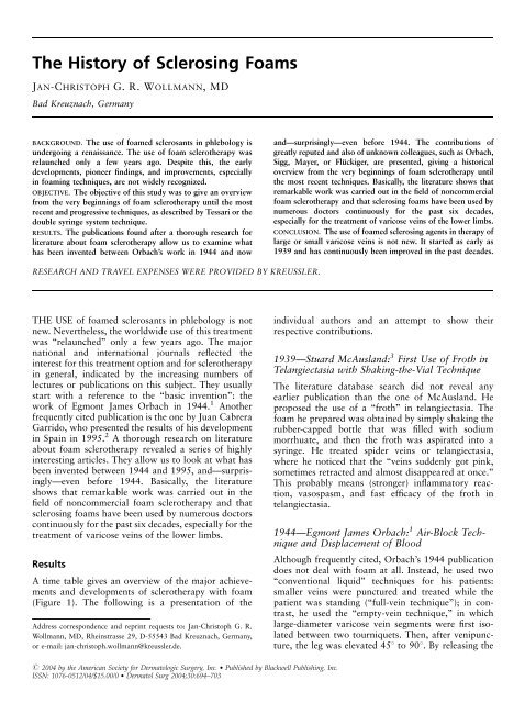
Results<br />
A time table gives an overview <strong>of</strong> the major achievements<br />
and developments <strong>of</strong> sclerotherapy with foam<br />
(Figure 1). <strong>The</strong> following is a presentation <strong>of</strong> the<br />
Address correspondence and reprint requests to: Jan-Christoph G. R.<br />
Wollmann, MD, Rheinstrasse 29, D-55543 Bad Kreuznach, Germany,<br />
or e-mail: jan-christoph.wollmann@kreussler.de.<br />
r 2004 by the American Society for Dermatologic Surgery, Inc. Published by Blackwell Publishing, Inc.<br />
ISSN: 1076-0512/04/$15.00/0 Dermatol Surg 2004;30:694–703<br />
and—surprisingly—even before 1944. <strong>The</strong> contributions <strong>of</strong><br />
greatly reputed and also <strong>of</strong> unknown colleagues, such as Orbach,<br />
Sigg, Mayer, or Flückiger, are presented, giving a historical<br />
overview from the very beginnings <strong>of</strong> foam sclerotherapy until<br />
the most recent techniques. Basically, the literature shows that<br />
remarkable work was carried out in the field <strong>of</strong> noncommercial<br />
foam sclerotherapy and that sclerosing foams have been used by<br />
numerous doctors continuously for the past six decades,<br />
especially for the treatment <strong>of</strong> varicose veins <strong>of</strong> the lower limbs.<br />
CONCLUSION. <strong>The</strong> use <strong>of</strong> foamed sclerosing agents in therapy <strong>of</strong><br />
large or small varicose veins is not new. It started as early as<br />
1939 and has continuously been improved in the past decades.<br />
individual authors and an attempt to show their<br />
respective contributions.<br />
1939—Stuard McAusland: 3 First Use <strong>of</strong> Froth in<br />
Telangiectasia with Shaking-the-Vial Technique<br />
<strong>The</strong> literature database search did not reveal any<br />
earlier publication than the one <strong>of</strong> McAusland. He<br />
proposed the use <strong>of</strong> a ‘‘froth’’ in telangiectasia. <strong>The</strong><br />
foam he prepared was obtained by simply shaking the<br />
rubber-capped bottle that was filled with sodium<br />
morrhuate, and then the froth was aspirated into a<br />
syringe. He treated spider veins or telangiectasia,<br />
where he noticed that the ‘‘veins suddenly got pink,<br />
sometimes retracted and almost disappeared at once.’’<br />
This probably means (stronger) inflammatory reaction,<br />
vasospasm, and fast efficacy <strong>of</strong> the froth in<br />
telangiectasia.<br />
1944—Egmont James Orbach: 1 Air-Block Technique<br />
and Displacement <strong>of</strong> Blood<br />
Although frequently cited, Orbach’s 1944 publication<br />
does not deal with foam at all. Instead, he used two<br />
‘‘conventional liquid’’ techniques for his patients:<br />
smaller veins were punctured and treated while the<br />
patient was standing (‘‘full-vein technique’’); in contrast,<br />
he used the ‘‘empty-vein technique,’’ in which<br />
large-diameter varicose vein segments were first isolated<br />
between two tourniquets. <strong>The</strong>n, after venipuncture,<br />
the leg was elevated 451 to 901. By releasing the
Dermatol Surg 30:5:May 2004 WOLLMANN: THE HISTORY OF SCLEROSING FOAMS 695<br />
Figure 1. Overview about the contributions to foam sclerotherapy.<br />
proximal tourniquet, blood could flow into the central<br />
or more proximal direction, while the distal tourniquet<br />
reduced or eliminated the supply <strong>of</strong> new blood from<br />
distally. This technique caused less dilution <strong>of</strong> the<br />
liquid sclerosing agent.<br />
Nevertheless, he sometimes noticed therapeutic<br />
failures, so he had the ‘‘famous idea’’: to intensify the<br />
contact between the sclerosant and the endothelium,<br />
the diameter <strong>of</strong> the vein should not only be reduced as<br />
far as possible prior to injection, but the vein should be<br />
rather free from any blood. <strong>The</strong>refore, he injected a<br />
small amount <strong>of</strong> air into the venous segment to be<br />
treated to completely displace the remaining blood (see<br />
Figure 2).<br />
Experimentally, he had shown that 1 cm 2 <strong>of</strong> air<br />
injected into a 6-mm infusion tube pending vertically<br />
and filled with water did not separate into several<br />
ascending bubbles but remained in the area <strong>of</strong><br />
injection as one large bubble, if the injection was not<br />
given too slowly. He then injected a colored solution<br />
into this air bubble, which, over a period <strong>of</strong> 3 to 5 s,<br />
did not get mixed with water but remained undiluted<br />
in the ‘‘air pocket.’’<br />
Clinically, he used the ‘‘air-block technique’’ only<br />
for smaller and medium-sized varicose veins; He<br />
recommended the conventional technique, without<br />
air injection, for larger veins. Unfortunately, Orbach’s<br />
article does not reveal the reasons for this recommendation.<br />
It remains unclear why the method that he<br />
considered to be more effective should not be suited<br />
for large varicose veins which usually were more<br />
difficult to treat. In 1970, Stemmer 4 et al. showed in an<br />
experiment that the air-block technique was only<br />
reliable with vessel diameters <strong>of</strong> up to 4 mm and was<br />
never successful with diameters <strong>of</strong> more than 8 mm.<br />
Sclerosis was rather impeded with such diameters: <strong>The</strong><br />
air bubble floating on the blood column protected the<br />
vessel from contact with the sclerosant at the upper<br />
Figure 2. <strong>The</strong> air-block technique: a small amount <strong>of</strong> air was injected<br />
before (top) the injection <strong>of</strong> sclerosant (bottom) to avoid dilution <strong>of</strong> the<br />
liquid.<br />
Figure 3. <strong>The</strong> air-block technique in large vessels: A floating air<br />
bubble (top) was able to prevent contact between the endothelium on<br />
the upper vessel wall and the sclerosant.<br />
circumference (see Figure 3), thus being effective only<br />
partially. <strong>The</strong> original air-block technique is basically<br />
no longer used today, but its development to a ‘‘foam<br />
block’’ (air block with large-bubbled foams) is still<br />
used today by some phlebologists under various names<br />
for smaller vessels. Small advantages in efficacy<br />
(increase by 20%) are believed to be outweighed by<br />
the disadvantages (plus 100% side effects). 5 <strong>The</strong><br />
maximum amount <strong>of</strong> air injected was 3 mL, a limit<br />
that became an orientation for most physicians using<br />
the air-block technique.<br />
1944—Robert Rowden Foote: 6 Foam in Feeder<br />
Veins with Shaking-the-Syringe Technique<br />
In the same year as Orbach’s publication, a book by<br />
Robert Rowden Foote was published in London. In<br />
addition to an overview <strong>of</strong> his ‘‘empty-vein technique,’’<br />
he wrote about the treatment <strong>of</strong> spider veins: ‘‘<strong>The</strong><br />
feeder vein should be dealt with first, whenever<br />
possible. <strong>The</strong> best injection fluid is the soapy froth<br />
obtained by shaking up 1 cc <strong>of</strong> ethamoline (ethanolamine<br />
oleate) in a 2-cc syringe. [It should be<br />
mentioned that the ratio <strong>of</strong> 1 plus 1 is, strictly<br />
speaking, not foam but an air/liquid dispersion.<br />
Usually a mixture is defined as foam if the gas portion<br />
is higher than 0.52. 7 A characteristic order <strong>of</strong><br />
magnitude <strong>of</strong> gas/fluid systems is the gas proportion<br />
j. Depending on the gas proportion, distinction is<br />
made between gas dispersion, spherical foam (wet<br />
foam), and polyhedral foam (dry foam). In gas<br />
dispersions, there are individual air bubbles in liquid;<br />
the gas proportion j is less than 0.52. In the spherical<br />
foam, the number <strong>of</strong> individual bubbles is higher—<br />
there is still a rather high amount <strong>of</strong> liquid; the gas
696 WOLLMANN: THE HISTORY OF SCLEROSING FOAMS Dermatol Surg 30:5:May 2004<br />
proportion is 0.524j40.74. In the polyhedral foam,<br />
in contrast, the amount <strong>of</strong> liquid between the bubbles<br />
is so small that the individual bubbles get closer and<br />
closer to each other, thus forming polyhedrons. <strong>The</strong><br />
gas proportion j is greater than 0.74. In a foaming<br />
fluid (e.g., beer), all types generally occur, the<br />
polyhedral foam being above the spherical foam and<br />
the gas dispersion and liquid below.] Once the needle<br />
is in the vein, the bubbles can be made to course along<br />
the minute veins and can be followed visually in their<br />
transit.’’ Nowadays, ethanolamine oleate is no longer<br />
available in most countries. <strong>The</strong> ‘‘agitation technique’’<br />
was refined in the following years and is no longer in<br />
use. <strong>The</strong> air:sclerosant ratio <strong>of</strong> 1 plus 1 described by<br />
Foote suggests that the dispersion was very fluid so<br />
that it could not be used for displacing the blood in<br />
larger diameter veins.<br />
1949—Karl Sigg: 8 Foam-Block Technique and<br />
Viscosity <strong>of</strong> Foam<br />
In 1949, Karl Sigg picked up the air-block technique<br />
described 5 years before: Also for other varices than<br />
spider veins, i.e., larger veins, he used the new<br />
technique and reported more than 4000 treatments<br />
performed ‘‘without problems.’’ Sigg describes the<br />
rationale for the use <strong>of</strong> the air-block technique in a<br />
similar manner as Orbach and Foote: ‘‘<strong>The</strong> purpose is<br />
to prevent the dilution <strong>of</strong> the solutions for injection in<br />
the vein by the blood and to ensure that the sclerosant<br />
gets in contact with the intima <strong>of</strong> the vein in a rather<br />
concentrated formy. Even with this small-scale use,<br />
the air bubble chased the blood before the injection<br />
fluid arrived and thus cleared the way for a closer<br />
contact <strong>of</strong> the sclerosant with the venous intima.’’<br />
Later, Sigg combined Orbach’s air-block technique<br />
and Foote’s foam application and thus introduced a<br />
new aspect into the therapeutic options known to that<br />
date: ‘‘Orbach’s method becomes even more beneficial<br />
if foam is injected instead <strong>of</strong> airy. This foam is less<br />
rapidly washed away in the varicose vein than it<br />
happens with the pure injection <strong>of</strong> air.’’ Thus Sigg<br />
introduced for the first time the idea (even if not<br />
pronounced) <strong>of</strong> increased viscosity <strong>of</strong> foam. With the<br />
use <strong>of</strong> foam, he improved the air-block technique<br />
(introducing a foam block), but without omitting the<br />
fluid sclerosant (see Figure 4). Sigg described his own<br />
procedure for the manufacture <strong>of</strong> foam; the glass<br />
syringe filled with fluid sclerosant was held with the<br />
opening pointing downward. Approximately 1 mL <strong>of</strong><br />
air was aspirated through it, pearls developed in the<br />
solution, and thus more or less large bubbles were<br />
generated in the syringe.<br />
Figure 4. <strong>The</strong> foam-block technique: First, foam was injected to<br />
maintain the displacing effect <strong>of</strong> the air block for a longer time (top),<br />
and then normal liquid sclerosant was injected (bottom).<br />
1950—Egmont James Orbach: 9,10 Vasospasm<br />
after Foam Sclerotherapy<br />
A later publication by Orbach reveals the first attempt<br />
to compare the efficacy <strong>of</strong> foam (administered as air<br />
block and foam) with the efficacy <strong>of</strong> fluid sclerosants.<br />
<strong>The</strong> end point was the length <strong>of</strong> the sclerothrombus<br />
generated by the injection <strong>of</strong> the respective substance.<br />
<strong>The</strong> efficacy <strong>of</strong> the foam generated by agitation <strong>of</strong> the<br />
syringe or drug vial was increased approximately 3.5to<br />
4-fold compared with the same amount <strong>of</strong> ‘‘conventional’’<br />
fluid. <strong>The</strong> observation <strong>of</strong> a ‘‘marked<br />
vasospasm’’ after injection <strong>of</strong> foam is nowadays<br />
considered to be an important immediate end point<br />
for the assessment <strong>of</strong> the efficacy <strong>of</strong> modern sclerotherapy.<br />
11–13 This (reversible) vasospasm, which is<br />
<strong>of</strong>ten clearly detectable in duplex-guided sclerotherapy,<br />
can be considered to be a sign <strong>of</strong> initial vascular<br />
damage after sclerotherapy. After administration <strong>of</strong> a<br />
viscous foam, vasospasm is more common and more<br />
pronounced than after sclerotherapy with conventional<br />
fluid. 42 An important factor in this respect is<br />
that a given volume <strong>of</strong> a blood-displacing foam can<br />
spread over a much longer venous segment after a<br />
vasospasm occurs and can even act at a certain<br />
distance from the site <strong>of</strong> administration. (For example,<br />
1 mL <strong>of</strong> a viscous foam completely fills a vein <strong>of</strong> 8 mm<br />
in diameter over a length <strong>of</strong> approximately 20 mm. A<br />
reduction <strong>of</strong> the vessel diameter owing to spasm to<br />
2 mm distributes the same foam volume to a length <strong>of</strong><br />
almost 32 cm. In vivo, a spasm <strong>of</strong> a mean length <strong>of</strong><br />
28 cm could be provoked with vessels <strong>of</strong> 4 to 8 mm in<br />
diameter and a foam application <strong>of</strong> 2 to 2.5 mL <strong>of</strong><br />
double-syringe-system foam.) As long as vasospasm<br />
exists, venipuncture at the same site is aggravated. <strong>The</strong><br />
use <strong>of</strong> small venous cannulae or catheters may help<br />
dealing with this.<br />
Because foams in current use are very different from<br />
those used by Orbach, it is impossible to draw<br />
conclusions about the general efficacy <strong>of</strong> foam from<br />
this experiment. Nevertheless, according to all findings<br />
and based on theoretical considerations, the efficacy <strong>of</strong><br />
foam is always higher than that <strong>of</strong> the same amount<br />
and concentration <strong>of</strong> fluid. <strong>The</strong> level <strong>of</strong> increased<br />
efficacy cannot be predicted in the individual case<br />
without sufficient standardization. <strong>The</strong> increased
Dermatol Surg 30:5:May 2004 WOLLMANN: THE HISTORY OF SCLEROSING FOAMS 697<br />
strength <strong>of</strong> foams needs to be respected if thinking<br />
about sclerosing smaller vessels with foam.<br />
1953—Arve Ree: 14 First Use <strong>of</strong> ‘‘Pure Foam’’<br />
<strong>The</strong> Norwegian doctor Ree introduced a new era in<br />
1953: He was the first to inject ‘‘pure’’ foam, not in<br />
addition to air and common fluid sclerosant but<br />
instead <strong>of</strong> the sclerosant and, in particular, without<br />
the previous injection <strong>of</strong> air. With his technique<br />
(agitation <strong>of</strong> a detergent solution in the vial and<br />
subsequent aspiration <strong>of</strong> the bubbles into the syringe)<br />
he first treated a series <strong>of</strong> 50 patients very successfully.<br />
He injected 2 to 7 mL <strong>of</strong> foam, depending on the vessel<br />
diameter, corresponding to an amount <strong>of</strong> air <strong>of</strong> up to<br />
6.6 mL. <strong>The</strong> bubble sizes seem to have been in the<br />
millimeter range with that technique. Owing to the<br />
higher kinetic energy (agitation <strong>of</strong> a vial and subsequent<br />
passage through a bottleneck with turbulent<br />
flow), it may be considered that a foam with smaller<br />
bubbles than with the previous techniques could be<br />
generated.<br />
1956—Peter Flückiger: 15 Retrograde Injection,<br />
Aspiration Technique, and Leg Elevation<br />
Flückiger recommended the technique known as<br />
‘‘retrograde sclerotherapy,’’ a technique characterized<br />
by leg elevation and sclerosant injection into saphenous<br />
veins from a proximal injection site in the distal<br />
direction. Thus, the sclerosant could reach all insufficient<br />
collaterals and the saphenous vein by one<br />
injection. He obtained better results by using sclerosing<br />
foam than with fluid sclerosants: ‘‘Basically, the<br />
procedure consists in injecting y Varsyl foam (ethanolamine<br />
oleate) into an appropriate vein. Due to the<br />
buoyancy, the foam has only a low tendency to move<br />
in central direction with the blood flowy. After<br />
removal <strong>of</strong> the needle, the location <strong>of</strong> the ‘‘foam seal’’<br />
can be determined by palpationy. <strong>The</strong> foam usually<br />
spreads in the surrounding <strong>of</strong> the injection site in distal<br />
and proximal direction. After injection, the intravascular<br />
foam depot y is displaced by manual massage<br />
against the periphery.’’ Flückiger additionally mentioned<br />
major aspects <strong>of</strong> modern foam sclerotherapy in<br />
his publication: He saw that the foam he used<br />
remained in the region <strong>of</strong> the injection site as a<br />
‘‘depot,’’ noticed that it was able to displace the blood<br />
in the proximal direction and was not washed away<br />
from the blood, and noticed that the foam could be<br />
directed to other regions by manual manipulation.<br />
Moreover, he postulated and discussed further treatment-relevant<br />
foam properties for the first time:<br />
Increased efficacy by increase <strong>of</strong> the effective surface<br />
<strong>of</strong> foam compared to fluid sclerosants;<br />
A stronger sclerosing effect with smaller amounts <strong>of</strong><br />
sclerosants;<br />
Postulation <strong>of</strong> a minimized bubble size; and<br />
Postulation <strong>of</strong> homogeneity <strong>of</strong> foam.<br />
It is obvious that a given amount <strong>of</strong> sclerosant in the<br />
form <strong>of</strong> foam has a much larger surface than it has in<br />
an unchanged, fluid state: the smaller the bubbles <strong>of</strong><br />
the foam are, the greater the surface. Since damage to<br />
the intima y is based on the contact <strong>of</strong> the intima<br />
with the sclerosant, the use <strong>of</strong> foam allows an<br />
extended sclerosation with a small amount <strong>of</strong> sclerosant.<br />
<strong>The</strong> preparation <strong>of</strong> a homogenous, fine-bubbled<br />
foam is an important condition for the success <strong>of</strong> the<br />
procedure.<br />
(<strong>The</strong> homogeneity <strong>of</strong> foam, i.e., a foam whose<br />
bubbles are as homogenous as possible, is an<br />
important prerequisite for its flow behavior (viscosity)<br />
and its stability. Ideally, a foam has only bubbles <strong>of</strong> the<br />
same diameter, which is, in reality, barely or never<br />
feasible. Foam is subjected to aging, which involves<br />
mainly two aspects: In contrast, the fluid that is<br />
between the gas bubbles in a young foam drains owing<br />
to the force <strong>of</strong> gravity, so that the upper bubbles slowly<br />
break down because <strong>of</strong> the ‘‘fluid deficiency.’’ In<br />
contrast, bubbles disappear because a gas diffusion<br />
takes place between the foam bubbles. <strong>The</strong> internal gas<br />
pressure is higher in small bubbles owing to the higher<br />
surface tension, so that gas moves from those bubbles<br />
into larger bubbles. Smaller bubbles <strong>of</strong> a foam get even<br />
smaller and large ones even larger—until they break<br />
down. In a homogenous foam, all bubbles would have<br />
the same inner gas pressure. A gas diffusion from one<br />
bubble to the adjacent bubble would not take place<br />
and the foam would remain stable until just the drain<br />
process would contribute to aging.)<br />
Flückiger considered the various known foam<br />
manufacturing procedures, and suggested his own<br />
new technique: ‘‘In order to obtain a homogenous finebubbled<br />
foam, the procedure consisting in the agitation<br />
<strong>of</strong> a syringe filled with air and sclerosant<br />
[Orbach’s technique 1950] is completely inappropriate.<br />
<strong>The</strong> drawing <strong>of</strong> air through a sclerosant in the<br />
syringe [Sigg’s technique 1949] does not yield a small<br />
fine-bubbled foam either. Moreover, none <strong>of</strong> these<br />
methods is able to convert the entire amount <strong>of</strong><br />
sclerosant into foam. <strong>The</strong>re is always a certain amount<br />
<strong>of</strong> remaining fluid, which cannot be used for retrograde<br />
sclerotherapy with the elevated leg. A perfect,<br />
fine-bubbled foam is obtained by the simultaneous<br />
aspiration <strong>of</strong> sclerosant y and air through a thin<br />
injection needley. <strong>The</strong> thin injection needle (the<br />
narrower the lumen, the finer the foam) y is
698 WOLLMANN: THE HISTORY OF SCLEROSING FOAMS Dermatol Surg 30:5:May 2004<br />
Figure 5. <strong>The</strong> aspiration technique: <strong>The</strong> tip <strong>of</strong> the needle was placed<br />
at the liquid–air interface, thus allowing coaspiration <strong>of</strong> air and<br />
sclerosant that would generate foam in one run.<br />
introduced into the ampoule in such a way that<br />
approximately two thirds <strong>of</strong> the opening <strong>of</strong> the bevel<br />
are imbibed in the fluid (see Figure 5). When drawing<br />
back the plunger, the syringe fills with Varsyl foam<br />
under a sizzling noise, the foam remaining stable for<br />
several minutes.’’<br />
1957: Heinz Mayer and Hans Brücke: 16 First<br />
Micr<strong>of</strong>oam Device (Double-Piston Syringe)<br />
A milestone regarding the improvement and standardization<br />
<strong>of</strong> sclerosing foam was the publication by two<br />
surgeons Mayer and Brücke: they described the first<br />
device that had been developed specifically for the<br />
preparation <strong>of</strong> viscous sclerosing foams, a syringe with<br />
a double plunger (see Figure 6). (A double-plunger<br />
syringe following the principle <strong>of</strong> Mayer-Brücke was<br />
‘‘reinvented’’ several times in the following decades. A<br />
very homogenous, extremely fine microbubble foam<br />
can be produced using a simple reproduction <strong>of</strong> that<br />
syringe—unfortunately, the original is no longer<br />
available. More than 40 years ago, Mayer and Brücke<br />
worked with foams that are now suggested to be<br />
classified as ‘‘micr<strong>of</strong>oam.’’ 17 )<br />
‘‘Before the main plunger, there is a second, thin<br />
plunger with numerous holes, whose piston leads to a<br />
central bore <strong>of</strong> the main plunger and protrudes it.<br />
When the main plunger is fixed, the plunger provided<br />
with holes can rapidly be moved forward and backward<br />
and the Phlebocid (ethanolamin oleate) can be<br />
mixed with the air contained in the syringe. A finebubbled,<br />
viscous foam developsy. We have tested<br />
Figure 6. <strong>The</strong> Mayer-Brücke device: <strong>The</strong> inner plunger with numerous<br />
tiny holes can rapidly be moved forward and backward to mix<br />
sclerosant and air contained in the syringe. <strong>The</strong> outer plunger was used<br />
for injection <strong>of</strong> the viscous and fine-bubbled foam.<br />
most sclerosants <strong>of</strong> similar composition and selected<br />
Phlebocid, which provides the stiffest foam upon<br />
agitation with a homogenous distribution <strong>of</strong> air<br />
bubblesy. We consider the greatest advantage <strong>of</strong> the<br />
use <strong>of</strong> foam y to be the homogenous distribution <strong>of</strong><br />
the sclerosanty.’’ Clinically, they stated after administration<br />
<strong>of</strong> foam: ‘‘<strong>The</strong> fibrous tissue <strong>of</strong> the venous<br />
lumen is so complete that in general, recanalizationydoes<br />
not occur. We never observed recurrent<br />
varicose veins after using Phlebocid foam. We never<br />
observed complications after foam filling such as air<br />
embolisms or skin necroses.’’<br />
1962—Peter Flückiger: 18 Turbulent Flow<br />
In 1962, Flückiger described another technique for the<br />
preparation <strong>of</strong> foam: pumping <strong>of</strong> air and sclerosant<br />
forward and backward between a drug vial and the<br />
attached syringe. Later, this technique was modernized<br />
and improved by Alessandro Frullini 19,20 by adding an<br />
adapter between the syringe and the bottle seal.<br />
Furthermore, Flückiger changed some points since<br />
his first publication in 1956: he maintained leg<br />
elevation not only during sclerotherapy but also for<br />
some minutes thereafter, to allow the foam to degrade.<br />
He warned therapists to be cautious while ‘‘stroking’’<br />
the foam seal in a peripheral direction to prevent any<br />
unintended movement <strong>of</strong> foam into the deep venous<br />
system. Today, it is recommended that you wait some<br />
minutes after foam sclerotherapy <strong>of</strong> large veins before<br />
applying compression. 42<br />
1963—Peter Lunkenheimer: 21 First Use <strong>of</strong><br />
Polidocanol Foam<br />
Even before polidocanol received its first marketing<br />
authorization, a German physician had the chance to<br />
use the novel sclerosing solution for research purposes<br />
for the first time. Kreussler’s archives revealed a letter<br />
from that phlebologist, which contains a written ‘‘case<br />
report’’ about the first patient treatment: ‘‘<strong>The</strong> first<br />
patient was treated with 2 mL Aethoxysklerol s<br />
foamy.’’
Dermatol Surg 30:5:May 2004 WOLLMANN: THE HISTORY OF SCLEROSING FOAMS 699<br />
1969—Walter Gillesberger: 22 Low-Pressure<br />
Technique<br />
Gillesberger’s technique was based on the generation<br />
<strong>of</strong> a negative pressure in a (glass) syringe, so that air<br />
could enter through the capillary gap between the<br />
syringe piston and the plunger and thus generate<br />
bubbles (see Figure 7). Gillesberger’s syringe was not<br />
closed hermetically like the Monfreux syringe tip later<br />
on, but the negative pressure was strong enough to<br />
create a foam and was not compensated by the inflow<br />
<strong>of</strong> the sclerosant from the vial. <strong>The</strong> technique by<br />
Gillesberger or its refinement by Monfreux, who<br />
generated foam with an ‘‘absolute’’ negative pressure,<br />
is very simple. What is problematic is that there is no<br />
standardized ratio between air and sclerosant. <strong>The</strong>refore,<br />
the foams produced in that manner are different,<br />
depending on the syringe type, needle, generated<br />
suction, and the resulting gas content; they may be<br />
more fluid one time and more viscous another time.<br />
This leads to different behavior in the vessel (degree<br />
and duration <strong>of</strong> the displacement <strong>of</strong> blood) and thus<br />
probably to an unpredictable efficacy in vivo. (<strong>The</strong><br />
ability <strong>of</strong> foam to displace blood depends on its quality<br />
or characteristics: foams obtained with low sclerosant<br />
concentrations, with small amounts <strong>of</strong> air, and/or with<br />
rather large bubbles have a more or less fluid behavior;<br />
i.e., they are more easily washed away by liquids<br />
(blood) and have no or just a small and short-term<br />
displacing effect. Viscous foams (higher concentration,<br />
higher gas content, and small bubbles), in contrast,<br />
basically have a displacing effect in larger vessels. 41 If<br />
sclerosing foam is to be used for small diameter veins,<br />
precaution is recommended owing to the enhancement<br />
<strong>of</strong> the efficacy. A viscous foam would probably have<br />
too a strong effect in small veins; moreover, the foam<br />
breaks down as a result <strong>of</strong> the passage through the<br />
required fine needles. 42<br />
Figure 7. <strong>The</strong> low-pressure technique: Pulling down the piston<br />
allowed air to enter the syringe through the tiny gap between the<br />
syringe piston and the plunger.<br />
1984—Gerald Hauer: 23 Twin-Syringe Technique<br />
Hauer patented a twin-syringe set for foam preparation:<br />
Two parallel syringes, one filled with air, the<br />
other one filled with sclerosing solution, are simultaneously<br />
emptied into a ‘‘mixing chamber’’ by pressure,<br />
which leads to the formation <strong>of</strong> bubbles. <strong>The</strong> ratio <strong>of</strong> 1<br />
plus 1 (sclerosing agent and air) suggests a dispersion<br />
that should have the effect <strong>of</strong> a foam block.<br />
1986—Michael Grigg: 24 Double Syringes and<br />
Connecting Tubes<br />
In 1986, Grigg demonstrated a new foam production<br />
procedure: the principle was the generation <strong>of</strong> a<br />
turbulent flow between two syringes, connected via a<br />
plastic infusion tube, so that fluid and air could be<br />
pumped forward and backward. Belcaro later improved<br />
this technique by the addition <strong>of</strong> a further<br />
strongly foaming detergent solution in small amounts<br />
(0.1–0.2 mL). (‘‘Improved’’ in this context means<br />
prolongation <strong>of</strong> half-life. Owing to the commonly<br />
relevant amount <strong>of</strong> silicone in disposables (syringes<br />
and infusion tubes, etc.), the half-life <strong>of</strong> the foam was<br />
evidently limited because silicone, which destroys the<br />
surfactant arrangement <strong>of</strong> the foam lamellae, is a very<br />
strong foam breaker. <strong>The</strong>refore, anything that contributes<br />
to the maintenance <strong>of</strong> foam bubbles supports<br />
the foam stability: this is, in addition to an increase <strong>of</strong><br />
the surfactant concentration, in particular the minimization<br />
<strong>of</strong> the amount <strong>of</strong> silicone.) According to him,<br />
the foam half-life was approximately 4 min. This socalled<br />
‘‘Irvine technique’’ (named after the laboratory<br />
in which the technique was demonstrated) can be<br />
considered to be a precursor <strong>of</strong> Tessari’s technique and<br />
the double-syringe system (DSS). Belcaro and coworkers<br />
24 first performed investigations on the safety <strong>of</strong><br />
the foam generated according to that technique: 5 to<br />
10 mL <strong>of</strong> the foam produced with that technique did<br />
not cause any change <strong>of</strong> the pulmonary ventilation<br />
scintigraphy or perfusion scintigraphy in 12 patients.<br />
1995—Juan Cabrera Garrido: 2 Rotating Brush<br />
Technique<br />
In 1995, Cabrera Garrido published data from the<br />
clinical use <strong>of</strong> ‘‘micr<strong>of</strong>oam.’’ He had treated venous<br />
malformations and saphenous veins including collaterals<br />
with high volumes <strong>of</strong> foam. His objective was to<br />
fill the complete venous lumen by injecting high doses<br />
<strong>of</strong> foam at one time. A new aspect <strong>of</strong> the production<br />
was the use <strong>of</strong> a high-speed rotating brush (a modified<br />
dental bur), so that foam was agitated—like cream<br />
with a food mixer—and the facultative addition <strong>of</strong><br />
CO2 as a carrier gas. In experiments, CO2—if
700 WOLLMANN: THE HISTORY OF SCLEROSING FOAMS Dermatol Surg 30:5:May 2004<br />
Figure 8. Foam prepared with 3% polidocanol solution according to<br />
the double-syringe system (DSS). Sterile air or CO2 was used as gas.<br />
<strong>The</strong> degradation <strong>of</strong> foam is faster if CO2 is used instead <strong>of</strong> air and even<br />
more pronounced if the ratio is changed from 1:5 to 1:10.<br />
employed as the only gas—led to foams with quickly<br />
degrading bubbles (see Figure 8). Regarding the highdose<br />
treatment, more recent publications by Cabrera<br />
Garridos mentioned that the foam could flow from the<br />
greater saphenous vein into the deep venous system via<br />
the saphen<strong>of</strong>emoral junction or other connections,<br />
where it may provoke thromboses. 25 <strong>The</strong> number <strong>of</strong><br />
deep venous thromboses in a first study on this foam<br />
(6% thromboses when administering 20 mL <strong>of</strong><br />
Varisolve foam and more) 26 does not lend support to<br />
the high-dose foam therapy and requires special safety<br />
measures.<br />
1997—Alain Monfreux: 27 Low-Pressure<br />
Technique and ‘‘Méthode MUS’’<br />
Monfreux’s technique, published in 1997 under the<br />
name <strong>of</strong> ‘‘Méthode MUS,’’ was picked up primarily by<br />
French phlebologists. <strong>The</strong> principle was based on<br />
Gillesberger’s technique: generation <strong>of</strong> a negative<br />
pressure in the glass syringe filled with sclerosing<br />
solution, thus inlet <strong>of</strong> air through the fine gap between<br />
syringe barrel and plunger with corresponding pearling<br />
<strong>of</strong> the solution and transformation into a foam.<br />
Unlike Gillesberger, Monfreux generated an ‘‘absolute’’<br />
negative pressure by placing a cap on the syringe.<br />
Although all concentrations <strong>of</strong> sclerosants can be used<br />
with this method, the efficacy cannot be predicted in<br />
each individual case: a defined ratio <strong>of</strong> gas and<br />
sclerosant or a defined bubble size cannot be determined,<br />
because many variables (e.g., the width <strong>of</strong> the<br />
capillary gap between the syringe barrel and plunger,<br />
force, and duration <strong>of</strong> traction at the plunger and thus<br />
the order <strong>of</strong> magnitude <strong>of</strong> the effective shearing force<br />
which is responsible for the pearling <strong>of</strong> the solution)<br />
depend on the brand and on the user.<br />
1998—Symon Sadoun/Jean-Patric Benigni: 5,28<br />
Advanced Low-Pressure Technique<br />
Both authors improved Monfreux’s technique by<br />
making it usable for plastic syringes: <strong>The</strong> principle—<br />
generation <strong>of</strong> a negative pressure in a syringe closed by<br />
a Luer stopper—remained similar: pulling down the<br />
piston to generate subatmospheric pressure was<br />
followed by quickly releasing the piston several times.<br />
As with Monfreux and his predecessors, a defined<br />
gas:sclerosant ratio could not be stated.<br />
1998—Miguel Santos Gaston: 29 Advanced<br />
Low-Pressure Technique<br />
Gaston basically adopted the Monfreux technique, but<br />
added some more steps to the preparation procedure:<br />
once the foam was prepared according to Monfreux,<br />
he emptied the foam into a glass container and then<br />
aspirated the foam again. This was repeated several<br />
times, making—on the one hand—the foam more fine,<br />
but—on the other hand—more dry.<br />
1999—Javier Garcia Mingo: 30,31 High-Pressure<br />
Technique and ‘‘Foam Medical System’’<br />
Mingo was the first to describe a sterilizable, reusable<br />
device for the preparation <strong>of</strong> foam, which generates<br />
foam by the introduction <strong>of</strong> various gases from a<br />
pressure-gas cylinder and subsequent passage <strong>of</strong> the<br />
mixture through a fine nozzle, the ‘‘foam medical<br />
system.’’ <strong>The</strong> handling and cleaning <strong>of</strong> the device,<br />
which appears a little complicated, has prevented its<br />
wide use so far, although Mingo’s clinical results are<br />
considered very promising.<br />
2000—Lorenzo Tessari: 32,33 Double Syringes,<br />
Three-Way Tap, and the ‘‘Tourbillon Technique’’<br />
Tessari’s Tourbillon technique is at present, together<br />
with the similar DSS technique, the most commonly<br />
used technique. It corresponds basically to Irvine’s<br />
technique 24 but has clear advantages over the latter.<br />
Tessari uses two syringes as well (various sizes being<br />
described), but Tessari’s technique does not require<br />
‘‘foaming aids’’: <strong>The</strong> two syringes are connected by a<br />
three-way stopcock, and the sclerosing solution and air<br />
are drawn back and forth by pump movements. Owing<br />
to the absence <strong>of</strong> a connecting tube, a lot <strong>of</strong> disturbing<br />
silicone is no longer present. A more detailed<br />
description <strong>of</strong> the pumping procedure (at least 10<br />
times) to improve the standardization <strong>of</strong> the foam was<br />
given in 2001. 34<br />
<strong>The</strong> three-way stopcock has an additional ‘‘technical<br />
finesse’’: it is possible to vary the size <strong>of</strong> the passage
Dermatol Surg 30:5:May 2004 WOLLMANN: THE HISTORY OF SCLEROSING FOAMS 701<br />
by turning the cock—a narrow passage generates high<br />
turbulences and generates smaller blubbles than a wide<br />
passage. This procedures uses 2 to 2.5 mL <strong>of</strong> air and<br />
0.5 mL <strong>of</strong> sclerosing solution (all concentrations,<br />
mainly 1 and 3% for large and very large vessels).<br />
Irrespective <strong>of</strong> the concentration, the gas proportion j<br />
is approximately 0.7 to 0.83; the gas bubbles are very<br />
fine, and the half-life depends on the concentration and<br />
on the syringe. <strong>The</strong> foam prepared according to the<br />
Tessari method has successfully been tested in clinical<br />
trials. 35,36<br />
2000—Alessandro Frullini: 17–19 Advanced<br />
Turbulent Flow<br />
In 2000, Frullini improved the technique developed by<br />
Flückinger in 1962 and made it usable for disposable<br />
syringes by adding an adapter 18 and in 2001 by<br />
optionally using sterile air. 17,19<br />
2001—Gilles Gachet: 37 Aspiration Technique<br />
<strong>The</strong> aspiration technique published by Gachet in 2001<br />
is, regarding foam preparation, very similar with the<br />
description <strong>of</strong> Flückiger’s method from 1956.<br />
2001—Double-Syringe System (DSS), 11,12,38–40<br />
Two-Way Connector, and Pressure<br />
When searching for a suitable technique for the<br />
preparation <strong>of</strong> foam, it was noticed that the methods<br />
available to date all had at least one disadvantage that<br />
aggravated experiments with sclerosing foam or even<br />
made it impossible from the scientific point <strong>of</strong> view:<br />
the foams were not reproducible, because important<br />
foam characteristics (e.g., the gas proportion, the<br />
sclerosant concentration, the bubble size, or others)<br />
were stated incompletely or not at all, because the<br />
required materials were not described in detail, or<br />
because the very preparation was not defined clearly.<br />
<strong>The</strong> foams prepared for laboratory experiments and<br />
preclinical experiments all had a different consistence<br />
and an inhomogeneous flow behavior, if minor<br />
changes were made. Various types, manufacturers,<br />
and sizes <strong>of</strong> syringes; changes to the needle diameters<br />
and lengths; different temperatures during preparation;<br />
and other factors led to completely diverging<br />
results. <strong>The</strong>refore, a preparation variant was searched<br />
for that was simple, fast, sterile and especially<br />
reproducible. In a laboratory experimental series, the<br />
various variables that may affect the stability <strong>of</strong> the<br />
foam were changed systematically and the best<br />
possible combination <strong>of</strong> syringe type, gas:fluid ratio,<br />
sclerosant concentration, and manufacturing instructions<br />
was looked for.<br />
<strong>The</strong> most stable and fine-bubbled foam was obtained<br />
according to the following instructions: the required<br />
materials were a 10-mL Omnifix syringe, a 10-mL<br />
Inject syringe (each with a Luer-Lock connection), one<br />
Combidyn adapter (to connect the syringes), and a 0.2mm<br />
filter (for sterilization <strong>of</strong> air) (see Figure 9).<br />
Exactly 8 mL air is drawn into the Inject syringe via<br />
the sterile filter; afterwards, after removal <strong>of</strong> the filter,<br />
exactly one ampoule (2 mL) <strong>of</strong> polidocanol 3%<br />
(Aethoxysklerol). <strong>The</strong> two syringes are connected to<br />
the adapter. First some pumping movements (5 )are<br />
performed against resistance (by thumb pressure onto<br />
the other syringe piston) until the two components are<br />
well mixed. Afterward, the foam is pumped again<br />
quickly forth and back seven times between the two<br />
syringes without resistance like in the Tessari technique,<br />
until a homogenous foam has formed (see Figure 10).<br />
<strong>The</strong> double-syringe-system foam has a fixed sclerosant:air<br />
ratio <strong>of</strong> 1:5 ( 5 1 plus 4), a half-life <strong>of</strong><br />
approximately 150 s, with an initial mean bubble size<br />
<strong>of</strong> 70 mm. Divergent syringes, concentrations, scler-<br />
Figure 9. Components used in the double-syringe technique.<br />
Figure 10. Double-syringe-system (DSS) foam.
702 WOLLMANN: THE HISTORY OF SCLEROSING FOAMS Dermatol Surg 30:5:May 2004<br />
osant:air ratios, or pump procedures make the foam<br />
less stable and less viscous.<br />
<strong>The</strong> first prospective, randomized multicenter study<br />
compared foam sclerotherapy with double-syringe<br />
system with conventional fluid sclerotherapy in 88<br />
patients with greater saphenous vein insufficiency. A<br />
single injection (the protocol did not include further<br />
injections) <strong>of</strong> 2 to 2.5 mL <strong>of</strong> double-syringe-system<br />
foam or 3% liquid polidocanol showed a 2-year<br />
occlusion rate <strong>of</strong> 84% in the foam group versus 40%<br />
in the fluid group. Vasospasm was clearly more<br />
frequent and more pronounced in the group treated<br />
with foam (mean length 28 cm) than in the control<br />
group, the rate <strong>of</strong> side effects being identical. 11,12<br />
Conclusion and Discussion<br />
<strong>The</strong> use <strong>of</strong> foamed sclerosing agents in therapy <strong>of</strong> large<br />
or small varicose veins is not new. It started as early as<br />
1939. <strong>The</strong> publications about foam sclerotherapy<br />
found and presented in this text allow us to look at<br />
what has been invented between the very beginnings<br />
and now. <strong>The</strong> contributions <strong>of</strong> widely known and also<br />
<strong>of</strong> less known colleagues show that remarkable work<br />
was performed in the field <strong>of</strong> noncommercial foam<br />
sclerotherapy continuously for the past six decades,<br />
especially for the treatment <strong>of</strong> varicose veins <strong>of</strong> the<br />
lower limbs. But they also show that there sometimes<br />
is a lack <strong>of</strong> accuracy in describing the particular foams<br />
used, which does not make it easy to reproduce the<br />
individual reported results and/or findings. <strong>The</strong> suggestion<br />
therefore is to describe any foam used in<br />
nonclinical or clinical trials according to the ‘‘definition<br />
<strong>of</strong> sclerosing foams’’: <strong>Sclerosing</strong> foam is characterized<br />
by (at least) the following variables: type and<br />
concentration <strong>of</strong> the tensioactive sclerosing agent, type<br />
<strong>of</strong> gas, ratio <strong>of</strong> liquid to gas, the method <strong>of</strong><br />
preparation, the time between processing and use,<br />
and bubble sizes. This could really help to make<br />
sclerosing foams, and clinical results, comparable.<br />
References<br />
1. Orbach EJ. Sclerotherapy <strong>of</strong> varicose veins—utilization <strong>of</strong> an<br />
intravenous air block. Am J Surg 1944;LXVI(3):362–6.<br />
2. Cabrera J, Cabrera Garcia-Olmedo JR. Nuevo método de esclerosis<br />
en las varices tronculares. Patol Vasc 1995;4:55–73.<br />
3. McAusland S. <strong>The</strong> modern treatment <strong>of</strong> varicose veins. Med Press<br />
Circular 1939;201:404–10.<br />
4. Stemmer R, Kopp C, Voglet P. Physikalische Studie der Sklerosierungsinjektion.<br />
Zentralbl Phlebol 1970;9:112–23.<br />
5. Benigni JP, Sadoun S, Thirion V, et al. Télangiectasies et varices<br />
réticulaires—traitement par la mousse d’Aetoxisclérol à 0.25%:<br />
présentation d’une étude pilote. Phlébologie 1999;52:283–9.<br />
6. Foote RR. <strong>The</strong> injection treatment. In: Foote RR, ed. Varicose<br />
Veins, Haemorrhoids and Other Conditions. London: Lewis, 1944:<br />
13–44.<br />
7. Pahl MH, Meinecke H. Schaumzerstörung mit arteigener Flüssigkeit.<br />
In: Dechema-Monographien Band 114. Weinheim: VCH-<br />
Verlagsgesellschaft, 1989.<br />
8. Sigg K. Neuere gesichtspunkte zur Technik der Varizenbehandlung.<br />
<strong>The</strong>r Umsch 1949;6:127–34.<br />
9. Orbach EJ, Petretti AK. <strong>The</strong> thrombogenic property <strong>of</strong> foam <strong>of</strong> a<br />
synthetic anionic detergent. Angiology 1950;1:237–43.<br />
10. Orbach EJ. Contributions to the therapy <strong>of</strong> the varicose complex. J<br />
Int Coll Surg 1950;6:765–71.<br />
11. Hamel-Desnos C, Desnos P, Wollmann JC, et al. Evaluation <strong>of</strong> the<br />
efficacy <strong>of</strong> polidocanol in form <strong>of</strong> foam compared to liquid form in<br />
sclerotherapy <strong>of</strong> the greater saphenous vein: intial results. Dermatol<br />
Surg 2003;29:1170–5.<br />
12. Hamel-Desnos C, Ouvry P, Desnos P, Mako S. Evaluation <strong>of</strong> the<br />
Efficacy <strong>of</strong> Polidocanol in the Form <strong>of</strong> Foam versus Liquid Form in<br />
Sclerotherapy <strong>of</strong> the Long Saphenous Vein. American College <strong>of</strong><br />
Phlebology 16th Annual Congress; 2002 Nov 8–10; Ft. Lauderdale,<br />
FL. Oakland, CA: ACP, 2003.<br />
13. Schadeck M, Allaert FA. Duplex scanning in the mechanism <strong>of</strong><br />
the sclerotherapy: importance <strong>of</strong> the spasm. Phlebol Suppl 1995;1:<br />
574–6.<br />
14. Ree A. Etamolin foam in the treatment <strong>of</strong> varicose veins. A new<br />
method. Acta Dermatovenerol 1953;33:435–6.<br />
15. Flückiger P. Nicht-operative retrograde Varicenverödung mit<br />
Varsylschaum. Schweiz Med Wochenschr 1956;48:1368–70.<br />
16. Mayer H, Brücke H. Angiologie—Zur Ätiologie und Behandlung<br />
der Varizen der unteren Extremitäten. Chir Prax 1957;4:521–8.<br />
17. Frullini A. <strong>Sclerosing</strong> foam in the treatment <strong>of</strong> recurrent varicose<br />
veins. In: Henriet JP, ed. Foam Sclerotherapy State <strong>of</strong> the Art. Paris:<br />
Editions Phlebologiques Françaises, 2001:73–8.<br />
18. Flückiger P. Beitrag Zur Technik der ambulanten Varizenbehandlung.<br />
Die Med Welt 1963;12:617–21.<br />
19. Frullini A. New technique in producing sclerosing foam in a<br />
disposable Syringe. Dermatol Surg 2000;26:705–6.<br />
20. Frullini A. <strong>The</strong> <strong>Sclerosing</strong> Foam in the Treatment <strong>of</strong> Teleangectasia<br />
and Varicose Veins: New Techniques for Production <strong>of</strong> a Foam with<br />
Sterile Air. American College <strong>of</strong> Phlebology 14th Annual Congress;<br />
2000 Nov 16–19; Atlanta, GA. Oakland, CA: ACP, 2001.<br />
21. Lunkenheimer E. Personal letter to Kreussler. Mainz (Germany):<br />
Institut für Beinleiden, 1967.<br />
22. Gillesberger W. Die Ausrüstung des phlebologisch tätigen Dermatologen.<br />
Z Hautkrank 1969;44:669–74.<br />
23. Hauer Gerald, inventor; German Patent DE 34 17 182 C2.<br />
Zwillings-Spritzen-Set; May 9, 1984.<br />
24. Belcaro G, Geroulakos C, Cesarone MR, et al. Comparison among<br />
treatment schemes for varicose veins: surgery, sclerotherapy, foamsclerotherapy<br />
and combined options—a 10-year, prospective,<br />
randomised, follow-up study the VEDICO trial and EST. (European<br />
Sclerotherapy Trial). In: Belcaro G, Geroulakos G, Cesarone MR,<br />
Nicolaides AN, eds. Sclerotherapy in Venous Disease. Torino, Italy:<br />
Edizioni Minerva Medica, 2002:96–104.<br />
25. Cabrera J. Application techniques for sclerosant in micr<strong>of</strong>oam<br />
form. In: Henriet JP, ed. Foam Sclerotherapy State <strong>of</strong> the Art. Paris:<br />
Editions Phlebologiques Françaises, 2001:39–44.<br />
26. Bhowmick A, Harper D, Wright D, McCollum CN. Polidocanol<br />
micr<strong>of</strong>oam sclerotherapy for long saphenous varicose veins.<br />
Phlebology 2001;16:41–50.<br />
27. Monfreux A. Traitement sclérosant des troncs saphéniens et leurs<br />
collatérales de gros calibre par la méthode mus. Phlébologie 1997;<br />
50:351–3.<br />
28. Sadoun S, Benigni JP. <strong>The</strong> Treatment <strong>of</strong> Varicosities and Telangiectases<br />
with TDS or Lauromacrogol Foam: Video Tape. UIP World<br />
Congress <strong>of</strong> Phlebology; 1998; Sydney.<br />
29. Santos Gaston M.Escleroterapia por ‘‘Mousse’’ (espuma). In:<br />
Santos Gaston M, Santos-Gaston Orus M, eds. Esclerotherapia de<br />
Varices. Madrid: Vegalon, 1999:73–7.<br />
30. Garcia-Mingo J. ‘Foam medical system’: a new technique to treat<br />
varicose veins with foam. In: Henriet JP, ed. Foam Sclerotherapy<br />
State <strong>of</strong> the Art. Paris: Editions Phlebologiques Francaises,<br />
2001:45–50.<br />
31. Garcia-Mingo J. Esclerosis Venosa con Espuma: Foam Medical<br />
System [Internet]. S. Benedetto del Tronto: Attilio Cavezzi, c2004<br />
[updated 2004 Feb 3]. Available from: http://www.cavezzi.it/.
Dermatol Surg 30:5:May 2004 WOLLMANN: THE HISTORY OF SCLEROSING FOAMS 703<br />
32. Tessari L. Extemporary sclerosing foam according to personal<br />
method: experimental clinical data and catheter usage. Int Angiol<br />
Suppl 2001;1:54.<br />
33. Tessari L. Nouvelle technique d’obtention de la scléro-mousse.<br />
Phlébologie 2000;53:129.<br />
34. Tessari L, Cavezzi A, Frullini A. Preliminary experience with a new<br />
sclerosing foam in the treatment <strong>of</strong> varicose veins. Dermatol Surg<br />
2001;27:58–60.<br />
35. Cavezzi A, Frullini A, Ricci S, Tessari L. Treatment <strong>of</strong> varicose<br />
veins by foam sclerotherapy: two clinical series. Phlebology 2002;<br />
17:13–8.<br />
36. Frullini A, Cavezzi A. Echosclérose par mousse de tétradécyl-sulfate<br />
de sodium et de polidocanol: deux années d’expérience. Phlébologie<br />
2000;53:431–5.<br />
37. Gachet G. Une nouvelle méthode simple et économique pour<br />
confectionner de la mousse pour sclérose échoguidée. Phlébologie<br />
2001;54:63–5.<br />
Commentary<br />
Many authors have claimed to have developed a technique for<br />
foaming detergent solutions to increase the efficacy <strong>of</strong><br />
sclerotherapy treatment <strong>of</strong> varicose veins. In this article, Dr.<br />
Wollmann has researched the literature to provide us with an<br />
accurate history <strong>of</strong> this therapeutic advance. As with ambulatory<br />
phlebectomy and many other surgical techniques, foaming<br />
sclerosing solutions is not new. This technique was first<br />
described in 1939 and has been reported using many different<br />
methods over the past six decades. Present ‘‘advances’’ in<br />
38. Ouvry P, Barrellier MT, Escalard JM, et al. Sclerotherapy <strong>of</strong> the<br />
long saphenous vein with foam <strong>of</strong> Lauromacrogol: a prospective<br />
duplex controlled randomized study protocol and first result. Int<br />
Angiol 2001;20(Suppl 1):343.<br />
39. Wollmann JC. Schaum zwischen Vergangenheit und Zukunft.<br />
Vasomed 2002;16:34–5.<br />
40. Hamel-Desnos C, Desnos P, Ouvry P, et al. Nouveautés thérapeutiques<br />
dans la prise en charge de la maladie variqueuse: échosclérothérapie<br />
et mousse. Phlébologie 2003;56:41–8.<br />
41. Wollmann JC. An Experimental Model to Pinpoint Properties and<br />
Behavior <strong>of</strong> <strong>Sclerosing</strong> <strong>Foams</strong>. 17th Annual Congress <strong>of</strong> the<br />
American College <strong>of</strong> Phlebology; 2003 Aug 27–31; San Diego,<br />
CA. Oakland, CA: ACP, 2003.<br />
42. Breu FX, Guggenbichler S. European Consensus Meeting on Foam<br />
Sclerotherapy April 4–6, 2003, Tegernsee, Gemany. Dermatol Surg<br />
2004;30:709–717.<br />
sclerotherapy technique represent minor modifications <strong>of</strong><br />
previously reported techniques and should not be named after<br />
their recent revivalists. One wonders whether a language barrier<br />
prevents a wider appreciation <strong>of</strong> the past achievements <strong>of</strong><br />
medicine. Perhaps a universal language <strong>of</strong> medical and scientific<br />
reports will better help to disseminate information. Certainly, in<br />
this, the ‘‘information–computer age’’ it is possible.<br />
MITCHEL GOLDMAN, MD<br />
La Jolla, California