Computational Biomechanics Computational Biomechanics
Computational Biomechanics Computational Biomechanics
Computational Biomechanics Computational Biomechanics

Create successful ePaper yourself
Turn your PDF publications into a flip-book with our unique Google optimized e-Paper software.
injury—a torn anterior cruciate ligament<br />
(ACL)—often requires invasive reconstructive<br />
surgery. But, mysteriously, some<br />
athletes can recover and perform well<br />
without it. To find out how “copers”<br />
manage to compensate for their damaged<br />
ACL, Buchanan and his collaborators<br />
are combining musculoskeletal models<br />
with patient data from MRI scans, electromyographic<br />
(EMG) recordings, and<br />
kinematic experiments. “Our hypothesis<br />
is that some people have a way of using<br />
their muscles that allows them to stabilize<br />
the knee joint without relying on<br />
that ligament,” Buchanan says. “The<br />
question is whether it’s possible to train<br />
other people to use the same technique.”<br />
Indeed, biomechanics simulations<br />
might be a boon for individualized<br />
physical therapy. B.J. Fregly, PhD, an<br />
associate professor of mechanical and<br />
aerospace engineering at the University<br />
of Florida, is interested in designing<br />
customized treatments for patients with<br />
knee osteoarthritis. In fact, Fregly himself<br />
suffers from early-stage osteoarthritis,<br />
a remnant from his days of soccer,<br />
basketball and track in high school and<br />
college. Treatment for osteoarthritis<br />
often involves replacing the damaged<br />
knee with an artificial joint or—only<br />
slightly less invasive—undergoing a high<br />
tibial osteotomy. The latter is a procedure<br />
in which a surgeon cuts the tibia<br />
close to the knee and adds or removes a<br />
wedge of bone, so that the patient then<br />
becomes slightly knock-kneed. The goal<br />
is to shift some of the load away from<br />
the diseased inner portion of the knee<br />
and place it on the still-healthy outer<br />
portions of the knee. Fregly originally<br />
wanted to use computational biomechanics<br />
to help surgeons better plan for<br />
high tibial osteotomies in individual<br />
patients. But when he ran up against a<br />
dearth of surgical patients to study,<br />
Fregly decided to ask a different question:<br />
Could he plan an individualized<br />
therapy program that would give him<br />
the same benefits as high tibial osteotomy<br />
surgery?<br />
First, Fregly and Jeff Reinbolt, PhD,<br />
now a postdoc at Stanford, built a computer<br />
model of Fregly’s gait. They took<br />
a general, full-body walking model and<br />
calibrated it to his own movement data.<br />
After an iterative process of adjusting<br />
the model to match each joint individually<br />
and then fine-tuning the entire<br />
model globally, Fregly had a walking<br />
simulation that closely resembled his<br />
own natural stride. Then the team used<br />
inverse dynamic optimization, a numerical<br />
technique often used in computer<br />
animation, to predict the overall joint<br />
loads and motions that would minimize<br />
the load on the inside of the knee. They<br />
used this optimizer to determine which<br />
small changes in Fregly’s gait would likely<br />
reduce the loading in his knees in<br />
much the same way that a high tibial<br />
osteotomy would.<br />
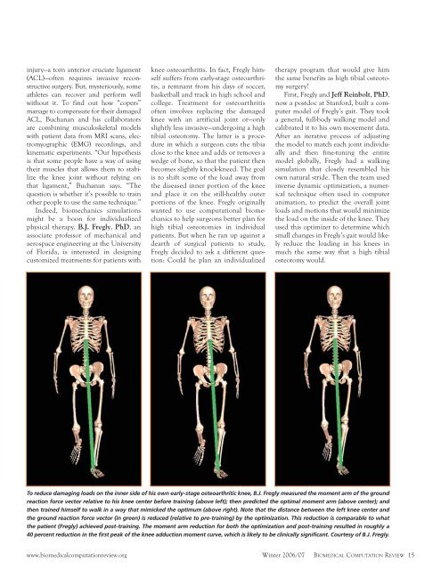
To reduce damaging loads on the inner side of his own early-stage osteoarthritic knee, B.J. Fregly measured the moment arm of the ground<br />
reaction force vector relative to his knee center before training (above left); then predicted the optimal moment arm (above center); and<br />
then trained himself to walk in a way that mimicked the optimum (above right). Note that the distance between the left knee center and<br />
the ground reaction force vector (in green) is reduced (relative to pre-training) by the optimization. This reduction is comparable to what<br />
the patient (Fregly) achieved post-training. The moment arm reduction for both the optimization and post-training resulted in roughly a<br />
40 percent reduction in the first peak of the knee adduction moment curve, which is likely to be clinically significant. Courtesy of B.J. Fregly.<br />
www.biomedicalcomputationreview.org<br />
Winter 2006/07 BIOMEDICAL COMPUTATION REVIEW 15