Computational Biomechanics Computational Biomechanics
Computational Biomechanics Computational Biomechanics
Computational Biomechanics Computational Biomechanics

You also want an ePaper? Increase the reach of your titles
YUMPU automatically turns print PDFs into web optimized ePapers that Google loves.
<strong>Computational</strong><br />
<strong>Biomechanics</strong><br />
BY REGINA NUZZO, PhD<br />
Making Strides<br />
Toward Patient Care<br />
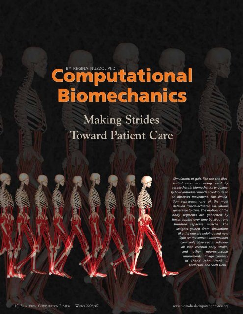
Simulations of gait, like the one illustrated<br />
here, are being used by<br />
researchers in biomechanics to quantify<br />
how individual muscles contribute to<br />
an observed movement. This simulation<br />
represents one of the most<br />
detailed muscle-actuated simulations<br />
generated to date. The motions of the<br />
body segments are generated by<br />
forces applied over time by about one<br />
hundred separate muscles. The<br />
insights gained from simulations<br />
like this one are helping shed new<br />
light on movement abnormalities<br />
commonly observed in individuals<br />
with cerebral palsy, stroke,<br />
and other neuromuscular<br />
impairments. Image courtesy<br />
of Chand John, Frank C.<br />
Anderson, and Scott Delp.<br />
10 BIOMEDICAL COMPUTATION REVIEW Winter 2006/07 www.biomedicalcomputationreview.org
Walk, run, bend, reach.<br />
The elements of human movement have<br />
fascinated research scientists for centuries.<br />
To understand how muscles contract<br />
and joints flex, researchers have dissected<br />
cadavers and experimented<br />
with animals. They can describe how<br />
bones, muscles, and tendons connect in a<br />
complicated geometry; how muscles<br />
exert forces on joints; and<br />
even how sparks in the brain can<br />
trigger a muscle’s contraction.<br />
Meanwhile, and mostly independently,<br />
clinicians have been<br />
treating people for sports<br />
injuries, stroke, and movement<br />
diseases such as cerebral palsy<br />
and osteoarthritis. Using trial<br />
and error, they’ve assessed which<br />
rehabilitative strategies and surgical<br />
interventions work best.<br />
Until recently, these two perspectives<br />
have not been well integrated.<br />
Clinical observations<br />
might miss the interplay of forces<br />
that lead to an injured knee,<br />
while static equations regarding<br />
the flexion of a dead man’s knee<br />
may by themselves be of little<br />
help in treating a torn ligament.<br />
Researchers were missing the cause-andeffect<br />
models that linked the physical<br />
forces with the clinical outcomes.<br />
Now, computational models are bridging<br />
the gap between micro and macro,<br />
between brain signals and gross human<br />
movement, and most recently, between<br />
research and clinic—helping to bring biomechanics<br />
knowledge from the bench all<br />
the way to the bedside. “<strong>Computational</strong><br />
biomechanics moves us beyond intuition,<br />
so that we’re able to understand how a<br />
basic cadaver study might relate to a<br />
movement disorder,” says Frank (Clay)<br />
Anderson, PhD, senior research associate<br />
at the Neuromuscular <strong>Biomechanics</strong><br />
Lab at Stanford University.<br />
Computer models can let surgeons<br />
explore different types of surgery on a<br />
patient before attempting the real<br />
thing. Or generate graphic simulations<br />
of athletes sprinting or cerebral palsy<br />
patients walking with a stiff-kneed gait.<br />
Some models can provide personalized<br />
recommendations for rehabilitation;<br />
others let researchers design new and<br />
When innovative engineers<br />
tried to apply standard<br />
mathematical and<br />
computational techniques<br />
to interesting biological<br />
problems, they soon learned<br />
that what works for planes<br />
and cars doesn’t always work<br />
for muscles and bones.<br />
improved implants that last longer than<br />
ever before. Flexible models allow<br />
researchers to tweak variables and ask<br />
“why?” and “what if?” in ways that bring<br />
the power of computational biomechanics<br />
straight to the patient—picking<br />
up where intuition and trial-and-error<br />
treatments leave off.<br />
BUILDING A<br />
SHARED FOUNDATION<br />
Unlike their forebears in the field of<br />
traditional biomechanics, many computational<br />
biomechanics researchers come<br />
from an engineering background. When<br />
innovative engineers tried to apply standard<br />
mathematical and computational<br />
techniques to interesting biological problems,<br />
they soon learned that what works<br />
for planes and cars doesn’t always work<br />
for muscles and bones. So the first challenge<br />
for these new “computational biomechanists”<br />
was to learn the language of<br />
biology. Starting in the 1970s, they dug<br />
into biology and began sharing their<br />
tools—multibody dynamics, control theory,<br />
and finite element modeling, for example—in<br />
journals and at conferences that<br />
had previously served only physical therapists,<br />
rehabilitation medicine experts,<br />
neurologists, and orthopedic surgeons. By<br />
the late 1980s, the rehabilitation commu-<br />
www.biomedicalcomputationreview.org<br />
Winter 2006/07 BIOMEDICAL COMPUTATION REVIEW 11
nity had welcomed these new approaches,<br />
and computational biomechanics as a<br />
field had come into its own.<br />
At the same time, advances in computing<br />
were infusing the field with newfound<br />
power. With the increases in computer<br />
speed in the late 1980s and 1990s,<br />
researchers could suddenly analyze their<br />
biomechanics models computationally<br />
with numerical algorithms rather than<br />
just analytically with pencil-and-paper<br />
mathematics. In the mid-1990s, parallel<br />
computing took off, and within the<br />
decade new and better algorithms, borrowed<br />
from control theory in robotics,<br />
were available. Ten years ago, a pair of<br />
simulated legs walking a single step would<br />
take approximately 1,000 hours to compute.<br />
Today, researchers can churn out<br />
similar calculations in about an hour.<br />
But even from the beginning, one<br />
important challenge faced computational<br />
biomechanics researchers: the need to<br />
develop ways to work with each other.<br />
With no standard modeling or software<br />
framework in the community, computational<br />
biomechanics labs were spending<br />
up to five years developing a single musculoskeletal<br />
model. Collaborating and<br />
building off of others’ progress was nearly<br />
impossible. “Everyone had to reinvent<br />
the wheel, one graduate student at a<br />
time,” says Scott Delp, PhD, professor<br />
and chair of bioengineering and head of<br />
the Neuromuscular <strong>Biomechanics</strong> Lab<br />
at Stanford University. “It really held up<br />
the field.”<br />
To help remedy that situation, in the<br />
early 1990s, Delp and Peter Loan, a biomechanist<br />
and software engineer, introduced<br />
a new modeling environment,<br />
called Software for Interactive<br />
Musculoskeletal Modeling (SIMM). This<br />
general software package let users create,<br />
alter, and evaluate models of many structures—all<br />
within a common platform.<br />
Since then, hundreds of labs have adopted<br />
SIMM to create and analyze computer<br />
models that range from a human<br />
climbing stairs to a Tyrannosaur nimbly<br />
trotting on hind limbs. The common<br />
tool also allowed researchers to more<br />
easily share models, and interdisciplinary<br />
studies started to focus on clinical<br />
applications of biomechanics.<br />
At the same time, funding agencies<br />
were taking notice. Biomedical computing<br />
tools—and the development of an<br />
infrastructure to support them—began to<br />
find a deserving place alongside traditional<br />
laboratory science. In 2004, NIH<br />
announced the award of $79.7 million in<br />
funding to establish four National<br />
Centers for Biomedical Computing.<br />
One of these was Simbios, the Center for<br />
Physics-Based Simulation of Biological<br />
Structures at Stanford University, which<br />
includes neuromuscular dynamics as a<br />
driving biological project.<br />
“<strong>Computational</strong> biomechanics moves us beyond intuition, so that<br />
we’re able to understand how a basic cadaver study might relate<br />
to a movement disorder,” says Frank “Clay” Anderson.<br />
The neuromuscular dynamics group<br />
at Simbios, in an effort led by Delp and<br />
Anderson, is now developing OpenSim,<br />
an open-source software system for biomechanical<br />
modeling and simulation.<br />
Freely available and incorporating modern<br />
plug-in technology, the new software<br />
could be a boon for researchers who need<br />
simulation software. “Having a common<br />
platform will radically change the field,”<br />
says Thomas Buchanan, PhD, a professor<br />
of mechanical engineering at<br />
University of Delaware. “This will really<br />
take the field in a whole new direction.”<br />
REPLACING AND<br />
REPAIRING JOINTS<br />
The design of joint replacements is<br />
one of the most mature applications of<br />
computational biomechanics. Ideally, artificial<br />
hips and knees need to withstand<br />
12 BIOMEDICAL COMPUTATION REVIEW Winter 2006/07 www.biomedicalcomputationreview.org
loads up to several times a person’s body<br />
weight and should function seamlessly<br />
over a lifetime. Forty years ago, however,<br />
joint replacements were far from perfect.<br />
Implants often wore down or broke outright<br />
under the rigors of everyday use.<br />
Then, with the help of advanced musculoskeletal<br />
computer models, researchers<br />
started to better understand the loads<br />
applied to natural joints under<br />
normal conditions. When they<br />
brought these models to the<br />
drafting table, they were able to<br />
design better artificial joints. By<br />
the 1980s, prosthetic joints had<br />
improved dramatically. Now, artificial<br />
joints far outperform their<br />
old-style predecessors in durability<br />
and reliability.<br />
Today, researchers continue to<br />
improve implant designs in<br />
increasingly refined ways. Donald<br />
Bartel, PhD, a professor of<br />
mechanical and aerospace engineering<br />
at Cornell University, is<br />
working to build real-world variability into<br />
the prosthetic design process. He uses biomechanical<br />
and statistical models to create<br />
artificial hips that are designed to function<br />
in a diverse set of situations—in patients<br />
ranging from frail to heavy-set, for activities<br />
from sitting down to climbing stairs.<br />
Bartel and his team build computational<br />
models that incorporate variables<br />
surgeons and implant designers<br />
can manipulate, such as implant materials<br />
and geometric design, as well as<br />
environmental variables beyond their<br />
“Having a common<br />
platform will radically<br />
change the field,” says<br />
Thomas Buchanan.<br />
“This will really take<br />
the field in a whole<br />
new direction.”<br />
control, such as a patient’s varying<br />
activity level and individual bone<br />
strength. Collaboration with Thomas<br />
Santner, PhD, a professor of statistics<br />
at Ohio State University, has led to<br />
robust statistical optimization methods<br />
that consider both the design variables<br />
and the environmental variables and<br />
then optimize the design of the<br />
implant. “The methodology allows us<br />
In 1995 it took 4,000 hours of parallel computing time to generate a half a second of walking<br />
in the image shown here. Contrast that with the transparent walker shown on the<br />
magazine cover and the previous pages: With twice the number of muscles, 2 seconds of<br />
walking took 20 minutes on an ordinary desktop PC. Advances in computing speed produced<br />
some of these improvements, but mostly they result from advances in algorithms.<br />
Images, courtesy of Frank (Clay) Anderson, come from Anderson’s dissertation work at the<br />
University of Texas, Austin, under Marcus G. Pandy.<br />
www.biomedicalcomputationreview.org<br />
Winter 2006/07 BIOMEDICAL COMPUTATION REVIEW 13
Finite element predictions of bone displacement in the direction of primary joint loading between a normal pelvis (left) and a pelvis with<br />
acetabular dysplasia (right) during simulated walking. The pelvic bone of the patient with hip dysplasia deformed much more than that of<br />
the normal hip joint, indicating that hip dysplasia may predispose the joint to early degeneration. Courtesy Jeffrey Weiss.<br />
gery,” says Jeffrey Weiss, PhD, associate<br />
professor of bioengineering and orthopedics<br />
at the University of Utah.<br />
To help surgeons make better decisions<br />
about where to cut, Weiss and his<br />
collaborators at University of Utah—<br />
Chris Peters, MD, professor of orthopedics,<br />
and Andrew Anderson, doctoral<br />
student in bioengineering—tailor computational<br />
biomechanical models to<br />
individual patients. They use CT images<br />
and biomechanical measurements to<br />
determine a patient’s specific hip geometry,<br />
standing loading patterns, and<br />
joint reaction forces. Using finite element<br />
analysis—a common way to model<br />
the mechanics of systems with complex<br />
geometries—the model predicts the contact<br />
stresses at the hip during inactivity<br />
as well as during everyday activities.<br />
Ultimately, this process might become<br />
routine. Weiss and his colleagues hope<br />
to say whether variation in the environmental<br />
variables will swamp out anything<br />
we do in the design,” Bartel says.<br />
“It helps us decide where to put our<br />
efforts and resources.”<br />
In addition to designers, orthopedic<br />
surgeons may benefit from computational<br />
biomechanics. A common surgery for<br />
hip dysplasia involves cutting out a portion<br />
of the hip socket and reorienting it<br />
to provide better alignment with the<br />
femoral head. The hope is that the procedure<br />
will distribute stresses in the hip<br />
more evenly, thus relieving the patient’s<br />
pain and also decreasing the wear and<br />
tear on the hip’s cartilage. But the surgery<br />
is tricky and not always successful.<br />
Surgeons have been guided mostly by<br />
their own experiences and intuition.<br />
“There’s little attention paid to the actual<br />
mechanics of the hip, either in its initial<br />
state or how it might be altered after surthat<br />
the standard surgical evaluation<br />
process for hip dysplasia patients will<br />
include a model of the patient’s individual<br />
stress-strain distribution, which predicts<br />
how the joint will change under<br />
increasing loads. That detailed information<br />
could then steer surgeons in deciding<br />
not only which patients are likely to<br />
benefit from surgery, but how to plan the<br />
surgery itself. Weiss and his team have<br />
already started to put this into practice:<br />
So far, they’ve collected CT scans from 15<br />
hip dysplasia patients as well as two<br />
healthy volunteers and are using the<br />
images to build models of their hips.<br />
PHYSICAL THERAPY<br />
APPLICATIONS<br />
Sometimes joint injuries or other<br />
joint problems are better resolved without<br />
surgical intervention. For example, a<br />
common, painful knee soft tissue<br />
14 BIOMEDICAL COMPUTATION REVIEW Winter 2006/07 www.biomedicalcomputationreview.org
injury—a torn anterior cruciate ligament<br />
(ACL)—often requires invasive reconstructive<br />
surgery. But, mysteriously, some<br />
athletes can recover and perform well<br />
without it. To find out how “copers”<br />
manage to compensate for their damaged<br />
ACL, Buchanan and his collaborators<br />
are combining musculoskeletal models<br />
with patient data from MRI scans, electromyographic<br />
(EMG) recordings, and<br />
kinematic experiments. “Our hypothesis<br />
is that some people have a way of using<br />
their muscles that allows them to stabilize<br />
the knee joint without relying on<br />
that ligament,” Buchanan says. “The<br />
question is whether it’s possible to train<br />
other people to use the same technique.”<br />
Indeed, biomechanics simulations<br />
might be a boon for individualized<br />
physical therapy. B.J. Fregly, PhD, an<br />
associate professor of mechanical and<br />
aerospace engineering at the University<br />
of Florida, is interested in designing<br />
customized treatments for patients with<br />
knee osteoarthritis. In fact, Fregly himself<br />
suffers from early-stage osteoarthritis,<br />
a remnant from his days of soccer,<br />
basketball and track in high school and<br />
college. Treatment for osteoarthritis<br />
often involves replacing the damaged<br />
knee with an artificial joint or—only<br />
slightly less invasive—undergoing a high<br />
tibial osteotomy. The latter is a procedure<br />
in which a surgeon cuts the tibia<br />
close to the knee and adds or removes a<br />
wedge of bone, so that the patient then<br />
becomes slightly knock-kneed. The goal<br />
is to shift some of the load away from<br />
the diseased inner portion of the knee<br />
and place it on the still-healthy outer<br />
portions of the knee. Fregly originally<br />
wanted to use computational biomechanics<br />
to help surgeons better plan for<br />
high tibial osteotomies in individual<br />
patients. But when he ran up against a<br />
dearth of surgical patients to study,<br />
Fregly decided to ask a different question:<br />
Could he plan an individualized<br />
therapy program that would give him<br />
the same benefits as high tibial osteotomy<br />
surgery?<br />
First, Fregly and Jeff Reinbolt, PhD,<br />
now a postdoc at Stanford, built a computer<br />
model of Fregly’s gait. They took<br />
a general, full-body walking model and<br />
calibrated it to his own movement data.<br />
After an iterative process of adjusting<br />
the model to match each joint individually<br />
and then fine-tuning the entire<br />
model globally, Fregly had a walking<br />
simulation that closely resembled his<br />
own natural stride. Then the team used<br />
inverse dynamic optimization, a numerical<br />
technique often used in computer<br />
animation, to predict the overall joint<br />
loads and motions that would minimize<br />
the load on the inside of the knee. They<br />
used this optimizer to determine which<br />
small changes in Fregly’s gait would likely<br />
reduce the loading in his knees in<br />
much the same way that a high tibial<br />
osteotomy would.<br />
To reduce damaging loads on the inner side of his own early-stage osteoarthritic knee, B.J. Fregly measured the moment arm of the ground<br />
reaction force vector relative to his knee center before training (above left); then predicted the optimal moment arm (above center); and<br />
then trained himself to walk in a way that mimicked the optimum (above right). Note that the distance between the left knee center and<br />
the ground reaction force vector (in green) is reduced (relative to pre-training) by the optimization. This reduction is comparable to what<br />
the patient (Fregly) achieved post-training. The moment arm reduction for both the optimization and post-training resulted in roughly a<br />
40 percent reduction in the first peak of the knee adduction moment curve, which is likely to be clinically significant. Courtesy of B.J. Fregly.<br />
www.biomedicalcomputationreview.org<br />
Winter 2006/07 BIOMEDICAL COMPUTATION REVIEW 15
Fregly’s customized simulation<br />
suggested he modify his<br />
walk in two ways: rotate his pelvis<br />
forward, and flex his knees a bit—<br />
both of which served to bring his<br />
knees toward the midline of his<br />
body. After nine months of training,<br />
Fregly managed a nearly 40<br />
percent reduction in a critical<br />
measure for knee loading. Since previous<br />
studies have suggested that the maximum<br />
reduction a high tibial osteotomy surgery<br />
can offer is 50 percent, Fregly is encouraged<br />
by these results, which have recently<br />
been accepted for publication. “These<br />
modifications were completely intuitive in<br />
retrospect, of course.” Fregly says. “But no<br />
one had ever come up with them before<br />
with just an experimental, trial-and-error<br />
approach.” Now, he’s conducting a pilot<br />
study of 10 patients with knee osteoarthritis<br />
to see if the same approach works for<br />
others as well as it worked for him. Early<br />
results are promising: The first subject<br />
learned the new motion in one training<br />
session and managed a 45 percent reduction<br />
in knee loading.<br />
Fregly and his team’s simulation and<br />
optimized suggestions can be computed<br />
for an individual in about a half-hour.<br />
The pilot study might help the<br />
researchers determine whether every<br />
patient needs to have a customized<br />
simulation built, or whether<br />
Fregly’s simple gait suggestions<br />
are generalizable to<br />
most knee osteoarthritis<br />
patients. Fregly eventually<br />
hopes to bring this kind<br />
of individualized rehabilitation<br />
to patients with a<br />
variety of other conditions<br />
as well, including patellofemoral<br />
pain and foot<br />
ulcers from diabetes.<br />
To extend the training<br />
rationale further,<br />
prevention of sports injuries<br />
might be an even more effective<br />
strategy than rehabilitation. Yet<br />
it’s not always clear exactly how<br />
and when injuries tend to<br />
occur. Darryl Thelen, PhD,<br />
associate professor of mechanical<br />
engineering at University of<br />
Wisconsin, uses computational<br />
biomechanics to analyze poten-<br />
“Determining how individual<br />
muscles influence injury risk<br />
is very difficult to answer<br />
experimentally,” Thelen says.<br />
“But these simulations give us a<br />
way to estimate the effects.”<br />
Thelen and his colleagues performed simulations of<br />
sprinters in order to estimate hamstring muscle force,<br />
work and fiber stretch during the swing phase of<br />
sprinting. Courtesy Darryl Thelen.<br />
tial mechanisms of injury during athletic<br />
activities. Thelen and Bryan<br />
Heiderscheit, PT, PhD, assistant professor<br />
in the department of orthopedics<br />
and rehabilitation at University of<br />
Wisconsin, recently investigated hamstring<br />
strain injuries in sprinters.<br />
They first used motion capture<br />
equipment to measure whole-body<br />
motion while athletes sprinted on a<br />
high-speed treadmill at speeds up to 20<br />
mph. They then fit scaled musculoskeletal<br />
models to the measured<br />
motion, which allowed them to estimate<br />
how the hamstring muscles are<br />
stretched during sprinting. They have<br />
shown that the greatest hamstring<br />
stretch—and potentially the time of<br />
greatest risk for injury—occurs late in<br />
the swing phase of the sprinting<br />
gait cycle. This is when the foot<br />
is still off the ground and the<br />
leg is being decelerated by the<br />
hamstrings before hitting the<br />
ground again.<br />
To better understand<br />
the dynamics of the<br />
movement, Thelen used<br />
an algorithm developed<br />
with Anderson to efficiently<br />
compute the muscle<br />
excitation patterns that<br />
produce the measured hip and<br />
knee motions of their sprinting<br />
volunteers. The muscle<br />
excitation patterns were<br />
inputs to a simulation of<br />
the crucial late sprinting<br />
stage. The model, like<br />
many other simulations<br />
of human movement,<br />
used a standard, computationally<br />
intensive technique called<br />
forward dynamics, in which<br />
the differential equations of a<br />
dynamic model are integrated<br />
forward in time. Thelen and<br />
his colleagues have used these<br />
sprinting simulations to compute<br />
the forces and work done by the<br />
hamstrings, and they’ve also examined<br />
how small changes in the system may<br />
affect injury risk. Their results show<br />
that strengthening core muscles<br />
attached to the pelvis, such as the hip<br />
flexors or abdominal obliques, have<br />
substantial influence on how the hamstring<br />
muscles are stretched and loaded.<br />
This suggests that exercises to improve<br />
coordination and strengthen core muscles<br />
might reduce injury risk.<br />
“Determining how individual muscles<br />
influence injury risk is very difficult to<br />
answer experimentally,” Thelen says.<br />
“But these simulations give us a way to<br />
estimate the effects.”<br />
NEUROMUSCULAR<br />
APPLICATIONS<br />
Neuroprostheses for patients with<br />
spinal cord injury represent one of the<br />
most dramatic success stories of computational<br />
biomechanics. For the past 30<br />
years, clinicians have performed experiments<br />
using electrodes to stimulate paralyzed<br />
muscles in coordinated patterns.<br />
Their work has led to the creation of<br />
implanted functional electrical stimulation<br />
(FES) systems that allow partially<br />
paralyzed patients to hold a fork and<br />
feed themselves, brush their teeth, or<br />
run a comb through their hair.<br />
Over time, biomechanical models<br />
have come to play a greater role in FES<br />
systems. “Basically, as the functions<br />
that we would like to restore have<br />
become more and more complex, our<br />
ability to implement this by intuition<br />
and trial and error is decreasing,” says<br />
Robert Kirsch, PhD, associate professor<br />
of biomedical engineering at Case<br />
Western Reserve University and a member<br />
of the Cleveland FES Center.<br />
“That’s why a number of years ago I<br />
began migrating toward the use of computational<br />
models to serve as a test bed<br />
for potential interventions.” He com-<br />
16 BIOMEDICAL COMPUTATION REVIEW Winter 2006/07 www.biomedicalcomputationreview.org
pares the high-stakes process to design<br />
in the aerospace industry. “If you’re<br />
building an airplane, you test it in a<br />
wind tunnel before you actually fly it.<br />
In our case, we’d like to test many different<br />
FES systems before we actually<br />
try them in human subjects.”<br />
Since there’s a limit to the number<br />
of electrodes that can be implanted in a<br />
patient, surgeons need to choose their<br />
of this work is not to build the most<br />
detailed possible model, but rather to<br />
create customized models that best predict<br />
surgical outcomes—and thus allow<br />
clinicians to circumvent the costly trialand-error<br />
process.<br />
Although individualized models<br />
like these are immensely helpful for<br />
planning patient treatment, clinicians<br />
can also glean useful knowledge from<br />
biomechanical models of an entire<br />
group of patients. Stroke survivors, for<br />
instance, often displaying a rather<br />
slow, asymmetrical shuffle. In order to<br />
best rehabilitate these patients, clinicians<br />
would like to know exactly what<br />
neuromuscular combinations are causing<br />
these changes in the first place. Jill<br />
Higginson, PhD, assistant professor of<br />
mechanical engineering at University<br />
This schematic elucidates the primary algorithms used for computational biomechanics simulations across multiple scales. Courtesy of<br />
Scott Delp and Clay Anderson.<br />
muscles for implantation carefully.<br />
Several combinations of muscles are<br />
often feasible candidates for implantation.<br />
Kirsch and his colleagues can customize<br />
simulations of each patient’s<br />
neuromuculoskeletal system so that clinicians<br />
can explore which system of<br />
electrodes and muscles would bring<br />
about the greatest clinical benefit.<br />
Kirsch, whose specialty is arm functionality,<br />
first measures the patient’s<br />
strength in various muscles and looks<br />
at restrictions in range of motion in the<br />
elbow and shoulder. Then he and his<br />
colleagues fit a musculoskeletal model<br />
of the upper arm to the patient’s individual<br />
data. Like many other<br />
researchers, Kirsch didn’t build this<br />
model from scratch; he works with an<br />
elbow-and-shoulder model originally<br />
developed by Frans Van der Helm,<br />
PhD, at Delft University of Technology<br />
in the Netherlands, which Kirsch has<br />
adapted for particular use in spinal<br />
cord injuries. Kirsch says that the goal<br />
“If you’re building<br />
an airplane, you test<br />
it in a wind tunnel<br />
before you actually<br />
fly it,” says Kirsch.<br />
“In our case, we’d<br />
like to test many<br />
different FES systems<br />
before we actually<br />
try them in<br />
human subjects.”<br />
of Delaware, is building musculoskeletal<br />
models of both stroke survivors and<br />
healthy older adults.<br />
Higginson and her colleagues want to<br />
understand the muscle excitation patterns—stemming<br />
from faulty neural communication—that<br />
lead to the characteristic<br />
stroke gait. Using dynamic optimization<br />
techniques, they’re building simulations<br />
that will help highlight exactly<br />
which patterns of muscle coordination<br />
are common to stroke patients.<br />
“Ultimately, we want to see what happens<br />
if you change the muscular excitation<br />
pattern,” Higginson says. “We can<br />
run that through the simulation.”<br />
The hope is that clinicians could<br />
then develop rehabilitation strategies<br />
that retrain the stroke patients’ neuromuscular<br />
system. To that end, Thomas<br />
Buchanan is working with colleagues at<br />
the University of Delaware to develop<br />
two new types of therapy programs.<br />
One, developed with help from Stuart<br />
Binder-MacLeod, PhD, a professor of<br />
www.biomedicalcomputationreview.org<br />
Winter 2006/07 BIOMEDICAL COMPUTATION REVIEW 17
physical therapy, would use electrodes to<br />
stimulate particular muscles, stepping in<br />
for the damaged brain until it regains<br />
more function. For the other, Sunil<br />
Agrawal, PhD, a professor of mechanical<br />
engineering, is helping to design<br />
robots to physically guide and reinforce<br />
patients’ movements during physical<br />
therapy. Both approaches use computational<br />
biomechanics methods to determine<br />
which particular muscles most<br />
need support.<br />
Biomechanical simulations are also<br />
helping guide individualized treatment<br />
for other disorders. People with cerebral<br />
palsy, for instance, often display characteristic<br />
abnormal movements that<br />
include stiff-knee gait, crouch gait, and<br />
excessive hip rotations. Clinicians prefer<br />
to intervene at a relatively young age,<br />
using a combination of strengthening<br />
exercises, braces, casts, and in severe<br />
cases, surgery. But common procedures<br />
such as detaching and rearranging tendons,<br />
or dividing and realigning bones<br />
in the leg are extremely invasive and not<br />
always successful.<br />
Scott Delp and his colleagues are<br />
developing musculoskeletal models of<br />
cerebral palsy patients based on MRI<br />
scans and kinematic measurements.<br />
The researchers want to identify the particular<br />
biomechanical causes of walking<br />
abnormalities in specific individuals.<br />
“Once we know the underlying cause,<br />
we can determine the best treatment,”<br />
Delp says. They can then work with surgeons<br />
to simulate the effects of different<br />
kinds of surgery on an individual<br />
patient, perhaps even devising new techniques.<br />
Recently, these cerebral palsy<br />
models helped Delp and his colleagues<br />
investigate ways to avoid a common<br />
weakening of the calf muscle after certain<br />
tendon-lengthening surgeries.<br />
Simulations had suggested a specific<br />
modification during surgery that might<br />
improve results. After turning to the literature,<br />
the researchers discovered that<br />
50 years ago a similar surgical technique<br />
had been published but had been largely<br />
ignored. Delp and his team published<br />
their findings, including simulation<br />
results and theoretical evidence.<br />
Surgeons are now reporting good results<br />
in treating cerebral palsy patients with<br />
the rediscovered procedure.<br />
A graphical representation of a shoulder and elbow musculoskeletal model, with muscles<br />
indicated by the red lines, and muscle wrapping surfaces represented by the blue<br />
ellipsoid (thorax), sphere (humeral head) and cylinders (humerus, ulna and radius).<br />
Courtesy of Dimitra Blana and Robert Kirsch.<br />
“With general<br />
models, you can<br />
uncover fundamental<br />
principles,” Delp says.<br />
“But it’s not clear to<br />
which patients those<br />
general principles<br />
actually apply. We<br />
need to customize<br />
treatment for the<br />
individuals.”<br />
CHALLENGES<br />
FOR THE FUTURE<br />
At present, most clinical applications<br />
of computational biomechanics rely on<br />
generic models rather than fully customized<br />
simulations. It’s a good first<br />
step, but future treatment decisions will<br />
demand greater specificity. “With general<br />
models, you can uncover fundamental<br />
principles,” Delp says. “But it’s not clear<br />
to which patients those general principles<br />
actually apply. We need to customize<br />
treatment for the individuals.”<br />
With data from individualized simulations<br />
and treatment outcomes,<br />
researchers could then turn from purely<br />
descriptive studies to analyses more<br />
focused on predicting clinical outcomes.<br />
Researchers currently try to scale musculoskeletal<br />
models to individual patient<br />
geometry as much as possible, but full<br />
individualization on a large scale is still<br />
prohibitive. “It would be nice to have a<br />
simple way to adjust the models to the<br />
individuals,” Kirsch says. “We can do it<br />
now through painstaking manual measurements,<br />
but that’s not practical for widespread<br />
deployment into the clinic.” Some<br />
researchers are working on ways to use<br />
MRI, CT and ultrasound data to more<br />
quickly and routinely scale flexible mus-<br />
18 BIOMEDICAL COMPUTATION REVIEW Winter 2006/07 www.biomedicalcomputationreview.org
culoskeletal models to individual patients.<br />
These imaging techniques might also<br />
prove useful in further refining existing<br />
musculoskeletal models with soft tissue<br />
dynamics in vivo. Researchers have thus<br />
far relied on static imaging work as well<br />
as extensive analysis of cadavers. But<br />
these methods are no substitute for<br />
examining soft tissue details in a living<br />
person. “With new work in imaging, we<br />
can see how a muscle is loaded during<br />
functional movement,” Thelen says. “A<br />
lot of macro models don’t capture the<br />
full soft tissue complexity. Imaging coupled<br />
with models could potentially<br />
improve our representation and understanding<br />
of soft tissue function.”<br />
As increasingly sophisticated biomechanical<br />
models produce prodigious<br />
amounts of data, researchers will<br />
also need to develop statistical methods<br />
to help interpret the results. “Like<br />
a lot of biology, computational biomechanics<br />
is overwhelmed by an incredible<br />
wealth of data,” Delp says. “A simulation<br />
can spit out 100 columns of<br />
data for each millisecond. We need a<br />
“The clinician’s work and the scientist’s work go hand in hand.<br />
Our results are still relatively in the beginning stages,” says Hoyen.<br />
“But it’s astonishing at present what we’ve been able to accomplish.”<br />
Delp and Allison Arnold, PhD, have modeled the various abnormal gaits observed in patients with<br />
cerebral palsy to help them evaluate appropriate treatments. Here, they used MRI images to create a<br />
musculoskeletal model of an individual with a crouched internal-rotation gait. The rotational moment<br />
arms of the medial hamstrings, adductors and other muscles were evaluated at the body positions corresponding<br />
to the subject’s internally rotated gait. Photo courtesy of Eugene Bleck; images previously<br />
published in Arnold and Delp (2004), The role of musculoskeletal models in patient assessment and<br />
treatment. In: Treatment of Gait Problems in Cerebral Palsy, Ed. Gage, MacKeith Press, London.<br />
way to quickly and objectively<br />
extract which factors are most<br />
important therapeutically.”<br />
As with any simulation, computational<br />
biomechanical models<br />
need to prove both their accuracy<br />
and their worth. Modelers<br />
need to work closely with experimentalists<br />
to demonstrate that<br />
their simulations are not only<br />
internally consistent but also<br />
accurately represent reality. And<br />
as clinical studies increase in<br />
number, researchers will gather<br />
evidence showing whether the<br />
use of simulations does in fact<br />
improve treatment outcomes.<br />
“Improving clinical outcomes is<br />
our grand challenge,” Delp says.<br />
“Achieving this goal requires<br />
that experts in biomechanical<br />
simulation stay connected to the<br />
realities of the clinic.”<br />
Finally, researchers must try<br />
to package simulations for everyday<br />
use and start to put them in<br />
the hands of biologists and clinicians.<br />
“Collaborations are essential,”<br />
says Harry Hoyen, MD,<br />
professor of orthopedics at Case<br />
Western Reserve University<br />
Medical School, who works with<br />
Robert Kirsch on FES systems to<br />
restore function to the shoulder<br />
in partially paralyzed patients.<br />
“The clinician’s work and the scientist’s<br />
work go hand in hand.<br />
Our results are still relatively in<br />
the beginning stages. But it’s<br />
astonishing at present what we’ve<br />
been able to accomplish.” ■<br />
www.biomedicalcomputationreview.org<br />
Winter 2006/07 BIOMEDICAL COMPUTATION REVIEW 19