Breath tests and airway gas exchange - Michael P. Hlastala
Breath tests and airway gas exchange - Michael P. Hlastala
Breath tests and airway gas exchange - Michael P. Hlastala

Create successful ePaper yourself
Turn your PDF publications into a flip-book with our unique Google optimized e-Paper software.
ARTICLE IN PRESS<br />
Pulmonary Pharmacology & Therapeutics 20 (2007) 112–117<br />
www.elsevier.com/locate/ypupt<br />
<strong>Breath</strong> <strong>tests</strong> <strong>and</strong> <strong>airway</strong> <strong>gas</strong> <strong>exchange</strong><br />
Joseph C. Anderson a, , <strong>Michael</strong> P. <strong>Hlastala</strong> a,b<br />
a Division of Pulmonary <strong>and</strong> Critical Care Medicine, Department of Medicine, Box 356522, University of Washington, Seattle, Washington 98195-6522, USA<br />
b Departments of Physiology <strong>and</strong> Biophysics, University of Washington, Seattle, Washington 98195, USA<br />
Received 2 December 2005; accepted 2 December 2005<br />
Abstract<br />
Measuring soluble <strong>gas</strong> in the exhaled breath is a non-invasive technique used to estimate levels of respiratory, solvent, <strong>and</strong> metabolic<br />
<strong>gas</strong>es. The interpretation of these measurements is based on the assumption that the measured <strong>gas</strong>es <strong>exchange</strong> in the alveoli. While the<br />
respiratory <strong>gas</strong>es have a low blood-solubility <strong>and</strong> <strong>exchange</strong> in the alveoli, high blood-soluble <strong>gas</strong>es <strong>exchange</strong> in the <strong>airway</strong>s. The effect of<br />
<strong>airway</strong> <strong>gas</strong> <strong>exchange</strong> on the interpretation of these exhaled breath measurements can be significant. We describe <strong>airway</strong> <strong>gas</strong> <strong>exchange</strong> in<br />
relation to exhaled measurements of soluble <strong>gas</strong>es that <strong>exchange</strong> in the alveoli. The mechanisms of <strong>airway</strong> <strong>gas</strong> <strong>exchange</strong> are reviewed <strong>and</strong><br />
criteria for determining if a <strong>gas</strong> <strong>exchange</strong>s in the <strong>airway</strong>s are provided. The effects of diffusion, perfusion, temperature <strong>and</strong> breathing<br />
maneuver on <strong>airway</strong> <strong>gas</strong> <strong>exchange</strong> <strong>and</strong> on measurement of exhaled soluble <strong>gas</strong> are discussed. A method for estimating the impact of<br />
<strong>airway</strong> <strong>gas</strong> <strong>exchange</strong> on exhaled breath measurements is presented. We recommend that investigators should carefully control the<br />
inspired air conditions <strong>and</strong> type of exhalation maneuver used in a breath test. Additionally, care should be taken when interpreting<br />
breath <strong>tests</strong> from subjects with pulmonary disease.<br />
r 2005 Elsevier Ltd. All rights reserved.<br />
Keywords: Bronchial circulation; Endogenous; Soluble; Mathematical modeling; Alveoli<br />
1. Introduction<br />
The primary function of the lung is as a <strong>gas</strong> <strong>exchange</strong><br />
organ. Most notably, the lung assists in exchanging oxygen<br />
<strong>and</strong> carbon dioxide between the atmosphere <strong>and</strong> the blood.<br />
However, the lung <strong>exchange</strong>s many more <strong>gas</strong>es than simply<br />
the respiratory <strong>gas</strong>es. Many non-respiratory <strong>gas</strong>es that<br />
<strong>exchange</strong> in the lung are soluble inert <strong>gas</strong>es such as ethane,<br />
ether, toluene, acetone <strong>and</strong> ethanol. Many times, these<br />
<strong>gas</strong>es can be found circulating in the blood as a result of<br />
environmental exposure, ingestion <strong>and</strong> metabolic production.<br />
When blood enters the lung, these <strong>gas</strong>es diffuse into<br />
the lung air <strong>and</strong> subsequently appear in the exhaled breath.<br />
Because the lungs transport soluble <strong>gas</strong> from the blood<br />
to the exhaled air, the exhaled breath is frequently used as a<br />
non-invasive surrogate for a blood sample. It is thought<br />
that the partial pressure of a soluble <strong>gas</strong> in the exhaled<br />
breath represents the partial pressure of that <strong>gas</strong> in the<br />
blood. In this sense, the lungs are thought to provide a<br />
Corresponding author. Tel.: +1 206 685 2929; fax: +1 206 685 8673.<br />
E-mail address: clarkja@u.washington.edu (J.C. Anderson).<br />
window into the blood <strong>and</strong>, by inference, the body.<br />
Because of its non-invasive nature, measurement of the<br />
exhaled breath is becoming increasingly common in many<br />
professions to infer soluble <strong>gas</strong> concentrations in the blood<br />
<strong>and</strong> by analogy to determine a medical condition or legal<br />
status of the subject. Industrial hygienists sample exhaled<br />
breath to estimate a worker’s exposure to chemical solvents<br />
such as toluene <strong>and</strong> methyl ethyl ketone. Toxicologists <strong>and</strong><br />
law enforcement agents depend on measurements of<br />
ethanol in the end-exhaled breath to estimate blood alcohol<br />
content in workers <strong>and</strong> automobile drivers. Currently,<br />
clinical scientists have become interested in sampling the<br />
end-exhaled breath for endogenous <strong>gas</strong>es such as ethane,<br />
isoprene <strong>and</strong> acetone. Recent literature suggests that<br />
absolute blood levels of these <strong>gas</strong>es may indicate common<br />
disorders such as lung cancer [1], acute myocardial<br />
infarction [2], <strong>and</strong> congestive heart failure [3].<br />
Although sampling the exhaled air is relatively simple,<br />
the relationship between partial pressure of soluble <strong>gas</strong> in<br />
the breath <strong>and</strong> blood can be complex depending on where<br />
<strong>gas</strong> <strong>exchange</strong> occurs. Location of <strong>gas</strong> <strong>exchange</strong>, <strong>airway</strong><br />
versus alveoli, depends primarily on the blood solubility of<br />
1094-5539/$ - see front matter r 2005 Elsevier Ltd. All rights reserved.<br />
doi:10.1016/j.pupt.2005.12.002
ARTICLE IN PRESS<br />
J.C. Anderson, M.P. <strong>Hlastala</strong> / Pulmonary Pharmacology & Therapeutics 20 (2007) 112–117 113<br />
the <strong>gas</strong> as described by the blood–air partition coefficient,<br />
l b:a . Low blood-soluble <strong>gas</strong>es like oxygen (l b:a 0:7) <strong>and</strong><br />
carbon dioxide (l b:a 3) <strong>exchange</strong> in the alveoli while high<br />
blood-soluble <strong>gas</strong>es like ethanol (l b:a 1756) <strong>exchange</strong> in<br />
the <strong>airway</strong>s. For low blood-soluble <strong>gas</strong>es, the relationship<br />
between the breath <strong>and</strong> blood is relatively simple <strong>and</strong><br />
depends predominately on two factors: the blood solubility<br />
of the <strong>gas</strong>, l b:a , <strong>and</strong> the distribution of ventilation-toperfusion<br />
in all <strong>gas</strong> <strong>exchange</strong> units. For these <strong>gas</strong>es, the<br />
partial pressure of soluble <strong>gas</strong> in the end-exhaled air<br />
depends on the breathing maneuver. For a tidal breath, the<br />
partial pressure of soluble <strong>gas</strong> in the end-exhaled air is<br />
equal to alveolar air. For these <strong>gas</strong>es, the partial pressure of<br />
soluble <strong>gas</strong> in the alveolar air is not equal to that in the<br />
blood. However, the partial pressure of soluble <strong>gas</strong> in this<br />
alveolar sample can be converted to an approximate blood<br />
value by estimating the ventilation-to-perfusion distribution<br />
in the lung <strong>and</strong> using the Farhi kernel [4]. For a singleexhalation<br />
breath test (i.e. the subject inhales to total lung<br />
capacity <strong>and</strong> exhales at a slow constant rate to residual<br />
volume), the partial pressure of soluble <strong>gas</strong> in the endexhaled<br />
air is larger than the alveolar air but generally<br />
smaller than that in the blood. This difference between the<br />
end-exhaled <strong>and</strong> blood partial pressure decreases as l b:a<br />
increases for these low blood-soluble <strong>gas</strong>es.<br />
For high blood-soluble <strong>gas</strong>es, <strong>gas</strong> <strong>exchange</strong> occurs in the<br />
<strong>airway</strong>s <strong>and</strong> the relationship between end-exhaled breath<br />
measurement <strong>and</strong> blood is complex. Airway <strong>gas</strong> <strong>exchange</strong><br />
depends on multiple factors such as <strong>airway</strong> temperature,<br />
bronchial blood flow <strong>and</strong> blood–air partition coefficient.<br />
For a particular <strong>gas</strong>, the specific <strong>airway</strong> generations<br />
participating in <strong>gas</strong> <strong>exchange</strong> affect the exhaled partial<br />
pressure. Additionally, the diffusion gradient for soluble<br />
<strong>gas</strong> <strong>exchange</strong> with the <strong>airway</strong>s reverses from inspiration to<br />
expiration. As a result, the partial pressure of these soluble<br />
<strong>gas</strong>es in the breath is always less than that in the blood <strong>and</strong><br />
the relationship between the two is difficult to precisely<br />
quantify.<br />
In this paper, we focus on <strong>airway</strong> <strong>gas</strong> <strong>exchange</strong>. First, we<br />
discuss the gross differences between <strong>gas</strong>es that <strong>exchange</strong> in<br />
the alveoli versus those that <strong>exchange</strong> in the <strong>airway</strong>s.<br />
Second, we discuss the specific mechanisms of <strong>airway</strong> <strong>and</strong><br />
alveolar <strong>gas</strong> <strong>exchange</strong> <strong>and</strong> show where soluble <strong>gas</strong>es<br />
<strong>exchange</strong> in the lung, alveoli versus <strong>airway</strong>. Third, we<br />
examine factors that most impact <strong>airway</strong> <strong>gas</strong> <strong>exchange</strong>.<br />
Fourth, we review how using a classical alveolar <strong>gas</strong><br />
<strong>exchange</strong> model can lead to errors in interpreting breath<br />
<strong>tests</strong> involving highly blood soluble <strong>gas</strong>es.<br />
2. Airway versus alveolar <strong>gas</strong> <strong>exchange</strong><br />
The differences between <strong>gas</strong>es that <strong>exchange</strong> in the<br />
alveoli <strong>and</strong> those that <strong>exchange</strong> in the <strong>airway</strong>s can be<br />
observed in their expirograms. For a <strong>gas</strong> that <strong>exchange</strong>s in<br />
the alveoli (e.g. carbon dioxide), the partial pressure of <strong>gas</strong><br />
in the exhaled breath as a function of exhaled volume is<br />
schematically shown as the parallel-lines tracing in Fig. 1.<br />
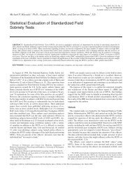
Fig. 1. Schematic identifying the phases of an expirogram from a low<br />
blood-soluble <strong>gas</strong> that <strong>exchange</strong>s in the alveoli (parallel-lines) <strong>and</strong> from a<br />
high blood-soluble <strong>gas</strong> that <strong>exchange</strong>s in the <strong>airway</strong>s (solid line). Phase I<br />
represents air emptying the anatomic dead space (i.e. the conducting<br />
<strong>airway</strong>s). Air emptying the <strong>exchange</strong> space, phase III, corresponds to air<br />
from the alveoli for low soluble <strong>gas</strong>es <strong>and</strong> <strong>airway</strong>s for high soluble <strong>gas</strong>es.<br />
For <strong>gas</strong>es exchanging in the <strong>airway</strong>s: (1) phase I is not present because the<br />
<strong>airway</strong>s participate in <strong>gas</strong> <strong>exchange</strong>; <strong>and</strong> (2) the end-exhaled breath<br />
concentration (solid circle) is always less than that in the alveoli.<br />
The initial volume of air does not contain soluble <strong>gas</strong> <strong>and</strong> is<br />
labeled as phase I. This volume of air is considered to<br />
represent air residing in the anatomic dead space (i.e.<br />
conducting <strong>airway</strong>s). The rapidly rising phase of the<br />
expirogram is labeled as phase II <strong>and</strong> represents the<br />
transition from air residing in the dead space to alveolar<br />
air. Soluble <strong>gas</strong> appearing at the mouth during phase III<br />
comes from the alveoli where <strong>gas</strong> <strong>exchange</strong> occurs. Thus,<br />
the final volume of air that exits the mouth represents<br />
alveolar air <strong>and</strong> measuring this end-exhaled breath<br />
provides an estimate of alveolar <strong>gas</strong> partial pressure (open<br />
circle). We contrast the expirogram of a <strong>gas</strong> that <strong>exchange</strong>s<br />
in the alveoli (parallel-lines curve) with a <strong>gas</strong> that<br />
<strong>exchange</strong>s in the <strong>airway</strong> (solid curve). For a <strong>gas</strong> that<br />
<strong>exchange</strong>s completely in the <strong>airway</strong>s (e.g. ethanol), phase I<br />
is not present in the expirogram because the <strong>airway</strong>s<br />
participate in <strong>gas</strong> <strong>exchange</strong> [5,6]. Therefore, an anatomical<br />
dead space cannot be defined for these <strong>gas</strong>es. Phase III does<br />
not represent air coming from the alveoli but rather air<br />
emptying the <strong>exchange</strong> space. For these highly soluble<br />
<strong>gas</strong>es, the <strong>exchange</strong> space is the conducting <strong>airway</strong>s. In<br />
contrast to <strong>gas</strong>es exchanging in the alveoli, the partial<br />
pressure of soluble <strong>gas</strong> in the end-exhaled breath (solid<br />
circle) is always less than that in the alveoli.<br />
The differences between these expirograms for these two<br />
types of pulmonary <strong>gas</strong> <strong>exchange</strong> result from the different<br />
mechanisms underlying their <strong>exchange</strong>. For alveolar <strong>gas</strong><br />
<strong>exchange</strong>, fresh air is inspired, fills the conducting <strong>airway</strong>s,<br />
<strong>and</strong> then enters the alveoli. Once in the alveoli, low blood<br />
soluble <strong>gas</strong> present in the blood moves into this inspired<br />
air. This movement of <strong>gas</strong> from the blood into the alveolar<br />
air continues throughout the entire breathing cycle,<br />
inspiration <strong>and</strong> expiration. On expiration, <strong>gas</strong> in the
114<br />
ARTICLE IN PRESS<br />
J.C. Anderson, M.P. <strong>Hlastala</strong> / Pulmonary Pharmacology & Therapeutics 20 (2007) 112–117<br />
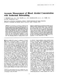
Fig. 2. The axial distribution of <strong>gas</strong> transport during inspiration (black<br />
columns) <strong>and</strong> expiration (gray columns) for a tidal breath of isopropanol<br />
(l b:a ¼ 848) shows that almost 100% of isopropanol <strong>exchange</strong>s occurs in<br />
the <strong>airway</strong>s. Each flux has been normalized by the total inspiratory soluble<br />
<strong>gas</strong> flux.<br />
conducting <strong>airway</strong>s leaves the lung first causing the phase I<br />
seen in the expirogram. Afterwards, alveolar air, containing<br />
high partial pressures of low blood soluble <strong>gas</strong>, exits the<br />
lung via the conducting <strong>airway</strong>s <strong>and</strong> appears as phase III in<br />
the expirogram. The mechanisms of <strong>airway</strong> <strong>gas</strong> <strong>exchange</strong><br />
are markedly different [5,7,8]. High blood-soluble <strong>gas</strong>es are<br />
present in large concentrations in the <strong>airway</strong> tissue <strong>and</strong><br />
mucus as compared with low blood-soluble <strong>gas</strong>es for a<br />
given partial pressure. Thus, as fresh air is inspired, it<br />
absorbs high blood-soluble <strong>gas</strong> from the mucus lining the<br />
<strong>airway</strong> wall (Fig. 2) thereby depleting the concentration of<br />
soluble <strong>gas</strong> in the <strong>airway</strong> wall. Because of the small<br />
bronchial blood flow <strong>and</strong> the significant tissue barrier<br />
between the bronchial circulation <strong>and</strong> mucus layer, the<br />
<strong>airway</strong> wall is not replenished with soluble <strong>gas</strong> before<br />
expiration begins. By the time the inspired air reaches the<br />
alveoli, the air is saturated with soluble <strong>gas</strong> <strong>and</strong> no<br />
additional <strong>gas</strong> <strong>exchange</strong> occurs within the alveoli. During<br />
expiration, the air, saturated with soluble <strong>gas</strong>, encounters a<br />
lower partial pressure of soluble <strong>gas</strong> in the mucus <strong>and</strong><br />
therefore a large driving force for the deposition of soluble<br />
<strong>gas</strong> on the mucus. This large air-to-mucus gradient<br />
promotes recovery of soluble <strong>gas</strong> by the mucous layer<br />
<strong>and</strong> delays the rise in soluble <strong>gas</strong> partial pressure at the<br />
mouth, thus accounting for the rising slope of phase III.<br />
These absorption–desorption phenomena cause the partial<br />
pressure of soluble <strong>gas</strong> in the end-exhaled breath to always<br />
be less that that in the alveoli (i.e. saturation) <strong>and</strong> is the<br />
major mechanism of pulmonary <strong>gas</strong> <strong>exchange</strong> for any <strong>gas</strong><br />
with a blood–air partition coefficient greater than 100 [7].<br />
3. Location of soluble <strong>gas</strong> <strong>exchange</strong> in the lung<br />
To determine the appropriate method of interpreting a<br />
breath test, it is important to underst<strong>and</strong> where a particular<br />
soluble <strong>gas</strong> <strong>exchange</strong>s within the lung. To determine<br />
location of <strong>gas</strong> <strong>exchange</strong>, we simulated pulmonary <strong>gas</strong><br />
<strong>exchange</strong> using a mathematical model of the <strong>airway</strong>s <strong>and</strong><br />
alveoli of the lung [7]. The model has a symmetrically<br />
bifurcating structure through 18 generations. The upper<br />
respiratory tract <strong>and</strong> intraparenchymal <strong>airway</strong>s are divided<br />
into 480 axial control volumes. Radially, the <strong>airway</strong>s are<br />
divided into six layers (see Fig. 1 in [7]): (1) the <strong>airway</strong><br />
lumen, (2) a thin mucous layer, (3) connective tissue<br />
(epithelium <strong>and</strong> mucosal tissue), (4) the bronchial circulation,<br />
(5) the adventitia, <strong>and</strong> (6) the pulmonary circulation.<br />
Cartilaginous <strong>airway</strong>s (generationo10), functionally, only<br />
have the first four layers. The respiratory bronchioles <strong>and</strong><br />
alveoli are lumped together into a single alveolar unit. The<br />
concentration of soluble <strong>gas</strong> in the alveolar air is allowed to<br />
vary with time <strong>and</strong> depends on the pulmonary blood flow,<br />
ventilation, blood solubility, <strong>and</strong> concentration of soluble<br />
<strong>gas</strong> in the incoming blood as described by a mass balance<br />
on the alveolar compartment. Within each radial layer,<br />
concentration <strong>and</strong> temperature values are bulk averages for<br />
the entire layer. Mass <strong>and</strong> energy are transported between<br />
control volumes by bulk convection <strong>and</strong> axial diffusion<br />
through the lumen. Radial transport between the <strong>gas</strong> phase<br />
<strong>and</strong> mucous layer is described with heat <strong>and</strong> mass transfer<br />
coefficients. Transport of water <strong>and</strong> soluble <strong>gas</strong> between<br />
layers occurs via filtration (from bronchial circulation to<br />
mucus) <strong>and</strong> diffusion (Fick’s law). Mass <strong>and</strong> energy<br />
balances around a control volume produce three partial<strong>and</strong><br />
9 ordinary-differential equations that are solved<br />
simultaneously in time <strong>and</strong> space. The solution of the<br />
mathematical model yields axial profiles of partial pressure<br />
within the <strong>airway</strong> lumen <strong>and</strong> <strong>airway</strong> wall (<strong>and</strong> all other<br />
layers as well) for any desired time during inspiration <strong>and</strong><br />
expiration.<br />
From these axial profiles of soluble <strong>gas</strong> partial pressure,<br />
the flow of soluble <strong>gas</strong> (mol/s) from the <strong>airway</strong> wall to the<br />
<strong>airway</strong> lumen for a given axial position (e.g. the trachea)<br />
<strong>and</strong> period of time can be calculated. For a given axial<br />
position such as an <strong>airway</strong> generation, these flows of<br />
soluble <strong>gas</strong> can be summed together over all time points<br />
throughout an inspiration <strong>and</strong>/or expiration. As an<br />
example of <strong>airway</strong> <strong>gas</strong> <strong>exchange</strong>, we simulated the<br />
<strong>exchange</strong> of isopropanol (l b:a ¼ 848 from [9]) during<br />
steady breath-to-breath tidal breathing <strong>and</strong> calculated the<br />
mass flows of isopropanol at each <strong>airway</strong> generation. The<br />
sum of these isopropanol flows from <strong>airway</strong> wall to lumen<br />
over an inspiration <strong>and</strong> expiration is plotted in Fig. 2 <strong>and</strong><br />
represents the axial distribution of isopropanol transport<br />
from the <strong>airway</strong> wall to the lumen (positive flow). The axial<br />
distribution of isopropanol transport on inspiration shows<br />
a bimodal distribution with a peak in the trachea <strong>and</strong> the<br />
12th generation. After the 12th generation, isopropanol<br />
flows during inspiration decrease per generation <strong>and</strong><br />
approach zero in the alveolar region. For isopropanol<br />
<strong>and</strong> other <strong>gas</strong>es that <strong>exchange</strong> in the <strong>airway</strong>s, almost 100%<br />
of <strong>exchange</strong> occurs within the <strong>airway</strong>s. For low bloodsoluble<br />
<strong>gas</strong>es (l b:a o10), their axial distribution of flow<br />
shows a concentrated spike in the alveoli. As the blood<br />
solubility of <strong>gas</strong> increases from low to high, the distribution<br />
shifts from a sharp concentrated peak in the alveolar region<br />
to a wider distribution that spreads throughout the <strong>airway</strong>s<br />
as seen for isopropanol (see Fig. 5 in Anderson et al. [7]).
ARTICLE IN PRESS<br />
J.C. Anderson, M.P. <strong>Hlastala</strong> / Pulmonary Pharmacology & Therapeutics 20 (2007) 112–117 115<br />
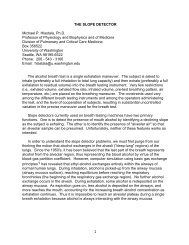
Fig. 3. The <strong>exchange</strong> ratio (ER) is the ratio of <strong>airway</strong> <strong>gas</strong> <strong>exchange</strong> to<br />
total pulmonary (<strong>airway</strong>+alveolar) <strong>gas</strong> <strong>exchange</strong> <strong>and</strong> is plotted versus l b:a<br />
for eleven soluble <strong>gas</strong>es as calculated from the <strong>airway</strong>–alveolar <strong>gas</strong><br />
<strong>exchange</strong> model during tidal breathing. Gases with l b:a o10 such as ethane<br />
<strong>and</strong> cyclopropane <strong>exchange</strong> predominately in the alveoli. Diethyl ether<br />
<strong>exchange</strong>s equally between the <strong>airway</strong> <strong>and</strong> alveoli. Gases with l b:a 4100<br />
such as acetone <strong>and</strong> ethanol <strong>exchange</strong> almost exclusively in the <strong>airway</strong>s<br />
(figure adapted from Anderson et al. [7]).<br />
In Anderson et al. [7], we constructed axial distributions<br />
of <strong>gas</strong> transport during tidal breathing for eleven <strong>gas</strong>es<br />
with l b:a ranging from 0.88 to 2709. To determine where<br />
the majority of <strong>gas</strong> <strong>exchange</strong> occurs, either <strong>airway</strong>s or<br />
alveoli, we defined an <strong>exchange</strong> ratio (ER) to be the ratio of<br />
<strong>airway</strong> <strong>gas</strong> <strong>exchange</strong> to total pulmonary (<strong>airway</strong>+alveolar)<br />
<strong>gas</strong> <strong>exchange</strong>. From the axial distribution, we calculated an<br />
ER for each <strong>gas</strong> <strong>and</strong> plotted ER against l b:a (Fig. 3). Fig. 3<br />
was adapted from Anderson et al. [7] <strong>and</strong> indicates where<br />
the majority of <strong>gas</strong> <strong>exchange</strong> occurs for a <strong>gas</strong> with a given<br />
blood–air partition coefficient. When ER ¼ 0orER¼ 1,<br />
100% of <strong>gas</strong> <strong>exchange</strong> occurs in the alveoli or <strong>airway</strong>s,<br />
respectively. Thus, soluble <strong>gas</strong>es with a blood–air partition<br />
coefficient less than ten (l b:a o10) <strong>exchange</strong> almost solely<br />
with the alveoli whereas <strong>gas</strong>es with a blood–air partition<br />
coefficient greater than 100 (l b:a 4100) <strong>exchange</strong> almost<br />
exclusively with the <strong>airway</strong>s. Gases in between these two<br />
extremes (10ol b:a o100) <strong>exchange</strong> partially with the<br />
<strong>airway</strong>s <strong>and</strong> partially with the alveoli. Although only<br />
eleven soluble <strong>gas</strong>es were identified in Fig. 3, many more<br />
soluble <strong>gas</strong>es <strong>exchange</strong> in the lung. The blood–air partition<br />
coefficients for some of these <strong>gas</strong>es have been summarized<br />
[10] <strong>and</strong> can be used to estimate where in the lung these<br />
<strong>gas</strong>es <strong>exchange</strong>.<br />
4. Factors influencing <strong>airway</strong> <strong>gas</strong> <strong>exchange</strong><br />
Perfusion. It is known that an increase in bronchial<br />
blood flow increases the amount of blood soluble <strong>gas</strong> in the<br />
exhaled breath. This relationship has been shown for many<br />
soluble <strong>gas</strong>es in the following experimental animal preparations:<br />
(1) a unidirectionally ventilated, in situ, isolated<br />
canine trachea [11]; (2) an intact, split lung sheep model<br />
with bronchial blood flow controlled via an aortic pouch<br />
preparation [12]; <strong>and</strong> (3) a sheep model where the left<br />
pulmonary artery was ligated for a period of weeks to years<br />
<strong>and</strong> caused the bronchial circulation to hypertrophy [13].<br />
Because of this correspondence between blood flow <strong>and</strong><br />
exhaled concentration, pathologies <strong>and</strong> drugs that increase<br />
bronchial blood flow will affect breath <strong>tests</strong> involving<br />
highly soluble <strong>gas</strong>es. A many-fold change in bronchial<br />
blood flow has been seen in some pathological conditions<br />
such as asthma (100% change) [14], bronchiectasis<br />
(500–1000% change) [15], smoke inhalation (500% change)<br />
[16], <strong>and</strong> cystic fibrosis [17].<br />
Diffusion. Soluble <strong>gas</strong> diffusion through the <strong>airway</strong> wall<br />
has been shown to affect <strong>airway</strong> <strong>gas</strong> <strong>exchange</strong>. Swenson et<br />
al. [18], using a unidirectionally ventilated, in situ, isolated<br />
canine trachea, demonstrated that <strong>gas</strong> <strong>exchange</strong> in the<br />
trachea is strongly dependent upon the diffusion of the <strong>gas</strong><br />
from the bronchial vasculature to the <strong>airway</strong> lumen.<br />
Schimmel et al. [12] showed that excretion from the<br />
bronchial circulation was inversely affected by molecular<br />
weight (MW), demonstrating a dependence on diffusion.<br />
From modeling experimentally measured exhalation profiles<br />
in healthy subjects, George et al. [5] found that ethanol<br />
diffusion through the <strong>airway</strong> wall affected the <strong>exchange</strong> of<br />
ethanol with the <strong>airway</strong>s. This diffusion dependence is best<br />
understood by examining the diffusing capacity of the<br />
<strong>airway</strong> tissue<br />
D L ¼ D tF c A s<br />
.<br />
‘ t<br />
This diffusing capacity is a function of the molecular<br />
diffusion coefficient through tissue, D t , the surface area for<br />
diffusion between the bronchial capillary <strong>and</strong> tissue, which<br />
is represented by the capillary area fraction, F c , multiplied<br />
by the <strong>airway</strong> surface area, A s , <strong>and</strong> divided by the mucosal<br />
tissue thickness, ‘ t . The diffusion coefficient of soluble <strong>gas</strong><br />
through tissue is generally assumed to be 33% of its value<br />
in water [19]. The capillary surface area, F c A s , <strong>and</strong> mucosal<br />
tissue thickness, ‘ t , can change as a result of disease or<br />
inflammation as might occur with asthma. These changes<br />
could impact exhaled measurement of soluble <strong>gas</strong>. For<br />
example, if the mucosal tissue thickness increases as a result<br />
of <strong>airway</strong> wall remodeling, then the diffusing capacity<br />
decreases. A diminished diffusing capacity hinders the<br />
speed at which the <strong>airway</strong> wall can be replenished with<br />
soluble <strong>gas</strong> from the bronchial circulation. Thus, during<br />
exhalation, more soluble <strong>gas</strong> will be deposited on an <strong>airway</strong><br />
wall from the airstream than would deposit on an <strong>airway</strong><br />
wall with a normal diffusing capacity. As a result the<br />
soluble <strong>gas</strong> partial pressure in the exhaled air would be less<br />
than that in a normal healthy lung.<br />
Temperature. We know that l b:a of a soluble <strong>gas</strong> is the<br />
most important factor for determining the location of<br />
pulmonary <strong>gas</strong> <strong>exchange</strong>. However, l b:a depends strongly<br />
on temperature [7], which can vary from 23 1C in the<br />
mouth during inspiration to 37 1C in the alveoli. Over this<br />
range, the blood–air partition coefficient can vary considerably.<br />
For example, methanol’s temperature coefficient
116<br />
ARTICLE IN PRESS<br />
J.C. Anderson, M.P. <strong>Hlastala</strong> / Pulmonary Pharmacology & Therapeutics 20 (2007) 112–117<br />
for l b:a is 5.9%/1C [20]. As a result, l b:a for methanol at<br />
the mouth during inspiration (T ¼ 23 1C) can be 120%<br />
larger than l b:a for ethanol in the alveoli (T ¼ 37 1C). The<br />
dependence of l b:a on temperature primarily affects soluble<br />
<strong>gas</strong> <strong>exchange</strong> occurring in the first 10 <strong>airway</strong> generations.<br />
These <strong>airway</strong>s heat <strong>and</strong> humidify the inspired air <strong>and</strong>, as a<br />
result, a large axial temperature gradient is present that will<br />
change the value of l b:a . When breathing room air at rest,<br />
this dependency will most likely only affect soluble <strong>gas</strong>es<br />
with l b:a 4800 like isopropanol that primarily <strong>exchange</strong> in<br />
the first ten <strong>airway</strong>s’ generations (Fig. 3). However, during<br />
exercise or breathing cold-dry air, a greater number of<br />
<strong>airway</strong> generations participate in heating <strong>and</strong> humidifying<br />
inspired air. As a result, the <strong>exchange</strong> of soluble <strong>gas</strong>es with<br />
much lower l b:a (T ¼ 37 1C) values than isopropanol will<br />
be significantly influenced by the axial temperature<br />
gradient. Because of the large impact <strong>airway</strong> temperature<br />
(via <strong>airway</strong> energy <strong>exchange</strong>) can have on soluble <strong>gas</strong><br />
<strong>exchange</strong>, it is important to control the inspired conditions<br />
<strong>and</strong> exertion of a subject when sampling the exhaled breath<br />
for soluble <strong>gas</strong> that interacts with the <strong>airway</strong>s.<br />
4.1. Exhalation maneuver<br />
<strong>Breath</strong> testing commonly requires either a tidal exhalation<br />
[21–23] or a prolonged exhalation [3,6,24]. During a tidal<br />
exhalation, subjects passively exhale 500 ml of air. The flow<br />
rate varies over the exhalation. During a prolonged exhalation,<br />
subjects inspire to total lung capacity <strong>and</strong> exhale<br />
5000 ml at a slow constant rate (200 ml/s) to residual<br />
volume. Between of the different exhaled volumes <strong>and</strong><br />
exhalation times, the type of exhalation maneuver affects<br />
the location of <strong>gas</strong> <strong>exchange</strong>. If we examine an inspiration as<br />
a sequence of small volumes of fresh air, the first volume of<br />
air absorbs soluble <strong>gas</strong> from the <strong>airway</strong> wall <strong>and</strong> decreases<br />
the concentration of soluble <strong>gas</strong> in the wall. Subsequent<br />
volumes of fresh air must each travel deeper into the lung<br />
than the preceding volumes to become saturated with soluble<br />
<strong>gas</strong>. Thus, compared to a tidal breath, a prolonged exhalation<br />
maneuver will shift <strong>gas</strong> <strong>exchange</strong> deeper into the lungs (i.e.<br />
towards the alveoli). For example, during a tidal breath 96%<br />
of acetone <strong>exchange</strong> occurs in the <strong>airway</strong>s (Fig. 3) but during<br />
a prolonged exhalation maneuver 73% of acetone <strong>exchange</strong><br />
occurs in the <strong>airway</strong>s [6]. In addition to the volume of the<br />
exhalation maneuver, the speed of the maneuver may affect<br />
the end-exhaled concentration. Recently, we found that the<br />
normalized end-exhaled acetone concentration was dependent<br />
on flow <strong>and</strong> was 0.7970.04 <strong>and</strong> 0.8570.04 for the slow<br />
(200 ml/s) <strong>and</strong> fast (350 ml/s) exhalation rates, respectively<br />
[6]. For these reasons, it is important to control the volume<br />
<strong>and</strong> speed of the exhalation maneuver to reduce systematic<br />
errors in breath testing within <strong>and</strong> between subjects.<br />
5. Potential errors from neglecting <strong>airway</strong> <strong>exchange</strong><br />
Model predictions of highly soluble <strong>gas</strong> <strong>exchange</strong><br />
demonstrate that the concentration of high soluble <strong>gas</strong> in<br />
a volume of air increases (decreases) as this volume travels<br />
from the mouth (alveoli) to the alveoli (mouth). The<br />
increase (decrease) in concentration is caused by absorption<br />
(desorption) from (into) the <strong>airway</strong> mucosa. To<br />
underst<strong>and</strong> how these absorption–desorption kinetics<br />
affect classic predictions of soluble <strong>gas</strong> <strong>exchange</strong>, we<br />
examined the <strong>exchange</strong> of 19 soluble <strong>gas</strong>es with l b:a<br />
ranging from 0.88 to 12382. We simulated <strong>gas</strong> <strong>exchange</strong><br />
during tidal breathing using our model of <strong>airway</strong> <strong>and</strong><br />
alveolar <strong>gas</strong> <strong>exchange</strong>. Each <strong>gas</strong> was assumed to originate<br />
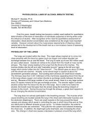
in the blood. For each <strong>gas</strong>, we normalized the end-exhaled<br />
partial pressure by the venous partial pressure <strong>and</strong> plotted<br />
these data (open circles) on a semi-log plot against l b:a<br />
(Fig. 4). This plot of soluble <strong>gas</strong> excretion is a reflection of<br />
a plot from Kumagai et al. [25] that summarized uptake<br />
data from the literature (Excretion ¼ 1 Uptake). Included<br />
on this plot are similar predictions from the classic model<br />
of <strong>gas</strong> <strong>exchange</strong> (solid line). This classic model [4] describes<br />
<strong>gas</strong> <strong>exchange</strong> in an alveolar unit, excludes <strong>airway</strong> <strong>gas</strong><br />
<strong>exchange</strong>, depends on the ventilation–perfusion ratio <strong>and</strong><br />
l b:a , <strong>and</strong> is used to interpret end-exhaled breath measurements.<br />
For l b:a o1, <strong>gas</strong> <strong>exchange</strong> occurs exclusively in the<br />
alveoli <strong>and</strong> thus, classic alveolar predictions accurately<br />
reflect pulmonary <strong>gas</strong> <strong>exchange</strong>. An algebraic transformation<br />
from Farhi [4] allows conversion of this end-exhaled<br />
measurement to an alveolar value. As l b:a exceeds 100, the<br />
classic alveolar model assumes that the end-exhaled<br />
concentration represents the blood <strong>and</strong> alveolar value.<br />
However, our <strong>airway</strong>–alveolar model predicts that the endexhaled<br />
partial pressure for <strong>gas</strong>es with l b:a 4100 is always<br />
less than the blood value. The difference between the endexhaled<br />
<strong>and</strong> blood partial pressures is a non-linear function<br />
of l b:a that reaches a maximum at l b:a 800. Currently,<br />
Fig. 4. End-exhaled inert <strong>gas</strong> partial pressure normalized by venous<br />
partial pressure versus blood–air partition coefficient during elimination<br />
of soluble <strong>gas</strong>es during tidal breathing. Predictions from two models are<br />
presented: (1) the classic alveolar <strong>gas</strong> <strong>exchange</strong> model [4] that excludes<br />
<strong>airway</strong> <strong>gas</strong> <strong>exchange</strong> (solid line); <strong>and</strong> (2) a model of <strong>gas</strong> <strong>exchange</strong> in<br />
humans [7] that includes both <strong>airway</strong> <strong>and</strong> alveolar <strong>gas</strong> <strong>exchange</strong> (open<br />
circles). When l b:a 4100, <strong>airway</strong> <strong>gas</strong> <strong>exchange</strong> dominates <strong>and</strong> the<br />
predictions from the two models deviate. The vertical difference between<br />
the solid line <strong>and</strong> each circle represents the underestimation of alveolar air<br />
if <strong>airway</strong> <strong>exchange</strong> is neglected. EGME, ethylene glycol monobutyl ether.
ARTICLE IN PRESS<br />
J.C. Anderson, M.P. <strong>Hlastala</strong> / Pulmonary Pharmacology & Therapeutics 20 (2007) 112–117 117<br />
a simple transformation is unavailable to adjust the endexhaled<br />
concentrations to blood values for <strong>gas</strong>es with<br />
l b:a 4100. Thus, end-exhaled concentrations of <strong>gas</strong>es with<br />
l b:a 4100 should be interpreted as an estimate of blood<br />
concentration that may be 30% less than the true blood<br />
value.<br />
6. Conclusion<br />
Airway <strong>gas</strong> <strong>exchange</strong> significantly impacts the interpretation<br />
of breath <strong>tests</strong>, particularly for <strong>gas</strong>es with a<br />
blood–air partition coefficient greater than 100. The<br />
absorption–desorption kinetics of <strong>airway</strong> <strong>gas</strong> <strong>exchange</strong><br />
cause the end-exhaled <strong>gas</strong> concentration to be less than the<br />
blood value by up to 30%. Additionally, factors such as<br />
<strong>airway</strong> perfusion <strong>and</strong> diffusion that govern <strong>airway</strong> <strong>gas</strong><br />
<strong>exchange</strong> are intrinsic to the lung <strong>and</strong> affected by lung<br />
disease. Other factors like inspired air temperature <strong>and</strong><br />
breathing maneuver should be carefully controlled to<br />
ensure accurate <strong>and</strong> repeatable breath measurements.<br />
Acknowledgments<br />
This work was supported, in part, by Grant<br />
T32EB001650 from the National Institute for Biomedical<br />
Imaging <strong>and</strong> Bioengineering <strong>and</strong> by Grants HL24163 <strong>and</strong><br />
HL073598 from the National Heart, Lung, <strong>and</strong> Blood<br />
Institute.<br />
References<br />
[1] Phillips M, Cataneo RN, Cummin AR, Gagliardi AJ, Gleeson K,<br />
Greenberg J, et al. Detection of lung cancer with volatile markers in<br />
the breath. Chest 2003;123:2115–23.<br />
[2] Mendis S, Sobotka PA, Euler DE. Expired hydrocarbons in patients<br />
with acute myocardial infarction. Free Radic Res 1995;23:117–22.<br />
[3] Kupari M, Lommi J, Ventila M, Karjalainen U. <strong>Breath</strong> acetone in<br />
congestive heart failure. Am J Cardiol 1995;76:1076–8.<br />
[4] Farhi LE. Elimination of inert <strong>gas</strong> by the lung. Respir Physiol<br />
1967;3:1–11.<br />
[5] George SC, Babb AL, <strong>Hlastala</strong> MP. Dynamics of soluble <strong>gas</strong><br />
<strong>exchange</strong> in the <strong>airway</strong>s. III. Single-exhalation breathing maneuver.<br />
J Appl Physiol 1993;75:2439–49.<br />
[6] Anderson JC, Lamm WJE, <strong>Hlastala</strong> MP. Measuring <strong>airway</strong> <strong>exchange</strong><br />
of endogenous acetone using a single exhalation breathing maneuver.<br />
J Appl Physiol 2006; 100, in press.<br />
[7] Anderson JC, Babb AL, <strong>Hlastala</strong> MP. Modeling soluble <strong>gas</strong><br />
<strong>exchange</strong> in the <strong>airway</strong>s <strong>and</strong> alveoli. Ann Biomed Eng 2003;31:<br />
1402–22.<br />
[8] Schrikker AC, de Vries WR, Zwart A, Luijendijk SC. Uptake of<br />
highly soluble <strong>gas</strong>es in the epithelium of the conducting <strong>airway</strong>s.<br />
Pflugers Arch—Eur J Physiol 1985;405:389–94.<br />
[9] Kaneko T, Wang PY, Sato A. Partition-coefficients of some acetate<br />
esters <strong>and</strong> alcohols in water, blood, olive oil, <strong>and</strong> rat-tissues. Occup<br />
Environ Med 1994;51:68–72.<br />
[10] Meulenberg CJ, Vijverberg HP. Empirical relations predicting human<br />
<strong>and</strong> rat tissue:air partition coefficients of volatile organic compounds.<br />
Toxicol Appl Pharmacol 2000;165:206–16.<br />
[11] Souders JE, George SC, Polissar NL, Swenson ER, <strong>Hlastala</strong> MP.<br />
Tracheal <strong>gas</strong> <strong>exchange</strong>: perfusion-related differences in inert <strong>gas</strong><br />
elimination. J Appl Physiol 1995;79:918–28.<br />
[12] Schimmel C, Bernard SL, Anderson JC, Polissar NL, Lakshminarayan<br />
S, <strong>Hlastala</strong> MP. Soluble <strong>gas</strong> <strong>exchange</strong> in the pulmonary <strong>airway</strong>s<br />
of sheep. J Appl Physiol 2004;97:1702–8.<br />
[13] Charan NB, Carvalho P. Angiogenesis in bronchial circulatory<br />
system after unilateral pulmonary artery obstruction. J Appl Physiol<br />
1997;82:284–91.<br />
[14] Kumar SD, Emery MJ, Atkins ND, Danta I, Wanner A. Airway<br />
mucosal blood flow in bronchial asthma. Am J Respir Crit Care Med<br />
1998;158:153–6.<br />
[15] Charan NB, Carvalho PG. The bronchial circulation in chronic lung<br />
infections. In: Butler J, editor. The Bronchial Circulation. New York:<br />
Marcel Dekker; 1992. p. 535–49.<br />
[16] Abdi S, Traber LD, Herndon DN, Rogers CS, Traber DL. Effects of<br />
ibuprofen on <strong>airway</strong> vascular response to cotton smoke injury. Eur J<br />
Pharmacol 1995;293:475–81.<br />
[17] Henig NR, Glenny RW, Aitken ML. A hypertrophied bronchial<br />
circulatory system may participate in <strong>gas</strong> <strong>exchange</strong>. Lancet 1998;<br />
351:113.<br />
[18] Swenson ER, Robertson HT, Polissar NL, Middaugh ME, <strong>Hlastala</strong><br />
MP. Conducting <strong>airway</strong> <strong>gas</strong> <strong>exchange</strong>: diffusion-related differences in<br />
inert <strong>gas</strong> elimination. J Appl Physiol 1992;72:1581–8.<br />
[19] George SC, Babb AL, Deffebach ME, <strong>Hlastala</strong> MP. Diffusion of<br />
nonelectrolytes in the canine trachea: effect of tight junction. J Appl<br />
Physiol 1996;80:1687–95.<br />
[20] Jones AW, Skagerberg S, Yonekura T, Sato A. Metabolic interaction<br />
between endogenous methanol <strong>and</strong> exogenous ethanol studied in<br />
human volunteers by analysis of breath. Pharmacol Toxicol 1990;66:<br />
62–5.<br />
[21] Smith D, Spanel P, Davies S. Trace <strong>gas</strong>es in breath of healthy<br />
volunteers when fasting <strong>and</strong> after a protein-calorie meal: a<br />
preliminary study. J Appl Physiol 1999;87:1584–8.<br />
[22] Musa-Veloso K, Rarama E, Comeau F, Curtis R, Cunnane S.<br />
Epilepsy <strong>and</strong> the ketogenic diet: assessment of ketosis in children<br />
using breath acetone. Pediatr Res 2002;52:443–8.<br />
[23] Moser B, Bodrogi F, Eibl G, Lechner M, Rieder J, Lirk P. Mass<br />
spectrometric profile of exhaled breath—field study by PTR-MS.<br />
Respir Physiol Neurobiol 2005;145:295–300.<br />
[24] Galassetti PR, Novak B, Nemet D, Rose-Gottron C, Cooper DM,<br />
Meinardi S, et al. <strong>Breath</strong> ethanol <strong>and</strong> acetone as indicators of serum<br />
glucose levels: an initial report. Diabetes Technol Ther 2005;7:<br />
115–23.<br />
[25] Kumagai S, Matsunaga I. A lung model describing uptake of organic<br />
solvents <strong>and</strong> roles of mucosal blood flow <strong>and</strong> metabolism in the<br />
bronchioles. Inhal Toxicol 2000;12:491–510.