Primary Thermosensory Events in Cells - Springer
Primary Thermosensory Events in Cells - Springer
Primary Thermosensory Events in Cells - Springer
You also want an ePaper? Increase the reach of your titles
YUMPU automatically turns print PDFs into web optimized ePapers that Google loves.
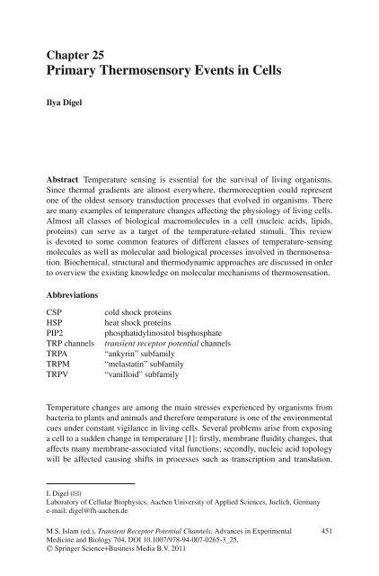
Chapter 25<br />
<strong>Primary</strong> <strong>Thermosensory</strong> <strong>Events</strong> <strong>in</strong> <strong>Cells</strong><br />
Ilya Digel<br />
Abstract Temperature sens<strong>in</strong>g is essential for the survival of liv<strong>in</strong>g organisms.<br />
S<strong>in</strong>ce thermal gradients are almost everywhere, thermoreception could represent<br />
one of the oldest sensory transduction processes that evolved <strong>in</strong> organisms. There<br />
are many examples of temperature changes affect<strong>in</strong>g the physiology of liv<strong>in</strong>g cells.<br />
Almost all classes of biological macromolecules <strong>in</strong> a cell (nucleic acids, lipids,<br />
prote<strong>in</strong>s) can serve as a target of the temperature-related stimuli. This review<br />
is devoted to some common features of different classes of temperature-sens<strong>in</strong>g<br />
molecules as well as molecular and biological processes <strong>in</strong>volved <strong>in</strong> thermosensation.<br />
Biochemical, structural and thermodynamic approaches are discussed <strong>in</strong> order<br />
to overview the exist<strong>in</strong>g knowledge on molecular mechanisms of thermosensation.<br />
Abbreviations<br />
CSP<br />
HSP<br />
PIP2<br />
TRP channels<br />
TRPA<br />
TRPM<br />
TRPV<br />
cold shock prote<strong>in</strong>s<br />
heat shock prote<strong>in</strong>s<br />
phosphatidyl<strong>in</strong>ositol bisphosphate<br />
transient receptor potential channels<br />
“ankyr<strong>in</strong>” subfamily<br />
“melastat<strong>in</strong>” subfamily<br />
“vanilloid” subfamily<br />
Temperature changes are among the ma<strong>in</strong> stresses experienced by organisms from<br />
bacteria to plants and animals and therefore temperature is one of the environmental<br />
cues under constant vigilance <strong>in</strong> liv<strong>in</strong>g cells. Several problems arise from expos<strong>in</strong>g<br />
a cell to a sudden change <strong>in</strong> temperature [1]: firstly, membrane fluidity changes, that<br />
affects many membrane-associated vital functions; secondly, nucleic acid topology<br />
will be affected caus<strong>in</strong>g shifts <strong>in</strong> processes such as transcription and translation.<br />
I. Digel (B)<br />
Laboratory of Cellular Biophysics, Aachen University of Applied Sciences, Juelich, Germany<br />
e-mail: digel@fh-aachen.de<br />
M.S. Islam (ed.), Transient Receptor Potential Channels, Advances <strong>in</strong> Experimental<br />
Medic<strong>in</strong>e and Biology 704, DOI 10.1007/978-94-007-0265-3_25,<br />
C○ Spr<strong>in</strong>ger Science+Bus<strong>in</strong>ess Media B.V. 2011<br />
451
452 I. Digel<br />
F<strong>in</strong>ally, the prote<strong>in</strong> function is directly affected both from structural and catalytic<br />
po<strong>in</strong>ts of view.<br />
Hence, liv<strong>in</strong>g cells need “devices” for sens<strong>in</strong>g environmental temperature<br />
changes <strong>in</strong> order to adapt their biochemical processes accord<strong>in</strong>gly. A successful<br />
adaptive thermotropic response cannot be performed only by correspond<strong>in</strong>g changes<br />
<strong>in</strong> the rate and equilibrium of enzymatic reactions. Such a mechanism of adaptive<br />
reaction is too unspecific and uncontrollable. To cope with temperature variation,<br />
liv<strong>in</strong>g organisms need sens<strong>in</strong>g temperature alterations and translat<strong>in</strong>g this sensory<br />
event <strong>in</strong>to a pragmatic gene response.<br />
While such regulatory cascades may ultimately be complicated, they conta<strong>in</strong><br />
primary sensor mach<strong>in</strong>ery at the top of the cascade. The functional core of such<br />
mach<strong>in</strong>ery is usually that of a temperature-<strong>in</strong>duced conformational or physicochemical<br />
change <strong>in</strong> the central constituents of the cell. Hence, a specific sensory<br />
transduction mechanism is needed, <strong>in</strong>clud<strong>in</strong>g, as a key element, a molecular sensor,<br />
transform<strong>in</strong>g certa<strong>in</strong> physical parameter (temperature) <strong>in</strong>to a biologically significant<br />
signal (change <strong>in</strong> membrane permeability, specific <strong>in</strong>hibition/stimulation of<br />
gene expression, etc.). In a sense, a liv<strong>in</strong>g organism can use structural alterations<br />
<strong>in</strong> its biomolecules as the primary thermometers or thermostats. Thus, sensory<br />
transduction is a complex biological process aimed at <strong>in</strong>tegrat<strong>in</strong>g and decod<strong>in</strong>g<br />
physical and chemical stimuli performed by primary sensory molecular devices.<br />
Furthermore, sensory perception of potentially harmful stimuli can function as a<br />
warn<strong>in</strong>g mechanism to avert potential tissue/organ damage.<br />
Among temperature-controlled processes <strong>in</strong> liv<strong>in</strong>g organisms, most well-known<br />
are the expression of heat-shock and cold-shock genes [2]. Relocation of a culture<br />
of Escherichia coli adapted to an optimal growth to a sudden temperature <strong>in</strong>crease,<br />
or decrease, by some 10–15 ◦ C results <strong>in</strong> adaptive shock responses. Such responses<br />
<strong>in</strong>volve a remodel<strong>in</strong>g of bacterial gene expression, aimed at adjust<strong>in</strong>g bacterial cell<br />
physiology to the new environmental demands [3, 4]. The response of prokaryotic<br />
and eukaryotic systems to heat-shock stress has been thoroughly <strong>in</strong>vestigated <strong>in</strong><br />
a large number of organisms and model cell systems. Notably, all organisms from<br />
prokaryotes to higher eukaryotes respond to cold and heat shock <strong>in</strong> a similar manner.<br />
The general response of cells to temperature stress (cold or heat) is the rapid overexpression<br />
of small groups of prote<strong>in</strong>s, the so-called CSPs (cold-shock prote<strong>in</strong>s)<br />
or HSPs (heat shock prote<strong>in</strong>s), respectively, but the <strong>in</strong>itial launch<strong>in</strong>g mechanism is<br />
different <strong>in</strong> both cases.<br />
In bacteria, the heat response generally <strong>in</strong>vokes some 20 heat-shock prote<strong>in</strong>s,<br />
mostly chaperones, whose functions are primarily to help deal<strong>in</strong>g with, and alleviate,<br />
the cellular stress imposed by heat [5]. Many of these prote<strong>in</strong>s participate <strong>in</strong><br />
reconstitut<strong>in</strong>g and stabiliz<strong>in</strong>g prote<strong>in</strong> structures and <strong>in</strong> remov<strong>in</strong>g misfolded ones.<br />
The expression of this special chaperone system, which <strong>in</strong>cludes the prote<strong>in</strong>s<br />
DnaK, DnaJ and GrpE, is activated by the appearence of misfolded, temperaturedenatured<br />
prote<strong>in</strong>s. Thus, one could implicate the b<strong>in</strong>d<strong>in</strong>g of partially unfolded<br />
prote<strong>in</strong>s by chaperones as the thermosensoric event regulat<strong>in</strong>g expression of heatshock<br />
prote<strong>in</strong>s, where the primary sensory element is constituted by some easily
25 <strong>Primary</strong> <strong>Thermosensory</strong> <strong>Events</strong> <strong>in</strong> <strong>Cells</strong> 453<br />
denatur<strong>in</strong>g prote<strong>in</strong>s. This, <strong>in</strong> turn, demonstrates that even bacteria can practically<br />
utilize destructive changes <strong>in</strong> prote<strong>in</strong> conformation as a means for temperature<br />
sens<strong>in</strong>g.<br />
In case of cold shock, the primary sens<strong>in</strong>g event is more obscure. Various reports<br />
have now shown that when <strong>in</strong> vitro cultivation temperature is lowered, the <strong>in</strong>crease<br />
of the cell membrane rigidity results <strong>in</strong> compromised membrane-associated cellular<br />
functions. Furthermore, cold stress dramatically h<strong>in</strong>ders membrane-bound enzymes,<br />
slows down diffusion rates and <strong>in</strong>duces cluster formation of <strong>in</strong>tegral membranous<br />
prote<strong>in</strong>s [6]. In mammalian cells, the five known mechanisms by which cold-shock<strong>in</strong>duced<br />
changes affect gene expression are: (i) a general reduction <strong>in</strong> transcription<br />
and translation, (ii) <strong>in</strong>hibition of RNA degradation, (iii) <strong>in</strong>creased transcription of<br />
specific target genes via elements <strong>in</strong> the promoter region of such genes, (iv) alternative<br />
pre-mRNA splic<strong>in</strong>g, and (v) via the presence of cold-shock specific <strong>in</strong>ternal<br />
ribosome entry segments <strong>in</strong> mRNAs that result <strong>in</strong> the preferential and enhanced<br />
translation of such mRNAs upon cold shock [7].<br />
It has been po<strong>in</strong>ted out that cold stress exposes cells not to one but to two major<br />
stresses: those related to changes <strong>in</strong> temperature and those related to changes <strong>in</strong> dissolved<br />
oxygen concentration at decreased temperature, and it is therefore necessary<br />
to study responses to each, either <strong>in</strong>dependently or as part of a coord<strong>in</strong>ated response.<br />
Separat<strong>in</strong>g the relative effects of temperature and oxygen as a result of decreased<br />
temperature is difficult and has not been extensively addressed to date. Both changes<br />
<strong>in</strong> dissolved oxygen and temperature reduction result <strong>in</strong> similar changes <strong>in</strong> cultured<br />
mammalian cells [7].<br />
The shock response systems briefly mentioned above belong to ultimate mechanisms<br />
aimed to survival under extreme temperature conditions. However, the ability<br />
to express certa<strong>in</strong> factors can be affected by reasonably small temperature changes.<br />
Less drastic changes <strong>in</strong> temperature may not <strong>in</strong>duce shock responses, but can be<br />
sufficient to modulate the expression of bacterial virulence genes, for example <strong>in</strong><br />
Shigellae [8] and Yers<strong>in</strong>iae [9]. While one might be surprised that organisms built<br />
on such m<strong>in</strong>imalist approaches as bacteria respond to temperature changes, the<br />
consequence of these observations is that even bacteria actually sense temperature<br />
shifts <strong>in</strong> order to control gene expression accord<strong>in</strong>gly. Investigators have now been<br />
study<strong>in</strong>g the moderate temperature sensation <strong>in</strong> a variety of organisms for at least<br />
several decades or more. Recently, a number of reports have shown that expos<strong>in</strong>g<br />
bacteria, yeasts or mammalian cells to sub-physiological temperatures <strong>in</strong>vokes a<br />
coord<strong>in</strong>ated cellular response manifest<strong>in</strong>g itself as alterations <strong>in</strong> transcription, translation,<br />
metabolism, the cell cycle and the cell cytoskeleton [7, 10–13]. Nevertheless,<br />
very little is known so far about the molecular mechanisms that govern <strong>in</strong>itial<br />
response on small thermal stimuli, particularly the primary sensory transduction<br />
mechanisms.<br />
Below, we have tried to uncover some aspects of the biophysical basis of<br />
temperature sens<strong>in</strong>g by biological molecular thermometers, summariz<strong>in</strong>g some<br />
most general ideas concern<strong>in</strong>g the primary components of temperature signal<br />
transduction.
454 I. Digel<br />
25.1 Temperature Sens<strong>in</strong>g Biomolecules<br />
In addition to specificity and sensitivity, successful thermoresponse should be one<br />
that is reversible and controlled. Thus, complexity of thermosens<strong>in</strong>g and thermoregulation<br />
on the organism level may reflect the demands to handle and f<strong>in</strong>e-tune<br />
responses to an important environmental factor <strong>in</strong> a dynamic fashion. However,<br />
ultimately, it seems that basic and rather simple (bio) chemical processes are<br />
serve as primary sensory events and, for that purpose thermotropic changes <strong>in</strong><br />
physico-chemical state of biological molecules appear highly suitable. While the<br />
<strong>in</strong>formation available is somewhat scant, the picture emerg<strong>in</strong>g shows that cells can<br />
use signals generated through changes <strong>in</strong> nucleic acid or prote<strong>in</strong> conformation, or<br />
changes <strong>in</strong> membrane lipid behavior, as sensory devices. Bellow we make a short<br />
overview of temperature-sens<strong>in</strong>g properties of most important groups of biological<br />
macro-molecules.<br />
It is worthy to note that probably even water alone could serve as a primitive<br />
temperature sensor. In the middle of the twentieth century Oppenheimer and Drost-<br />
Hansen [14] reported that a number of more or less abrupt changes <strong>in</strong> the properties<br />
of water and aqueous solutions occur when the temperature is <strong>in</strong>creased from 0<br />
to 60 ◦ C. These changes or “k<strong>in</strong>ks” occured with<strong>in</strong> a rather narrow temperature<br />
range (±2 ◦ C) near 15, 30, 45, and 60 ◦ C, respectively and most probably caused<br />
by changes <strong>in</strong> the hydrogen bond network of the water. The authors argued that the<br />
temperature-<strong>in</strong>duced structural changes <strong>in</strong> water and aqueous solutions exert a direct<br />
<strong>in</strong>fluence on biological phenomena. In a later work W. Drost-Hansen [15] suggested<br />
some mechanisms how these structural changes happen<strong>in</strong>g with vic<strong>in</strong>al (adjacent<br />
to surfaces) water can affect the behavior or activity of biological systems. It was<br />
argued that optimal conditions for a complex physiological activity (such as, for<br />
<strong>in</strong>stance, growth) will occur somewhere near the middle of the <strong>in</strong>terval between two<br />
consecutive k<strong>in</strong>ks. This issue has been discussed <strong>in</strong> literature very controversially<br />
and has not received wide recognition.<br />
25.2 Membrane Lipids Fluidity<br />
The physical state of phospholipid membranes does change <strong>in</strong> response to temperature<br />
shifts <strong>in</strong> phase-transition manner [16], but the temperature-<strong>in</strong>duced changes<br />
<strong>in</strong> real biological membranes are not sharp because many k<strong>in</strong>ds of fatty acids<br />
and cholesterol-like molecules present, hav<strong>in</strong>g different characteristic temperature<br />
po<strong>in</strong>ts of phase transition. Thus, it would not be surpris<strong>in</strong>g if cells (even<br />
those of bacteria) could utilize the changes <strong>in</strong> membrane fluidity as a thermometer<br />
device, assisted by prote<strong>in</strong> helpers, play<strong>in</strong>g a role of switchers, “sharpen<strong>in</strong>g”<br />
the temperature response. Microorganisms counteract the membrane propensity<br />
to rigidify at lower temperature and are able to ma<strong>in</strong>ta<strong>in</strong> a more-or-less constant<br />
degree of membrane fluidity (homeoviscous adaptation). The cyanobacterium<br />
Synecocystis responds to decreased temperature by <strong>in</strong>creas<strong>in</strong>g the cis-unsaturation
25 <strong>Primary</strong> <strong>Thermosensory</strong> <strong>Events</strong> <strong>in</strong> <strong>Cells</strong> 455<br />
of membrane-lipid fatty acids through express<strong>in</strong>g acyl-lipid desaturases [17–19].<br />
Lipid unsaturation would then restore membrane fluidity at the lower temperature.<br />
In B. subtilis, this lipid modification is <strong>in</strong>itiated through the activity of a so-called<br />
“two-component regulatory system” consist<strong>in</strong>g of the DesK and DesR prote<strong>in</strong>s [17].<br />
Prokaryotic two-component regulatory systems usually consist of prote<strong>in</strong> pairs: a<br />
sensor k<strong>in</strong>ase and a regulatory prote<strong>in</strong> [20].<br />
It appears that it is a comb<strong>in</strong>ation of membrane physical state and prote<strong>in</strong> conformation<br />
that is able to sense temperature and even to translate this sens<strong>in</strong>g event <strong>in</strong>to<br />
proper gene expression. However, sens<strong>in</strong>g of temperature through direct alteration<br />
<strong>in</strong> nucleic acid conformation might be more efficient temperature-mediated mechanism<br />
of gene expression.<br />
25.3 RNA and DNA Thermotropic Reactions<br />
Theoretically, RNA molecules have a strong potential as temperature sensors, <strong>in</strong> that<br />
they can form pronounced secondary and tertiary structures [21], and through their<br />
ability to form <strong>in</strong>termolecular RNA: RNA hybrids [22]. Both of these processes<br />
greatly depend on the formation of complementary base pair<strong>in</strong>g, and consequently<br />
one would anticipate these to be dependent on environmental temperature. Indeed,<br />
messenger RNAs, apart from carry<strong>in</strong>g their cod<strong>in</strong>g <strong>in</strong>formation for prote<strong>in</strong> generation<br />
are also rapidly emerg<strong>in</strong>g as regulators of expression of the encoded message.<br />
With unique chemical and structural properties, sensory RNAs perform vital regulatory<br />
roles <strong>in</strong> gene expression by detect<strong>in</strong>g changes <strong>in</strong> the cellular environment either<br />
alone or through <strong>in</strong>teractions with small ligands [23, 24] and prote<strong>in</strong>s [25, 26].<br />
Regulatory RNA elements, “riboswitches”, have been reported recently, respond<strong>in</strong>g<br />
to <strong>in</strong>tracellular signals by conformational changes. Riboswitches are conceptually<br />
divided <strong>in</strong>to two parts: an aptamer and an expression platform. The aptamer<br />
directly b<strong>in</strong>ds the small molecule, and the expression platform undergoes structural<br />
changes <strong>in</strong> response to the changes <strong>in</strong> the aptamer. The expression platform is what<br />
regulates gene expression. Riboswitches demonstrate that naturally occurr<strong>in</strong>g RNA<br />
can specifically response on versatile physical and chemical stimuli, a capability<br />
that many previously believed was the doma<strong>in</strong> of prote<strong>in</strong>s or artificially constructed<br />
RNAs [27].<br />
RNA thermometers operate at the post-transcriptional level to sense selectively<br />
the temperature and transduce a signal to the translation mach<strong>in</strong>ery via a conformational<br />
change. They have usually a highly structured 5 ′ -end that shields the<br />
ribosome b<strong>in</strong>d<strong>in</strong>g site at physiological temperatures [28–31]. Changes <strong>in</strong> temperature<br />
are manifested by the liberation of the Sh<strong>in</strong>e–Dalgarno (SD) sequence, thereby<br />
facilitat<strong>in</strong>g ribosome b<strong>in</strong>d<strong>in</strong>g and translation <strong>in</strong>itiation.<br />
It is known that both <strong>in</strong> prokaryotic and eukaryotic cells the geometry and tension<br />
of DNA is highly dynamic and corresponds to its functional activity. In the<br />
bacterial cell, chromosome and plasmid DNAs are conta<strong>in</strong>ed <strong>in</strong> a “twisted” superhelical<br />
conformation [32, 33], where the degree of supercoil<strong>in</strong>g varies <strong>in</strong> response to
456 I. Digel<br />
changes <strong>in</strong> the ambient temperature. The expression of many genes is dependent on<br />
DNA conformation, and temperature-dependent gene regulation is mastered through<br />
changes <strong>in</strong> DNA supercoil<strong>in</strong>g [3, 34, 35].<br />
Examples of pure DNA-related temperature sensitivity are rare if ever reported.<br />
In most cases, genomic thermo-sensitivity appears to be a result of certa<strong>in</strong> <strong>in</strong>terplay<br />
among DNA, RNA and prote<strong>in</strong>s. Some bacteria carry a DNA-plasmid which shows<br />
a controlled constant plasmid copy number at one temperature and a much higher or<br />
totally uncontrolled copy number at a different temperature The high copy number<br />
phenotype of pLO88 plasmid ma<strong>in</strong>ta<strong>in</strong>ed <strong>in</strong> Escherichia coli (HB101) is observed<br />
only at elevated temperatures, (above 37 ◦ C), and is due to the precise position of a<br />
Tn5 <strong>in</strong>sertion <strong>in</strong> DNA, but the exact mechanism rema<strong>in</strong>s obscure [36].<br />
Recent experiments show [37] that artificial thermoresponsive devices may be<br />
constructed based on the temperature-dependence of the relative populations of<br />
left- and right-handed nucleic acid helical conformations. The authors reported that<br />
“upon an <strong>in</strong>crease <strong>in</strong> temperature, particular sequences of DNA oligonucleotide<br />
duplexes <strong>in</strong> high salt conditions switch from a left-handed (Z) form to a righthanded<br />
(B) one, while RNA responds <strong>in</strong>versely by switch<strong>in</strong>g from a right- (A)<br />
to a left-handed (Z) form ...Calculations revealed a complex <strong>in</strong>terplay between<br />
configurational, water, and ionic entropies, which, comb<strong>in</strong>ed with the sequencedependence,<br />
rationalize the experimentally observed transitions from A- to Z-RNA<br />
and Z- to B-DNA <strong>in</strong> high salt concentrations and provide <strong>in</strong>sight that may aid<br />
future developments of the use of nucleic acids oligomers for thermal sens<strong>in</strong>g at<br />
the nanoscale <strong>in</strong> physiological conditions.” [37]<br />
The role of DNA-b<strong>in</strong>d<strong>in</strong>g prote<strong>in</strong>s has been established for plant thermosensitivity<br />
too. Kumar and Wigge [38] have revealed that eviction of the histone<br />
H2A.Z from nucleosomes performs a central role <strong>in</strong> plant thermosensory perception.<br />
Us<strong>in</strong>g purified nucleosomes, they showed that H2A.Z displays dist<strong>in</strong>ct responses to<br />
temperature <strong>in</strong> vivo, <strong>in</strong>dependently of transcription events.<br />
Apparently, the temperature-<strong>in</strong>duced conformational changes <strong>in</strong> DNA are ma<strong>in</strong>ly<br />
controlled through the presence of “nucleotid-associated” prote<strong>in</strong>s, of which H–NS<br />
is the best characterized [32, 39]. In E. coli, creat<strong>in</strong>g and ma<strong>in</strong>ta<strong>in</strong><strong>in</strong>g conformational<br />
structures <strong>in</strong> the DNA molecule are ma<strong>in</strong>ly regulated through the balance of<br />
two oppos<strong>in</strong>g topoisomerase activities, ma<strong>in</strong>ly those of topoisomerases II and I [40,<br />
41]. The abovementioned examples of membrane- and nucleic acid-based temperature<br />
sensitivity imply that these systems often <strong>in</strong>clude prote<strong>in</strong>s as a key regulatory<br />
component. Therefore, from the po<strong>in</strong>t of view of molecular temperature sensation,<br />
prote<strong>in</strong>-based molecular “thermometers” represent an extremely <strong>in</strong>terest<strong>in</strong>g group.<br />
25.4 Prote<strong>in</strong> Thermometers<br />
Many sensory pathways <strong>in</strong> liv<strong>in</strong>g organisms use structural changes <strong>in</strong> prote<strong>in</strong>s as<br />
a primary perceptive event, activat<strong>in</strong>g further signal<strong>in</strong>g cascades. E. coli be<strong>in</strong>g<br />
exposed to an oxidative agent such as hydrogen peroxide, responds by the activation
25 <strong>Primary</strong> <strong>Thermosensory</strong> <strong>Events</strong> <strong>in</strong> <strong>Cells</strong> 457<br />
of a transcriptional regulator prote<strong>in</strong> OxyR [42]. Activation of OxyR is achieved<br />
through the formation of a disulphide bound with<strong>in</strong> the prote<strong>in</strong>, upon which OxyR<br />
<strong>in</strong>duces the expression of a set of genes adapt<strong>in</strong>g the bacterial cell to oxidative stress.<br />
This illustrates how it is possible both to “sense” and respond to an abrupt change<br />
<strong>in</strong> a specific environmental factor <strong>in</strong> a simple, yet elegant mode.<br />
One would expect the organisms and cells to be similarly elegant when sens<strong>in</strong>g<br />
temperature shifts. Indeed, a strik<strong>in</strong>g example is the temperature-controlled<br />
switch<strong>in</strong>g of the flagellar rotary motor of E. coli between the two rotational states,<br />
clockwise (CW) and counterclockwise (CCW) [43]. The molecular mechanism for<br />
switch<strong>in</strong>g rema<strong>in</strong>s unknown, but seems to be connected to the response regulator<br />
prote<strong>in</strong> CheY-P. Two models of CheY-P action proposed so far expla<strong>in</strong> shift<strong>in</strong>g the<br />
difference <strong>in</strong> free energy between CW and CCW states <strong>in</strong> terms of (1) conformationrelated<br />
differential b<strong>in</strong>d<strong>in</strong>g [44, 45] and (2) thermodynamic changes <strong>in</strong> dissociation<br />
constants [46].<br />
Further studies on the thermosensory transduc<strong>in</strong>g system <strong>in</strong> E. coli revealed that<br />
two major chemoreceptors, Tar and Tsr, which detect aspartate and ser<strong>in</strong>e, respectively,<br />
also function as thermoreceptors, together with Trg and Tap receptors [47].<br />
Interest<strong>in</strong>gly, <strong>in</strong> spite of different specificity and sensitivity, am<strong>in</strong>o acid sequences<br />
of all these four chemoreceptors have a significant homology. They are transmembrane<br />
prote<strong>in</strong>s hav<strong>in</strong>g two functional doma<strong>in</strong>s act<strong>in</strong>g as chemoreceptors: one is a<br />
ligand-b<strong>in</strong>d<strong>in</strong>g doma<strong>in</strong> located <strong>in</strong> the periplasm and the other is a signal<strong>in</strong>g doma<strong>in</strong><br />
located <strong>in</strong> the cytoplasm. Thus, it is suggested that a temperature change <strong>in</strong>duces a<br />
conformational change <strong>in</strong> these two receptors and that this conformational change<br />
triggers the signal<strong>in</strong>g for thermoresponse. In the simplest model of thermoreception<br />
by these receptors, two conformational states of these receptors are assumed:<br />
a low-temperature state and a high-temperature state [48]. The swimm<strong>in</strong>g pattern<br />
of the Trg- and Tap-conta<strong>in</strong><strong>in</strong>g cells is determ<strong>in</strong>ed simply by the temperature of the<br />
medium, <strong>in</strong>dicat<strong>in</strong>g that these cells under nonadaptive conditions sense the absolute<br />
temperature as the thermal stimulus, and not the relative change <strong>in</strong> temperature.<br />
The understand<strong>in</strong>g of prote<strong>in</strong> thermotropic sensory transductions <strong>in</strong> terms of<br />
their underly<strong>in</strong>g molecular mechanism is fast-advanc<strong>in</strong>g thanks to the discovery and<br />
functional characterization of the transient receptor potential (TRP) channels. This<br />
prote<strong>in</strong> family, first identified <strong>in</strong> Drosophila, is at the forefront of the sensory stem,<br />
respond<strong>in</strong>g to both physical and chemical stimuli and, thus hav<strong>in</strong>g diverse functions<br />
[49, 50].<br />
The family of TRP channels currently comprises around 30 members grouped<br />
<strong>in</strong>to seven related subfamilies: TRPC, TRPV, TRPA, TRPP, TRPM, TRPN and<br />
TRPML. In higher organisms, TRPV channels are important polymodal <strong>in</strong>tegrators<br />
of noxious stimuli, mediat<strong>in</strong>g among all, thermosensation and nociception (pa<strong>in</strong><br />
sensation) [51].<br />
To characterize thermal sensitivity of cells, molecules and processes, the Q 10<br />
(temperature coefficient) is used. Q 10 reflects the rate of change of a biological<br />
or chemical system as a consequence of <strong>in</strong>creas<strong>in</strong>g the temperature by 10 ◦ C. This<br />
coefficient is used, for example, for the characterization of the nerve conduction<br />
velocity.
458 I. Digel<br />
The Q 10 is calculated as:<br />
Q 10 =<br />
(<br />
R2<br />
R 1<br />
) 10/(T2 −T 1 )<br />
where R is the rate of change and T is the temperature.<br />
For biological systems, the Q 10 value is generally between 1 and 3 but a subset<br />
of TRP channels, the thermo-TRPs, characterized by their unusually high temperature<br />
sensitivity (Q 10 >10): TRPV1–TRPV4 are heat activated [52–54], whereas<br />
TRPM8 [54, 55] and TRPA1 [56] are activated by cold. With a Q 10 of about 26 for<br />
TRPV1 [57] and about 24 for TRPM8 [58, 59], they far surpass the temperature<br />
dependence of the gat<strong>in</strong>g processes characterized by other ion channels (Q 10 ≈ 3)<br />
[57]. In spite of the great advances made <strong>in</strong> last years the molecular basis for regulation<br />
by temperature rema<strong>in</strong>s mostly obscure because of the lack of native structural<br />
<strong>in</strong>formation. Nevertheless, deeper understand<strong>in</strong>g of dynamics and thermodynamics<br />
of these prote<strong>in</strong>s will br<strong>in</strong>g us closer to revelation of universal pr<strong>in</strong>ciples of thermal<br />
sensation.<br />
25.5 Biophysical Aspects of Prote<strong>in</strong> Thermosensitivity<br />
It appears from the above mentioned examples of prote<strong>in</strong> participation <strong>in</strong> temperature<br />
sens<strong>in</strong>g events that sudden conformational changes, “structural transitions”<br />
play essential role on the primary conversion of physical stimulus <strong>in</strong>to biologically<br />
relevant signal.<br />
Phase transitions and other “critical” phenomena cont<strong>in</strong>ue to be the subject of<br />
<strong>in</strong>tensive experimental and theoretical <strong>in</strong>vestigation. In this context, systems consist<strong>in</strong>g<br />
primarily of well characterized prote<strong>in</strong>s and water can serve as particularly<br />
valuable objects of study. The importance of studies of specific phase transitions<br />
<strong>in</strong> prote<strong>in</strong>/water solutions derives also from their physiological relevance to the<br />
supramolecular organization of normal tissues and to certa<strong>in</strong> pathological states. For<br />
example, such phase transitions play the ma<strong>in</strong> role <strong>in</strong> the deformation of the erythrocyte<br />
<strong>in</strong> sickle-cell disease [25, 60] and <strong>in</strong> the cryoprecipitation of immunoglobul<strong>in</strong>s<br />
<strong>in</strong> cryoglobul<strong>in</strong>emia and rheumatoid arthritis [61].<br />
Discussions about prote<strong>in</strong> stability and temperature-<strong>in</strong>duced structural transitions<br />
are usually limited to the stability of the native state aga<strong>in</strong>st denaturation.<br />
Yet the native state may <strong>in</strong>clude different functionally relevant conformations characterized<br />
by different Gibbs energies and therefore different stabilities (e.g. the R<br />
and T states of hemoglob<strong>in</strong>). At biological temperatures, prote<strong>in</strong>s alternate between<br />
well-def<strong>in</strong>ed, dist<strong>in</strong>ct conformations. In order to those conformational states to be<br />
dist<strong>in</strong>ct, there must be a free-energy barrier separat<strong>in</strong>g them (Fig. 25.1). Adaptive<br />
alterations of prote<strong>in</strong> conformation <strong>in</strong> response to signal<strong>in</strong>g events might reflect correspond<strong>in</strong>g<br />
changes <strong>in</strong> free-energy profile. From this po<strong>in</strong>t of view, temperature as<br />
a stimulus does not differ physically from, for example, a ligand-b<strong>in</strong>d<strong>in</strong>g event. The<br />
experimental observation of dist<strong>in</strong>ct conformational populations by IR-spectroscopy<br />
is possible but usually requires the existence of at least two spectrally different but<br />
overlapp<strong>in</strong>g components of the amide I band [62].
25 <strong>Primary</strong> <strong>Thermosensory</strong> <strong>Events</strong> <strong>in</strong> <strong>Cells</strong> 459<br />
Fig. 25.1 A hypothetical model of a free energy profile of a globular prote<strong>in</strong>. The threshold<br />
character of the prote<strong>in</strong> reaction means that the rest<strong>in</strong>g states and the active states are different<br />
thermodynamic states of the system, separated by an energy barrier. Often, several prote<strong>in</strong> states<br />
are thermodynamically stable and prevail <strong>in</strong> appropriate conditions. Transitions between different<br />
prote<strong>in</strong> states take place <strong>in</strong> the cell <strong>in</strong> response to external stimuli<br />
The motions <strong>in</strong>volved to get from one state to another are usually much more<br />
complex than the oscillation of atoms and groups about their average positions. In<br />
prote<strong>in</strong>s, because most of the forces that stabilize the native state are non-covalent,<br />
there is enough thermal energy at physiological temperature for weak <strong>in</strong>teractions<br />
to break and reform frequently. Thus a prote<strong>in</strong> molecule is more flexible than a<br />
molecule <strong>in</strong> which only covalent forces dictate the structure.<br />
Recently, it became clear that natively unfolded prote<strong>in</strong>s also play an important<br />
role <strong>in</strong> the cell. Dunker et al. [63] proposed to widen the notion of functional prote<strong>in</strong><br />
types <strong>in</strong> the cell: to the “classical” prote<strong>in</strong>s with well def<strong>in</strong>ed tertiary structure,<br />
they added molten globules and prote<strong>in</strong>s with unfolded conformations. Uversky<br />
[64] has suggested to supplement this list with a fourth, relatively stable prote<strong>in</strong><br />
conformation – the premolten globule, which might be called the boil<strong>in</strong>g globule,<br />
as <strong>in</strong> the coord<strong>in</strong>ates of the unfold<strong>in</strong>g reaction it follows the globule and molten<br />
globule and precedes the completely unfolded conformation. Apparently, all these<br />
prote<strong>in</strong> states are thermodynamically stable, although to different degrees.<br />
Even when the native prote<strong>in</strong> does not undergo a conformational change, it is<br />
still characterized by the occurrence of a large number of local unfold<strong>in</strong>g events that<br />
give rise to many sub-states. Thus, the native state itself needs to be considered as<br />
a statistical ensemble of conformations rather than unique entity. These dist<strong>in</strong>ctions<br />
are very important from the functional po<strong>in</strong>t of view s<strong>in</strong>ce different conformations<br />
are usually characterized by different functional properties.
460 I. Digel<br />
The stabiliz<strong>in</strong>g contributions that arise from the hydrophobic effect and hydrogen<br />
bond<strong>in</strong>g are largely offset by the destabiliz<strong>in</strong>g configurational entropy. The<br />
hydrophobic effect is strongly temperature-dependent, and is considerably weaker<br />
and perhaps even destabiliz<strong>in</strong>g at low temperatures than at elevated temperatures.<br />
The contribution of various <strong>in</strong>teractions for a “typical” prote<strong>in</strong> is reported <strong>in</strong> many<br />
works [65–69]. Apparently, the transition from stabiliz<strong>in</strong>g to destabiliz<strong>in</strong>g conditions<br />
is achieved by relatively small changes <strong>in</strong> the environment. These can be<br />
changes <strong>in</strong> temperature, pH, addition of substrates or stabiliz<strong>in</strong>g co-solvents. While<br />
the exact contribution of different <strong>in</strong>teractions to the stability of globular prote<strong>in</strong>s<br />
rema<strong>in</strong>s a question, our understand<strong>in</strong>g seems to be ref<strong>in</strong>ed enough to allow for the<br />
reasonable prediction of the overall fold<strong>in</strong>g thermodynamics apply<strong>in</strong>g the second<br />
law of thermodynamics for free energy changes between folded and unfolded states<br />
[68, 69]. Important to mention that both the enthalpy end entropy changes are not<br />
constant but <strong>in</strong>creas<strong>in</strong>g functions of temperature, and that the Gibbs (free) energy<br />
stabilization of a prote<strong>in</strong> can be written as:<br />
G(T) = H(T R ) + C P (T − T R ) − TS(T R ) + C P ln(T/T R )<br />
where T R is a reference temperature. C p is the heat capacity change, and<br />
H(T R ) and S(T R ) are the enthalpy and entropy values at that temperature, correspond<strong>in</strong>gly.<br />
The temperature dependency of H and S is important because it<br />
transforms the Gibbs energy function from a l<strong>in</strong>ear <strong>in</strong>to a parabolic function of temperature.<br />
This equation has only limited applicability s<strong>in</strong>ce it does not consider the<br />
change of the solvent’s entropy, which is, without doubt, an important contributor<br />
to the thermodynamics of prote<strong>in</strong> behavior <strong>in</strong> solution.<br />
For large values of C p , the Gibbs energy crosses zero po<strong>in</strong>t twice – one at high<br />
temperature (heat denaturation) and one at low temperature (cold denaturation). The<br />
native state is thermodynamically stable between those two temperatures and G<br />
exhibits a maximum at the temperature at which S = 0. The peculiar shape of the<br />
free energy function of a prote<strong>in</strong> does not permit a unique def<strong>in</strong>ition of prote<strong>in</strong> stability.<br />
For example, hav<strong>in</strong>g a higher denaturation temperature does not necessarily<br />
imply that a prote<strong>in</strong> will be more stable at room temperature.<br />
With<strong>in</strong> the context of the structural parameterization of the energetics, the free<br />
energy of prote<strong>in</strong> stabilization is approximated by the equation:<br />
G = G gen + G ion + G tr + G other<br />
where G gen conta<strong>in</strong>s the contributions typically associated with the formation<br />
of secondary and tertiary structure (van der Waals <strong>in</strong>teractions, hydrogen bond<strong>in</strong>g,<br />
hydration, and conformational entropy), G ion comprises the electrostatic and<br />
ionization effects, and G tr reflects the contribution of the change <strong>in</strong> translational<br />
degrees of freedom exist<strong>in</strong>g <strong>in</strong> oligomeric prote<strong>in</strong>s. The term G other <strong>in</strong>cludes<br />
<strong>in</strong>teractions unique to specific prote<strong>in</strong>s that cannot be classified <strong>in</strong> a general way<br />
(e.g. prosthetic groups, metals, and ligands) and must be treated on a case-by-case<br />
basis.
25 <strong>Primary</strong> <strong>Thermosensory</strong> <strong>Events</strong> <strong>in</strong> <strong>Cells</strong> 461<br />
B. Nilius and co-workers have recently applied this simple thermodynamic<br />
formalism to describe the shifts <strong>in</strong> voltage dependence of prote<strong>in</strong> channels due<br />
to changes <strong>in</strong> temperature [70, 71], where the probability of the open<strong>in</strong>g of a<br />
channel is def<strong>in</strong>ed as a function of temperature, Faraday’s constant, the gat<strong>in</strong>g<br />
charge, and the free energy difference between open and closed states of the<br />
channel.<br />
In order to understand the nature of dynamic transitions <strong>in</strong> prote<strong>in</strong>s, it is also<br />
important to consider solvent effects. Solvent can affect prote<strong>in</strong> dynamics by<br />
modify<strong>in</strong>g the effective characteristics of the prote<strong>in</strong> surface and/or by frictional<br />
damp<strong>in</strong>g. Changes <strong>in</strong> the structure and <strong>in</strong>ternal dynamics of prote<strong>in</strong>s <strong>in</strong> dependency<br />
on solvent conditions at physiological temperatures have been found by us<strong>in</strong>g<br />
several experimental techniques [72–74]. It follows from the works of G. Büldt,<br />
G. Artmann, J. Zaccai, A. Stadler and others that solvent affects prote<strong>in</strong> dynamics<br />
differently at different temperatures and salt concentrations [74–77] Therefore,<br />
a solvent dependence of the dynamic transition might be expected. Indeed, measurements<br />
on ligand b<strong>in</strong>d<strong>in</strong>g to myoglob<strong>in</strong> <strong>in</strong>dicated that dynamic behavior of the<br />
prote<strong>in</strong> is correlated with a glass transition <strong>in</strong> the surround<strong>in</strong>g solvent [78]. Recent<br />
molecular dynamics analysis of hydrated myoglob<strong>in</strong> also <strong>in</strong>dicates a major solvent<br />
role <strong>in</strong> prote<strong>in</strong> dynamic transition behavior [79].<br />
One <strong>in</strong>terest<strong>in</strong>g aspect of thermosensation is conversion of the code conformational<br />
changes <strong>in</strong>to the code of cellular signal<strong>in</strong>g. In our op<strong>in</strong>ion, strong<br />
methodological basis for the understand<strong>in</strong>g of these events was provided by studies<br />
by the scientific schools of Dmitrii Nasonov and Gilbert L<strong>in</strong>g that have ga<strong>in</strong>ed new<br />
appreciation over the last 20–30 years ow<strong>in</strong>g to advances <strong>in</strong> prote<strong>in</strong> physics, and<br />
thank to series of works by Vladimir Matveev [80, 81]. The latter has postulated<br />
that when an action (for <strong>in</strong>stance, temperature change) on a cell or a cell structure<br />
exceeds the threshold, (i) formation of secondary structures beg<strong>in</strong>s <strong>in</strong> natively<br />
unfolded prote<strong>in</strong>s (or unfolded regions of prote<strong>in</strong>s), while (ii) secondary structures<br />
of molten globules start to become accessible for <strong>in</strong>teraction with secondary structures<br />
of other prote<strong>in</strong>s and with nucleic acids. Such secondary structures <strong>in</strong>duced<br />
by the external action were called by the author centers (sites) of native aggregation<br />
(personal communication). Thus, the first event <strong>in</strong> the activated cell is the<br />
appearance of new secondary structures able to <strong>in</strong>teract selectively with each other<br />
to form tertiary, quaternary, etc. structures. Prote<strong>in</strong>s whose secondary structures<br />
appear under such circumstances lose their previous <strong>in</strong>ertia and become reactioncapable.<br />
This po<strong>in</strong>t of view on understand<strong>in</strong>g the mechanisms of cellular reactions poses<br />
the question of native and denatured prote<strong>in</strong> states <strong>in</strong> a new way. Accord<strong>in</strong>g to it,<br />
<strong>in</strong> the native state the key cell prote<strong>in</strong>s are <strong>in</strong>ert, non-reaction-capable; they do<br />
not <strong>in</strong>teract with each other or with other biopolymers. Loss of the state of <strong>in</strong>ertia<br />
is partial denaturation, where new secondary structures can appear, or can be<br />
modified, or can “float up” to the surface from the hydrophobic nucleus. In all<br />
cases the secondary structures are ready to <strong>in</strong>teract. Numerous <strong>in</strong>termediate prote<strong>in</strong><br />
species, correspond<strong>in</strong>g to different free energy m<strong>in</strong>ima provide basis for native<br />
aggregation.
462 I. Digel<br />
Fig. 25.2 Some possible strategies <strong>in</strong> convert<strong>in</strong>g prote<strong>in</strong> structural <strong>in</strong>formation (shape, hydrophobicity,<br />
charge, doma<strong>in</strong> organization) <strong>in</strong>to cellular signal<strong>in</strong>g events. Thermal stimulus provides<br />
energy for transferr<strong>in</strong>g prote<strong>in</strong> molecule from the one energy state to the other, which results <strong>in</strong><br />
chang<strong>in</strong>g prote<strong>in</strong> surface. The <strong>in</strong>duced appearance/disappearance of recognition sites on the prote<strong>in</strong><br />
surface leads to establish<strong>in</strong>g <strong>in</strong>ter- and <strong>in</strong>tramolecular prote<strong>in</strong> contacts. These native prote<strong>in</strong><br />
aggregation/disaggregation events can be <strong>in</strong>terpreted as a key mechanism of signal propagation <strong>in</strong><br />
the cell<br />
Together with native aggregation, several other possibilities can be visualized to<br />
expla<strong>in</strong> how thermo-<strong>in</strong>duced conformational changes <strong>in</strong> prote<strong>in</strong>s can be converted<br />
<strong>in</strong>to signal<strong>in</strong>g event (Fig. 25.2).<br />
Kim et al. [82] and later Sourjik [83] studied the dynamics of the cytoplasmic<br />
doma<strong>in</strong>s of the E. coli chemotaxis receptor on <strong>in</strong>teraction with repellent and<br />
attractant. It was concluded that an attractant decreases the number of secondary<br />
structures <strong>in</strong> the doma<strong>in</strong>, which blocks signal transmission <strong>in</strong>to the cytoplasm. A<br />
repellent produces the opposite effect: it <strong>in</strong>creases the amount of secondary structures<br />
<strong>in</strong> the doma<strong>in</strong>, which makes the signal function of the receptor possible. In<br />
terms of the hypothesis of native aggregation, repellent converts the doma<strong>in</strong> <strong>in</strong>to the<br />
excited state, enabl<strong>in</strong>g <strong>in</strong>teractions necessary for signal transmission.<br />
S<strong>in</strong>ce native aggregation results <strong>in</strong> the appearance of signal<strong>in</strong>g and regulatory<br />
structures, it is obvious that as biological organization becomes more complicated<br />
dur<strong>in</strong>g evolution, where novel mechanisms of regulation of the cell activity are<br />
needed.<br />
25.6 Structural Features of Prote<strong>in</strong> Thermometers<br />
From the po<strong>in</strong>t of view of structural biophysics, thermosensation can be regarded<br />
a special case of mechanosensation and therefore many theoretical models and<br />
considerations developed for prote<strong>in</strong> mechanosensors are also applicable for thermosensors.<br />
The difference between mechanosensitive channels and thermosensitive
25 <strong>Primary</strong> <strong>Thermosensory</strong> <strong>Events</strong> <strong>in</strong> <strong>Cells</strong> 463<br />
molecules is only the size and the organization of the “excit<strong>in</strong>g” agents – a lot<br />
of non-coord<strong>in</strong>ated events (thermal stimuli) versus a net stretch (mechanical stimuli).<br />
Therefore, nor surpris<strong>in</strong>gly, many members of thermosens<strong>in</strong>g TRPV family<br />
are also known as osmo- and mechano-sensors. Because mechanical stimuli are<br />
omnipresent, mechanosensation could represent one of the oldest sensory transduction<br />
processes that have evolved <strong>in</strong> liv<strong>in</strong>g organisms. Similarly to thermal sensors,<br />
what exactly makes these channels respondent to membrane tension is unclear. The<br />
answer will not be simple because both thermal- and mechano-sensors are very<br />
diverse [84, 85]. However, there are <strong>in</strong>terest<strong>in</strong>g parallels <strong>in</strong> structural composition of<br />
different classes of known temperature-sensory prote<strong>in</strong>s, pend<strong>in</strong>g comprehension.<br />
Despite significant evolutionary distances and apparent differences of primary<br />
structure, all temperature-sensitive prote<strong>in</strong>s known so far display some remarkable<br />
similarities <strong>in</strong> their tertiary/quaternary structure. The ability of a big prote<strong>in</strong><br />
TlpA responsible <strong>in</strong> S. typhimurium for temperature regulation of transcription<br />
undoubtedly resides <strong>in</strong> its peculiar structural design. Two-thirds of the C-term<strong>in</strong>al<br />
portion of TlpA is folded <strong>in</strong> an alpha-helical-coiled-coil structure that constitutes<br />
an oligomerization doma<strong>in</strong>. The sensory capacity is consealed <strong>in</strong> this coiled-coil<br />
structure, which illustrates the means of sens<strong>in</strong>g temperature through changes <strong>in</strong><br />
prote<strong>in</strong> conformation. As the temperature <strong>in</strong>creases, the proportion of DNA-b<strong>in</strong>d<strong>in</strong>g<br />
oligomers decreases, lead<strong>in</strong>g to a de-repression of the target gene. At moderate temperatures,<br />
the concentration of TlpA <strong>in</strong>creases, shift<strong>in</strong>g the balance to the formation<br />
of DNA-b<strong>in</strong>d<strong>in</strong>g oligomers and, <strong>in</strong> part, restor<strong>in</strong>g the repression potential of TlpA.<br />
Thus, TlpA undergoes a reversible conformational shift <strong>in</strong> response to temperature<br />
alteration, lead<strong>in</strong>g to an alteration <strong>in</strong> the oligomeric structure [48].<br />
The coiled-coil prote<strong>in</strong> structure is a very versatile and a flexible motif <strong>in</strong> mediat<strong>in</strong>g<br />
prote<strong>in</strong>-prote<strong>in</strong> <strong>in</strong>teractions. In vertebrates, the thermosensitive elements of<br />
transcriptional mechanism typically conta<strong>in</strong> such coiled-coil fold<strong>in</strong>g motifs, like<br />
those <strong>in</strong> leuc<strong>in</strong>e zipper family.<br />
TRPV channel subunits have a common topology of six transmembrane segments<br />
(S1–S6) with a pore region between the fifth and sixth segment, and<br />
cytoplasmic N- and C-term<strong>in</strong>i. In these thermo-TRP channels, it has been proposed<br />
that the structural rearrangement leads to a change <strong>in</strong> tension on the helical l<strong>in</strong>ker<br />
connect<strong>in</strong>g the C-term<strong>in</strong>al doma<strong>in</strong>s with S6 segment. This tension on the l<strong>in</strong>ker provides<br />
the energy necessary to move the S6 <strong>in</strong>ner helix to the open conformation<br />
[58, 59]. Indeed, partial deletions performed <strong>in</strong> the C-term<strong>in</strong>al doma<strong>in</strong> of TRPV1<br />
resulted <strong>in</strong> functional channels with attenuated heat sensitivity, whereas truncation<br />
of the whole TRPV1 C-term<strong>in</strong>al doma<strong>in</strong> completely h<strong>in</strong>dered channel expression<br />
[57]. Another possibility could be that temperature affects the <strong>in</strong>teraction between a<br />
particular portion of the proximal C-term<strong>in</strong>al and some other region of the channel,<br />
probably an <strong>in</strong>tracellular loop. F<strong>in</strong>ally, it might be that <strong>in</strong>dependent arrangements<br />
<strong>in</strong>duced by temperature on C-term<strong>in</strong>al doma<strong>in</strong>s directly promote gate open<strong>in</strong>g [57].<br />
Bernd Nilius’ group <strong>in</strong> their study on the voltage dependence of TRP channel<br />
gat<strong>in</strong>g by temperature po<strong>in</strong>ted out that the small gat<strong>in</strong>g charge of TRP channels<br />
compared to that of classical voltage-gated channels could lie at the basis of the large<br />
shifts of their voltage-dependent activation curves, and may be essential for their
464 I. Digel<br />
gat<strong>in</strong>g versatility [70, 71]. Thus, small changes of the free energy of activation of<br />
these channels can result <strong>in</strong> large shifts of their voltage-dependent activation curves,<br />
and concomitant gat<strong>in</strong>g of these channels.<br />
In membrane, TRP channels assemble <strong>in</strong>to tetramers of identical subunits [86].<br />
Recently obta<strong>in</strong>ed data <strong>in</strong>dicate that the homo-oligomer, modular nature of the structures<br />
<strong>in</strong>volved <strong>in</strong> activation processes allow different stimuli (voltage, temperature,<br />
and agonists) to promote thermo-TRP channel open<strong>in</strong>g by different <strong>in</strong>terrelated<br />
mechanisms, for example, <strong>in</strong> the form of allosteric <strong>in</strong>teraction [58, 59, 87].<br />
The very <strong>in</strong>terest<strong>in</strong>g aspect resides <strong>in</strong> the observation that some bacterial<br />
prote<strong>in</strong>s like H–NS and StpA may form not only homo-oligomers but also heterooligomers<br />
exactly the same way as TRPV thermosensory channels of higher animals<br />
sometimes do [32, 58]. In this context, it is important to note that the temperaturesensitive<br />
H–NS function is also associated with oligomerization and that the<br />
H–NS oligomerization doma<strong>in</strong> most evidently relies on the formation of coiled-coil<br />
oligomers [33, 75].<br />
Together with polymerization, an <strong>in</strong>terest<strong>in</strong>g but still pend<strong>in</strong>g problem is modulation<br />
of thermotropic reactions by low-molecular weight compounds. A common<br />
feature shared by many TRPM8 channels is b<strong>in</strong>d<strong>in</strong>g of phosphatidyl<strong>in</strong>ositol bisphosphate<br />
(PIP2), that leads to channel activation [88]. B<strong>in</strong>d<strong>in</strong>g and activation by<br />
capsaic<strong>in</strong>, ADP-ribose, menthol, eucalyptol etc. are classical examples of polymodality<br />
of temperature-sensitive prote<strong>in</strong>s but the field still lacks systematical study.<br />
A pool of these and related questions will be generally addressed by a quickly develop<strong>in</strong>g<br />
discipl<strong>in</strong>e, chemical genetics, whose subject can be def<strong>in</strong>ed as “a selective<br />
<strong>in</strong>teraction of a small molecule with a prote<strong>in</strong> that may be regarded as functionally<br />
equivalent to mutation of the prote<strong>in</strong>” [89].<br />
The molecular dynamics and organization of the temperature-sens<strong>in</strong>g prote<strong>in</strong>s<br />
signal<strong>in</strong>g complexes are still elusive, although fast-advanc<strong>in</strong>g progress <strong>in</strong> this arena<br />
is uncover<strong>in</strong>g the molecular identity of these elements. A series of papers published<br />
by G. Artmann and coworkers, revealed <strong>in</strong>trigu<strong>in</strong>g temperature-related structural<br />
transitions phenomena <strong>in</strong> hemoglob<strong>in</strong>s (Hb) and myoglob<strong>in</strong>s of different species<br />
[65, 90, 91]. The reported non-l<strong>in</strong>earity <strong>in</strong> hemoglob<strong>in</strong> temperature behavior is<br />
determ<strong>in</strong>ed by physiological body temperature of the given species, is strongly <strong>in</strong>fluenced<br />
by many small molecules (ATP, PIP2 etc.) and therefore might surpris<strong>in</strong>gly<br />
imply the role of Hb as a molecular thermometer [92].<br />
Acknowledgments The author thanks Prof. Dr. Georg Büldt (Research Center Juelich, Germany),<br />
Dr. Prof. G. Artmann (AcUAS, Aachen, Germany), Dr. A. Stadler (Research Center Juelich,<br />
Germany), Dr. V. Matveev (Institute of Cytology, Sankt-Petersburg, Russia) and Dr. G. Zaccai<br />
(ILL, Grenoble, France) for many fruitful and <strong>in</strong>spirational discussions.<br />
References<br />
1. Yamanaka K, Fang L, Inouye M (1998) The CspA family <strong>in</strong> Escherichia coli: multiple gene<br />
duplication for stress adaptation. Mol Microbiol 27:247–255<br />
2. Narberhaus F, Waldm<strong>in</strong>ghaus T, Chowdhury S (2006) RNA thermometers. FEMS Microbiol<br />
Rev 30:3–16
25 <strong>Primary</strong> <strong>Thermosensory</strong> <strong>Events</strong> <strong>in</strong> <strong>Cells</strong> 465<br />
3. Hurme R, Rhen M (1998) Temperature sens<strong>in</strong>g <strong>in</strong> bacterial gene regulation–what it all boils<br />
down to. Mol Microbiol 30:1–6<br />
4. Ramos L (2001) Scal<strong>in</strong>g with temperature and concentration of the nonl<strong>in</strong>ear rheology of a<br />
soft hexagonal phase. Phys Rev E Stat Nonl<strong>in</strong> Soft Matter Phys 64:061502<br />
5. Lemaux PG, Herendeen SL, Bloch PL, Neidhardt FC (1978) Transient rates of synthesis of<br />
<strong>in</strong>dividual polypeptides <strong>in</strong> E. coli follow<strong>in</strong>g temperature shifts. Cell 13:427–434<br />
6. Hazel JR (1995) Thermal adaptation <strong>in</strong> biological membranes: is homeoviscous adaptation<br />
the explanation? Annu Rev Physiol 57:19–42<br />
7. Al Fageeh MB, Smales CM (2006) Control and regulation of the cellular responses to cold<br />
shock: the responses <strong>in</strong> yeast and mammalian systems. Biochem J 397:247–259<br />
8. Maurelli AT, Sansonetti PJ (1988) Identification of a chromosomal gene controll<strong>in</strong>g<br />
temperature-regulated expression of Shigella virulence. Proc Natl Acad Sci USA 85:<br />
2820–2824<br />
9. Straley SC, Perry RD (1995) Environmental modulation of gene expression and pathogenesis<br />
<strong>in</strong> Yers<strong>in</strong>ia. Trends Microbiol 3:310–317<br />
10. Coll<strong>in</strong>s AC, Smolen A, Wayman AL, Marks MJ (1984) Ethanol and temperature effects on<br />
five membrane bound enzymes. Alcohol 1:237–246<br />
11. Ferrer-Montiel A, Garcia-Mart<strong>in</strong>ez C, Morenilla-Palao C, Garcia-Sanz N, Fernandez-Carvajal<br />
A, Fernandez-Ballester G, Planells-Cases R (2004) Molecular architecture of the vanilloid<br />
receptor. Insights for drug design. Eur J Biochem 271:1820–1826<br />
12. G<strong>in</strong>sberg L, Gilbert DL, Gershfeld NL (1991) Membrane bilayer assembly <strong>in</strong> neural tissue of<br />
rat and squid as a critical phenomenon: <strong>in</strong>fluence of temperature and membrane prote<strong>in</strong>s. J<br />
Membr Biol 119:65–73<br />
13. Mandal M, Breaker RR (2004) Gene regulation by riboswitches. Nat Rev Mol Cell Biol<br />
5:451–463<br />
14. Oppenheimer CH, Drost-Hansen W (1960) A relationship between multiple temperature<br />
optima for biological systems and the properties of water. J Bacteriol 80:21–24<br />
15. Drost-Hansen W (2001) Temperature effects on cell-function<strong>in</strong>g–a critical role for vic<strong>in</strong>al<br />
water. Cell Mol Biol (Noisy-le-grand) 47:865–883<br />
16. Vigh L, Maresca B, Harwood JL (1998) Does the membrane’s physical state control the<br />
expression of heat shock and other genes? Trends Biochem Sci 23:369–374<br />
17. Aguilar PS, Cronan JE Jr., de Mendoza D (1998) A Bacillus subtilis gene <strong>in</strong>duced by cold<br />
shock encodes a membrane phospholipid desaturase. J Bacteriol 180:2194–2200<br />
18. Aguilar PS, Hernandez-Arriaga AM, Cybulski LE, Erazo AC, de Mendoza D (2001)<br />
Molecular basis of thermosens<strong>in</strong>g: a two-component signal transduction thermometer <strong>in</strong><br />
Bacillus subtilis. EMBO J 20:1681–1691<br />
19. Suzuki T, Nakayama T, Kurihara T, Nish<strong>in</strong>o T, Esaki N (2001) Cold-active lipolytic activity<br />
of psychrotrophic Ac<strong>in</strong>etobacter sp. stra<strong>in</strong> no. 6. J Biosci Bioeng 92:144–148<br />
20. Dutta R, Q<strong>in</strong> L, Inouye M (1999) Histid<strong>in</strong>e k<strong>in</strong>ases: diversity of doma<strong>in</strong> organization. Mol<br />
Microbiol 34:633–640<br />
21. Andersen J, Forst SA, Zhao K, Inouye M, Delihas N (1989) The function of micF RNA. micF<br />
RNA is a major factor <strong>in</strong> the thermal regulation of OmpF prote<strong>in</strong> <strong>in</strong> Escherichia coli. J Biol<br />
Chem 264:17961–17970<br />
22. Lease RA, Belfort M (2000) A trans-act<strong>in</strong>g RNA as a control switch <strong>in</strong> Escherichia coli:<br />
DsrA modulates function by form<strong>in</strong>g alternative structures. Proc Natl Acad Sci USA 97:<br />
9919–9924<br />
23. Mandal M, Breaker RR (2004) Aden<strong>in</strong>e riboswitches and gene activation by disruption of a<br />
transcription term<strong>in</strong>ator. Nat Struct Mol Biol 11:29–35<br />
24. W<strong>in</strong>kler WC, Breaker RR (2005) Regulation of bacterial gene expression by riboswitches.<br />
Annu Rev Microbiol 59:487–517<br />
25. Brunel C, Romby P, Sacerdot C, de Smit M, Graffe M, Dondon J, van Du<strong>in</strong> J, Ehresmann B,<br />
Ehresmann C, Spr<strong>in</strong>ger M (1995) Stabilised secondary structure at a ribosomal b<strong>in</strong>d<strong>in</strong>g site<br />
enhances translational repression <strong>in</strong> E. coli. J Mol Biol 253:277–290
466 I. Digel<br />
26. Kaempfer R (2003) RNA sensors: novel regulators of gene expression. EMBO Rep 4:<br />
1043–1047<br />
27. Tucker BJ, Breaker RR (2005) Riboswitches as versatile gene control elements. Curr Op<strong>in</strong><br />
Struct Biol 15:342–348<br />
28. Johansson J, Mand<strong>in</strong> P, Renzoni A, Chiarutt<strong>in</strong>i C, Spr<strong>in</strong>ger M, Cossart P (2002) An<br />
RNA thermosensor controls expression of virulence genes <strong>in</strong> Listeria monocytogenes. Cell<br />
110:551–561<br />
29. Morita MT, Tanaka Y, Kodama TS, Kyogoku Y, Yanagi H, Yura T (1999) Translational <strong>in</strong>duction<br />
of heat shock transcription factor sigma32: evidence for a built-<strong>in</strong> RNA thermosensor.<br />
Genes Dev 13:655–665<br />
30. Nocker A, Hausherr T, Balsiger S, Krstulovic NP, Hennecke H, Narberhaus F (2001) A<br />
mRNA-based thermosensor controls expression of rhizobial heat shock genes. Nucleic Acids<br />
Res 29:4800–4807<br />
31. Yamanaka K (1999) Cold shock response <strong>in</strong> Escherichia coli. J Mol Microbiol Biotechnol<br />
1:193–202<br />
32. Dorman CJ (1996) Flexible response: DNA supercoil<strong>in</strong>g, transcription and bacterial adaptation<br />
to environmental stress. Trends Microbiol 4:214–216<br />
33. Dorman CJ, H<strong>in</strong>ton JC, Free A (1999) Doma<strong>in</strong> organization and oligomerization among H-<br />
NS-like nucleoid-associated prote<strong>in</strong>s <strong>in</strong> bacteria. Trends Microbiol 7:124–128<br />
34. Dorman CJ (1991) DNA supercoil<strong>in</strong>g and environmental regulation of gene expression <strong>in</strong><br />
pathogenic bacteria. Infect Immun 59:745–749<br />
35. Grau R, Gardiol D, Glik<strong>in</strong> GC, de Mendoza D (1994) DNA supercoil<strong>in</strong>g and thermal<br />
regulation of unsaturated fatty acid synthesis <strong>in</strong> Bacillus subtilis. Mol Microbiol 11:933–941<br />
36. Lupski JR, Projan SJ, Ozaki LS, Godson GN (1986) A temperature-dependent pBR322<br />
copy number mutant result<strong>in</strong>g from a Tn5 position effect. Proc Natl Acad Sci USA 83:<br />
7381–7385<br />
37. Wereszczynski J, Andricioaei I (2010) Conformational and solvent entropy contributions to<br />
the thermal response of nucleic acid-based nanothermometers. J Phys Chem B 114:2076–2082<br />
38. Kumar SV, Wigge PA (2010) H2A.Z-conta<strong>in</strong><strong>in</strong>g nucleosomes mediate the thermosensory<br />
response <strong>in</strong> Arabidopsis. Cell 140:136–147<br />
39. Williams RM, Rimsky S (1997) Molecular aspects of the E. coli nucleoid prote<strong>in</strong>, H-NS: a<br />
central controller of gene regulatory networks. FEMS Microbiol Lett 156:175–185<br />
40. Drlica K (1992) Control of bacterial DNA supercoil<strong>in</strong>g. Mol Microbiol 6:425–433<br />
41. Tse-D<strong>in</strong>h YC, Qi H, Menzel R (1997) DNA supercoil<strong>in</strong>g and bacterial adaptation: thermotolerance<br />
and thermoresistance. Trends Microbiol 5:323–326<br />
42. Carmel-Harel O, Storz G (2000) Roles of the glutathione- and thioredox<strong>in</strong>-dependent reduction<br />
systems <strong>in</strong> the Escherichia coli and saccharomyces cerevisiae responses to oxidative<br />
stress. Annu Rev Microbiol 54:439–461<br />
43. Turner L, Samuel AD, Stern AS, Berg HC (1999) Temperature dependence of switch<strong>in</strong>g of<br />
the bacterial flagellar motor by the prote<strong>in</strong> CheY(13DK106YW). Biophys J 77:597–603<br />
44. Alon U, Camarena L, Surette MG, Arcas B, Liu Y, Leibler S, Stock JB (1998) Response<br />
regulator output <strong>in</strong> bacterial chemotaxis. EMBO J 17:4238–4248<br />
45. Monod J, Wyman J, Changeux JP (1965) On the nature of allosteric transitions: a plausible<br />
model. J Mol Biol 12:88–118<br />
46. Scharf BE, Fahrner KA, Turner L, Berg HC (1998) Control of direction of flagellar rotation<br />
<strong>in</strong> bacterial chemotaxis. Proc Natl Acad Sci USA 95:201–206<br />
47. Nara T, Lee L, Imae Y (1991) Thermosens<strong>in</strong>g ability of Trg and Tap chemoreceptors <strong>in</strong><br />
Escherichia coli. J Bacteriol 173:1120–1124<br />
48. Eriksson S, Hurme R, Rhen M (2002) Low-temperature sensors <strong>in</strong> bacteria. Philos Trans R<br />
Soc Lond B Biol Sci 357:887–893<br />
49. Harteneck C, Plant TD, Schultz G (2000) From worm to man: three subfamilies of TRP<br />
channels. Trends Neurosci 23:159–166<br />
50. Montell C (2005) The TRP superfamily of cation channels. Sci STKE 2005:re3
25 <strong>Primary</strong> <strong>Thermosensory</strong> <strong>Events</strong> <strong>in</strong> <strong>Cells</strong> 467<br />
51. Yang XR, L<strong>in</strong> MJ, Sham JS (2010) Physiological functions of transient receptor potential<br />
channels <strong>in</strong> pulmonary arterial smooth muscle cells. Adv Exp Med Biol 661:109–122<br />
52. Cater<strong>in</strong>a MJ (2007) Transient receptor potential ion channels as participants <strong>in</strong> thermosensation<br />
and thermoregulation. Am J Physiol Regul Integr Comp Physiol 292:R64–R76<br />
53. Moran MM, Xu H, Clapham DE (2004) TRP ion channels <strong>in</strong> the nervous system. Curr Op<strong>in</strong><br />
Neurobiol 14:362–369<br />
54. Peier AM, Reeve AJ, Andersson DA, Moqrich A, Earley TJ, Hergarden AC, Story GM,<br />
Colley S, Hogenesch JB, McIntyre P, Bevan S, Patapoutian A (2002) A heat-sensitive TRP<br />
channel expressed <strong>in</strong> kerat<strong>in</strong>ocytes. Science 296:2046–2049<br />
55. McKemy DD (2005) How cold is it? TRPM8 and TRPA1 <strong>in</strong> the molecular logic of cold<br />
sensation. Mol Pa<strong>in</strong> 1:16<br />
56. Story GM, Peier AM, Reeve AJ, Eid SR, Mosbacher J, Hricik TR, Earley TJ, Hergarden AC,<br />
Andersson DA, Hwang SW, McIntyre P, Jegla T, Bevan S, Patapoutian A (2003) ANKTM1,<br />
a TRP-like channel expressed <strong>in</strong> nociceptive neurons, is activated by cold temperatures. Cell<br />
112:819–829<br />
57. Liu B, Hui K, Q<strong>in</strong> F (2003) Thermodynamics of heat activation of s<strong>in</strong>gle capsaic<strong>in</strong> ion<br />
channels VR1. Biophys J 85:2988–3006<br />
58. Brauchi S, Orio P, Latorre R (2004) Clues to understand<strong>in</strong>g cold sensation: thermodynamics<br />
and electrophysiological analysis of the cold receptor TRPM8. Proc Natl Acad Sci USA<br />
101:15494–15499<br />
59. Brauchi S, Orta G, Salazar M, Rosenmann E, Latorre R (2006) A hot-sens<strong>in</strong>g cold receptor:<br />
C-term<strong>in</strong>al doma<strong>in</strong> determ<strong>in</strong>es thermosensation <strong>in</strong> transient receptor potential channels. J<br />
Neurosci 26:4835–4840<br />
60. Huang HW (1976) Allosteric l<strong>in</strong>kage and phase transition. Physiol Chem Phys 8:143–150<br />
61. DeLucas LJ, Moore KM, Long MM (1999) Prote<strong>in</strong> crystal growth and the International Space<br />
Station. Gravit Space Biol Bull 12:39–45<br />
62. Leeson DT, Gai F, Rodriguez HM, Gregoret LM, Dyer RB (2000) Prote<strong>in</strong> fold<strong>in</strong>g and<br />
unfold<strong>in</strong>g on a complex energy landscape. Proc Natl Acad Sci USA 97:2527–2532<br />
63. Dunker AK, Oldfield CJ, Meng J, Romero P, Yang JY, Chen JW, Vacic V, Obradovic Z,<br />
Uversky VN (2008) The unfoldomics decade: an update on <strong>in</strong>tr<strong>in</strong>sically disordered prote<strong>in</strong>s.<br />
BMC Genomics 9(Suppl 2):S1<br />
64. Uversky VN (2010) The mysterious unfoldome: structureless, underappreciated, yet vital part<br />
of any given proteome. J Biomed Biotechnol 2010:568068<br />
65. Artmann GM, Kelemen C, Porst D, Buldt G, Chien S (1998) Temperature transitions of<br />
prote<strong>in</strong> properties <strong>in</strong> human red blood cells. Biophys J 75:3179–3183<br />
66. Ip SH, Ackers GK (1977) Thermodynamic studies on subunit assembly <strong>in</strong> human hemoglob<strong>in</strong>.<br />
Temperature dependence of the dimer-tetramer association constants for oxygenated and<br />
unliganded hemoglob<strong>in</strong>s. J Biol Chem 252:82–87<br />
67. Valdes R Jr., Ackers GK (1977) Thermodynamic studies on subunit assembly <strong>in</strong> human<br />
hemoglob<strong>in</strong>. Calorimetric measurements on the reconstitution of oxyhemoglob<strong>in</strong> from isolated<br />
cha<strong>in</strong>s. J Biol Chem 252:88–91<br />
68. Bowler BE (2007) Thermodynamics of prote<strong>in</strong> denatured states. Mol Biosyst 3:88–99<br />
69. Levy Y, Onuchic JN (2006) Water mediation <strong>in</strong> prote<strong>in</strong> fold<strong>in</strong>g and molecular recognition.<br />
Annu Rev Biophys Biomol Struct 35:389–415<br />
70. Nilius B, Talavera K, Owsianik G, Prenen J, Droogmans G, Voets T (2005) Gat<strong>in</strong>g of TRP<br />
channels: a voltage connection? J Physiol 567:35–44<br />
71. Nilius B, Owsianik G, Voets T, Peters JA (2007) Transient receptor potential cation channels<br />
<strong>in</strong> disease. Physiol Rev 87:165–217<br />
72. Rodger A, Marr<strong>in</strong>gton R, Geeves MA, Hicks M de Alwis, L, Halsall, DJ, Dafforn, TR (2006)<br />
Look<strong>in</strong>g at long molecules <strong>in</strong> solution: what happens when they are subjected to Couette flow?<br />
Phys Chem Phys 8:3161–3171<br />
73. Urry DW (1988) Entropic elastic processes <strong>in</strong> prote<strong>in</strong> mechanisms. I. Elastic structure due<br />
to an <strong>in</strong>verse temperature transition and elasticity due to <strong>in</strong>ternal cha<strong>in</strong> dynamics. J Prote<strong>in</strong><br />
Chem 7:1–34
468 I. Digel<br />
74. Artmann GM, Digel I, Zerl<strong>in</strong> KF, Maggakis-Kelemen C, L<strong>in</strong>der P, Porst D, Kayser P, Stadler<br />
AM, Dikta G, Temiz AA (2009) Hemoglob<strong>in</strong> senses body temperature. Eur Biophys J 38:<br />
589–600<br />
75. Smith JC, Merzel F, Bondar AN, Tournier A, Fischer S (2004) Structure, dynamics and<br />
reactions of prote<strong>in</strong> hydration water. Philos Trans R Soc Lond B Biol Sci 359:1181–1189<br />
76. Stadler AM, Digel I, Embs JP, Unruh T, Tehei M, Zaccai G, Buldt G, Artmann GM (2009)<br />
From powder to solution: hydration dependence of human hemoglob<strong>in</strong> dynamics correlated<br />
to body temperature. Biophys J 96:5073–5081<br />
77. Zaccai G (2004) The effect of water on prote<strong>in</strong> dynamics. Philos Trans R Soc Lond B Biol<br />
Sci 359:1269–1275<br />
78. Doster W (2010) The prote<strong>in</strong>-solvent glass transition. Biochim Biophys Acta 1804:3–14<br />
79. Muthuselvi L, Dhathathreyan A (2009) Understand<strong>in</strong>g dynamics of myoglob<strong>in</strong> <strong>in</strong> heterogeneous<br />
aqueous environments us<strong>in</strong>g coupled water fractions. Adv Colloid Interface Sci<br />
150:55–62<br />
80. Matveev VV (2005) Protoreaction of protoplasm. Cell Mol Biol (Noisy-le-grand) 51:715–723<br />
81. Matveev VV, Wheatley DN (2005) “Fathers” and “sons” of theories <strong>in</strong> cell physiology: the<br />
membrane theory. Cell Mol Biol (Noisy-le-grand) 51:797–801<br />
82. Kim SH, Wang W, Kim KK (2002) Dynamic and cluster<strong>in</strong>g model of bacterial chemotaxis<br />
receptors: structural basis for signal<strong>in</strong>g and high sensitivity. Proc Natl Acad Sci USA<br />
99:11611–11615<br />
83. Sourjik V (2004) Receptor cluster<strong>in</strong>g and signal process<strong>in</strong>g <strong>in</strong> E. coli chemotaxis. Trends<br />
Microbiol 12:569–576<br />
84. Hamill OP, Mart<strong>in</strong>ac B (2001) Molecular basis of mechanotransduction <strong>in</strong> liv<strong>in</strong>g cells. Physiol<br />
Rev 81:685–740<br />
85. Sachs F, Morris CE (1998) Mechanosensitive ion channels <strong>in</strong> nonspecialized cells. Rev<br />
Physiol Biochem Pharmacol 132:1–77<br />
86. Kedei N, Szabo T, Lile JD, Treanor JJ, Olah Z, Iadarola MJ, Blumberg PM (2001) Analysis<br />
of the native quaternary structure of vanilloid receptor 1. J Biol Chem 276:28613–28619<br />
87. McKemy DD (2007) Temperature sens<strong>in</strong>g across species. Pflugers Arch 454:777–791<br />
88. Rohacs T, Nilius B (2007) Regulation of transient receptor potential (TRP) channels by<br />
phospho<strong>in</strong>ositides. Pflugers Arch 455:157–168<br />
89. Stockwell BR (2000) Frontiers <strong>in</strong> chemical genetics. Trends Biotechnol 18:449–455<br />
90. Artmann GM, Burns L, Canaves JM, Temiz-Artmann A, Schmid-Schonbe<strong>in</strong> GW, Chien S,<br />
Maggakis-Kelemen C (2004) Circular dichroism spectra of human hemoglob<strong>in</strong> reveal a<br />
reversible structural transition at body temperature. Eur Biophys J 33:490–496<br />
91. Digel I, Maggakis-Kelemen C, Zerl<strong>in</strong> KF, L<strong>in</strong>der P, Kasischke N, Kayser P, Porst D, Temiz<br />
AA, Artmann GM (2006) Body temperature-related structural transitions of monotremal and<br />
human hemoglob<strong>in</strong>. Biophys J 91:3014–3021<br />
92. Zerl<strong>in</strong> KF, Kasischke N, Digel I, Maggakis-Kelemen C, Temiz AA, Porst D, Kayser P,<br />
L<strong>in</strong>der P, Artmann GM (2007) Structural transition temperature of hemoglob<strong>in</strong>s correlates<br />
with species’ body temperature. Eur Biophys J 37:1–10


