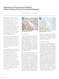
HercepTest⢠Interpretation Manual - Breast - Dako
HercepTest⢠Interpretation Manual - Breast - Dako
HercepTest⢠Interpretation Manual - Breast - Dako

Create successful ePaper yourself
Turn your PDF publications into a flip-book with our unique Google optimized e-Paper software.
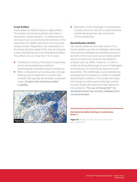
Crush Artifact<br />
Crush artifact is related closely to edge artifact.<br />
This artifact may be encountered more often in<br />
stereotactic needle biopsies. It is presumed that<br />
the tissue injury occurs during the extraction of the<br />
tissue from the needle rather than from the actual<br />
biopsy process. Regardless, the compression of<br />
the tissues along the edges of the core can produce<br />
a linear staining that has to be interpreted as artifact.<br />
This artifact occurs in less than 1% of cases.<br />
• Inadvertent crushing of the tissue occasionally<br />
occurs during sectioning resulting in<br />
morphologically distorted cellular architecture.<br />
• When compared to surrounding cells, stronger<br />
staining may be observed in crushed cells.<br />
Crushed cells typically demonstrate condensed<br />
nuclei. Crushed cells should be avoided<br />
in grading.<br />
• Deposition of the chromogen is characteristic<br />
in areas where the cells are crushed while the<br />
central well-preserved cells are devoid<br />
of immunoreactivity.<br />
Decalcification Artifact<br />
The spinal vertebrae and other areas of the<br />
human skeleton are sites of metastatic carcinoma.<br />
Interventional radiology has facilitated access to<br />
domains of the body and has provided another<br />
source of specimens that can be tested for<br />
analytes such as HER2. However, in order to<br />
render the tissue soft enough to cut on a histologist’s<br />
microtome (at 4-5 microns) the tissue has to be<br />
decalcified. This traditionally is accomplished by<br />
exposing the bony tissue to a variety of available<br />
decalcification solutions. This renders the tissue<br />
soft enough to obtain good histologic section<br />
but also renders the tissue less than optimal for<br />
immunostains. The use of HercepTest TM on<br />
decalcified tissues has not been validated and is<br />
not recommended.<br />
Carcinoma has darker staining on crushed areas.<br />
Score 1+<br />
Figure 37<br />
Figure 37: <strong>Breast</strong> carcinoma showing crush artifact.<br />
(40x magnification).<br />
HercepTest TM <strong>Interpretation</strong> <strong>Manual</strong> – <strong>Breast</strong> Cancer<br />
ROW Version<br />
27