A New Flavonol Glycoside Gallate Ester from Acer ... - Pharmanet
A New Flavonol Glycoside Gallate Ester from Acer ... - Pharmanet
A New Flavonol Glycoside Gallate Ester from Acer ... - Pharmanet

You also want an ePaper? Increase the reach of your titles
YUMPU automatically turns print PDFs into web optimized ePapers that Google loves.
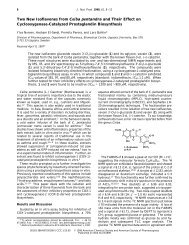
146 Journal of Natural Products, 1998, Vol. 61, No. 1 Notes<br />
Table 1. Chemical Shifts and HMBC NMR Correlations of 1<br />
in CD 3OD<br />
position δ H δ<br />
a C HMBC<br />
2 158.1 (s)<br />
3 134.8 (s)<br />
4 179.0 (s)<br />
5 162.7 (s)<br />
6 6.08 (s, 1H) 100.1 (d) C-10<br />
7 167.1 (s)<br />
8 6.22 (s, 1H) 95.1 (d)<br />
9 158.2 (s)<br />
10 105.2 (s)<br />
1′ 123.0 (s)<br />
2′ 7.55 (d, 2.2) 117.1 (d) C-2,C-3′,C-4′,C-6′<br />
3′ 145.9 (s)<br />
4′ 149.8 (s)<br />
5′ 6.73 (d, 8.5) 116.3 (d) C-1′,C-3′,C-4′<br />
6′ 7.42 (dd, 2.2, 8.5) 123.2 (d) C-2, C-2′,C-4′<br />
1′′ 5.55 (d, 8.0) 101.2 (d)<br />
2′′ 5.44 (dd, 8.0, 9.9) 74.4 (d) C-1′′,C-3′′,C ) O<br />
3′′ 3.84 (dd, 3.5, 9.9) 73.3 (d) C- 2′′<br />
4′′ 3.93 (t, 3.4) 70.5 (d) C-3′′,C-5′′<br />
5′′ 3.89 (t, 6.9) 74.6 (d) C-4′′,C-6′′<br />
6′′′ 4.24 (dd, 5.9, 11.2) 63.6 (t) C-5′′,C ) O<br />
4.43 (dd, 7.1, 11.2)<br />
1′′′ 121.2 (s)<br />
1′′′′ 120.8 (s)<br />
2′′′, 6′′′ 7.18 (s, 2H) 110.6 (d) C-3′′′,C-4′′′,C-5′′′<br />
2′′′′, 6′′′′ 6.92 (s, 2H) 110.1 (d) C-3′′′′,C-4′′′′,C-5′′′′<br />
3′′′, 5′′′ 146.4 (s)<br />
3′′′′, 5′′′′ 146.3 (s)<br />
4′′′, 4′′′′ 140.2 (s)<br />
C ) O<br />
168.0 (s)<br />
C ) O<br />
168.2 (s)<br />
a Multiplicities were established using the DEPT pulse sequence.<br />
shifts at C-2 (δ 158.1), C-3 (δ 134.8), and C-4 (δ 179.0)<br />
in the 13 C NMR spectrum supported the above assignment.<br />
8 The 1 H NMR spectrum of compound 1, analyzed<br />
with the aid of a COSY spectrum, showed characteristic<br />
signals assignable to an anomeric proton at δ 5.55 (d,<br />
J ) 8.0 Hz) and methylene protons adjacent to an ester<br />
group at δ 4.24 (dd, J ) 5.9, 11.2 Hz) and 4.43 (dd, J )<br />
7.1, 11.2 Hz). Attachment of another galloyl group<br />
through an ester linkage at C-2 in galactose was<br />
suggested by the downfield shift of H-2′′ (δ 5.44) in the<br />
1 H NMR spectrum. Three oxygenated methine protons<br />
at δ 3.84 (dd, J ) 3.5, 9.9 Hz), 3.89 (t, J ) 6.9 Hz), and<br />
3.93 (t, J ) 3.4 Hz), together with aromatic protons at<br />
δ 6.92 (2H, s) and 7.18 (2H, s) that were assignable to<br />
a galloyl group, suggested the presence of a 2,6-Odigalloyl<br />
galactoside residue in compound 1. The HMBC<br />
NMR spectrum of compound 1 indicated that the carbon<br />
signal (δ 168.2, C-7′′′) of the galloyl carbonyl units<br />
showed 1 H- 13 C long-range correlations with the H-2′′<br />
and H-2′′′, H-6′′′ signals (δ 5.44 and 7.18). The carbon<br />
signal (δ 168.0, C-7′′′′) of the other galloyl carbonyl was<br />
correlated with the proton signals at δ 4.24 (H-6′′), 4.43<br />
(H-6′′), and 6.92 (H-2′′′′ and 6′′′′), respectively (Table 1).<br />
These data indicated that the galloyl groups were<br />
located at C-2′′ and C-6′′ of galactose. On the basis of<br />
the foregoing observations, compound 1 was assigned<br />
as quercetin 3-O-(2′′,6′′-digalloyl)-β-D-galactopyranoside.<br />
The anti-HIV-1 integrase activities of the A. okamotoanum<br />
isolates were investigated. As shown in Table<br />
2, compounds 6 and 1 showed strong inhibitory activity<br />
against HIV-1 integrase (IC 50 values of 18.1 ( 1.3 and<br />
24.2 ( 6.6 µg/mL, respectively).<br />
Table 2. HIV-1 Integrase Inhibitory Activities of Compounds<br />
1-10 a<br />
compd IC 50 (µg/mL) compd IC 50 (µg/mL)<br />
1 24.2 ( 6.6 6 18.1 ( 1.3<br />
2 64.6 ( 3.9 7 27.9 ( 2.4<br />
3 75.2 ( 8.1 8 38.5 ( 5.1<br />
4 >100 9 28.3 ( 10.2<br />
5 >100 10 28.0 ( 2.2<br />
a IC 50 values with standard deviations are <strong>from</strong> at least three<br />
independent experiments.<br />
Experimental Section<br />
General Experimental Procedures. Melting points<br />
were determined on a Thomas-Hoover capillary melting<br />
apparatus and are uncorrected. Optical rotations were<br />
determined on a Autopol III automatic polarimeter<br />
(Rudolph Research Co., Flanders, <strong>New</strong> Jersey). UV<br />
spectra were taken with a Shimazu UV 240 UV-vis<br />
recording spectrometer. IR spectra were recorded on a<br />
Perkin-Elmer 16F-PC FT-IR and a Midac 101025 instrument<br />
using potassium bromide pellets. 1 H NMR<br />
spectra were recorded on a Gemini Varian-300 (300<br />
MHz) spectrometer, using TMS as internal standard.<br />
13<br />
C NMR spectra were recorded on a Gemini Varian-<br />
300 (75 MHz) spectrometer. 1 H- 1 H COSY, HMQC, and<br />
HMBC NMR spectra were obtained with the usual pulse<br />
sequences, and data processing was performed with the<br />
standard Gemini and Bruker software. EIMS were<br />
determined on a HP 5890 GC/5988 mass spectometer<br />
at 70 eV, electrospray, mass spectra were determined<br />
on an Api ES/MS (HP 59987A ES/5989A MS) instrument,<br />
and HRFABMS were determined on a JEOL<br />
JMS-HX 110/100A (Japan) mass spectometer. Preparative<br />
HPLC was performed on a Waters pump (model<br />
510) with UV detector (λ 254 nm, Waters model 486)<br />
using a LiChrosorb RP-18 (10 mm i.d. x 25 cm, Merck)<br />
column. Cellulose TLC was carried out on precoated<br />
cellulose F TLC plates (Merck, art. 5718).<br />
Plant Material. The leaves of A. okamotoanum<br />
Nakai were collected <strong>from</strong> Ullung Island, Korea, in July<br />
1995. Voucher specimens (565-12A) have been deposited<br />
in the laboratory of Korea Institute of Science &<br />
Technology.<br />
Extraction and Isolation. Dried leaves (2.1 kg)<br />
were cut into small pieces and percolated three times<br />
with MeOH at room temperature to afford 296 g of a<br />
dark-green residue on removal of solvent under reduced<br />
pressure. The methanol extract was suspended in water<br />
and then partitioned in turn with dichloromethane,<br />
ethyl acetate, and butanol. The combined EtOAc extract<br />
was evaporated under reduced pressure to yield<br />
75 g of a residue. This residue was divided into six fractions<br />
by column chromatography on Sephadex LH-20<br />
with a CHCl 3 -MeOH gradient system. Active fraction<br />
2 was further purified by column chromatography over<br />
silica gel with a CH 2 Cl 2 -EtOAc-MeOH-H 2 O gradient<br />
system to give nine subfractions. Subfraction 2C was<br />
further purified by column chromatography over RP-<br />
18 using 50% MeOH as eluent and finally purified by<br />
preparative HPLC (LiChrosorb 250-10, RP-18, Merck)<br />
eluted with 40% MeOH, followed by increasing percentages<br />
of MeOH in H 2 O to afford 13 mg of compound 1 as<br />
a yellow amorphous powder. Fractions 5, 2C, 2E, 2G,<br />
2H, and 2I were also further purified by column chromatography<br />
in a similar manner to yield nine known