A New Flavonol Glycoside Gallate Ester from Acer ... - Pharmanet
A New Flavonol Glycoside Gallate Ester from Acer ... - Pharmanet
A New Flavonol Glycoside Gallate Ester from Acer ... - Pharmanet

Create successful ePaper yourself
Turn your PDF publications into a flip-book with our unique Google optimized e-Paper software.
J. Nat. Prod. 1998, 61, 145-148<br />
145<br />
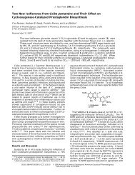
A <strong>New</strong> <strong>Flavonol</strong> <strong>Glycoside</strong> <strong>Gallate</strong> <strong>Ester</strong> <strong>from</strong> <strong>Acer</strong> okamotoanum and Its<br />
Inhibitory Activity against Human Immunodeficiency Virus-1 (HIV-1) Integrase<br />
Hyoung Ja Kim, † Eun-Rhan Woo,* ,† Cha-Gyun Shin, ‡ and Hokoon Park †<br />
Division of Applied Science, Korea Institute of Science & Technology, P.O. Box No. 131, Cheongryang, Seoul 130-650, Korea,<br />
and Department of Biotechnology, Chung-Ang University, An-Sung 456-756, Korea<br />
Received March 14, 1997 X<br />
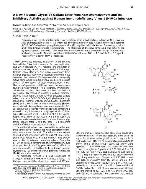
Bioassay-directed chromatographic fractionation of an ethyl acetate extract of the leaves of<br />
<strong>Acer</strong> okamotoanum using HIV-1 integrase afforded a new acylated flavonol glycoside, quercetin<br />
3-O-(2′′,6′′-O-digalloyl)-β-D-galactopyranoside (1), together with six known flavonol glycosides<br />
and three known phenolic compounds. The structure of the new compound was determined<br />
by spectroscopic methods. The most active compounds were quercetin 3-O-(2′′-galloyl)-R-Larabinopyranoside<br />
(6) and 1, which exhibited IC 50 values of 18.1 ( 1.3 and 24.2 ( 6.6 µg/mL,<br />
respectively, against HIV-1 integrase.<br />
HIV-1 integrase mediates insertion of viral DNA into<br />
host cellular DNA that is essential for viral replication<br />
and virion production. 1-3 Therefore, the inhibition of<br />
this enzyme may be efficacious in anti-AIDS therapy.<br />
Despite many efforts to find active compounds <strong>from</strong><br />
natural products, few HIV-1 integrase inhibitors have<br />
been described to date. 4 During a search for biologically<br />
active compounds <strong>from</strong> traditional medicines, a crude<br />
extract of the leaves of <strong>Acer</strong> okamotoanum Nakai<br />
(<strong>Acer</strong>aceae) growing on Ullung Island in Korea was<br />
found to potently inhibit HIV-1 integrase. Phytochemical<br />
studies on this plant have not been carried out<br />
previously. By means of bioassay-directed chromatographic<br />
fractionation, a new flavonol glycoside gallate<br />
ester, quercetin 3-O-(2′′,6′′-O-digalloyl)-β-D-galactopyranoside<br />
(1) together with six known flavonol glycosides<br />
(2-7) and three known phenolic compounds (8-10),<br />
were isolated. Among these compounds, quercetin 3-O-<br />
(2′′-galloyl)-R-L-arabinopyranoside (6) 5 and compound 1<br />
showed strong inhibitory activity against HIV-1 integrase<br />
(IC 50 values of 18.1 ( 1.3 and 24.2 ( 6.6 µg/mL,<br />
respectively) in our assay system. Herein we report the<br />
isolation and characterization of the new flavonol glycoside<br />
gallate ester 1 and the anti-HIV-1 integrase<br />
activity of the A. okamotoanum isolates.<br />
The methanol extract was suspended in water and<br />
then consecutively partitioned with dichloromethane,<br />
ethyl acetate, and butanol. The ethyl acetate extract<br />
showed strong inhibitory activity against HIV-1 integrase<br />
(IC 50 values of 13.6 ( 5.6 µg/mL). The extract<br />
was purified by column chromatography on Sephadex<br />
LH-20, as well as a combination of chromatography over<br />
silica gel, RP-18, and Toyopearl HW-40C to afford the<br />
new compound 1. In addition to 1, six known flavonol<br />
glycosides (2-7) and three known phenolic compounds<br />
(8-10) were also isolated.<br />
The molecular formula of 1 was established as<br />
C 35 H 28 O 20 by ES and HRFAB mass spectrometry. The<br />
UV spectrum exhibited absorption maxima at 267 and<br />
* To whom correspondence should be addressed. Phone: +82-2-958-<br />
5147. Fax: +82-2-958-5189. E-mail: wooer@kistmail.kist.re.kr.<br />
†<br />
Korea Institute of Science & Technology.<br />
‡<br />
Chung-Ang University.<br />
X<br />
Abstract published in Advance ACS Abstracts, December 15, 1997.<br />
355 nm that are characteristic absorption bands of a<br />
flavone skeleton. 6 In the IR spectrum, along with the<br />
signal for an ester carbonyl (1710 cm -1 ), signals for a<br />
hydroxyl group (3414 cm -1 ), a conjugated carbonyl (1638<br />
cm -1 ), and a phenyl group (1618,1498 cm -1 ) were<br />
apparent. The 1 H NMR spectrum suggested that 1 has<br />
a quercetin aglycon. Signals at δ 6.73 (1H, d, J ) 8.5<br />
Hz), 7.42 (1H, dd, J ) 2.2 and 8.5 Hz), and 7.55 (1H, d,<br />
J ) 2.2 Hz) are characteristic for a 3,4-disubstituted B<br />
ring of a quercetin unit. Singlets at δ 6.92 (2H, s) and<br />
7.18 (2H, s) originated <strong>from</strong> the galloyl groups. The<br />
placement of the sugar residue at C-3 in 1 was suggested<br />
<strong>from</strong> characteristic UV shifts. After addition of<br />
NaOAc and AlCl 3 + HCl, absorption maxima at 267 and<br />
355 nm shifted to 273, 325, and 415 and to 270, 360,<br />
and 405 nm, respectively. 7 Furthermore, the chemical<br />
S0163-3864(97)00171-7 CCC: $15.00<br />
© 1998 American Chemical Society and American Society of Pharmacognosy<br />
Published on Web 01/23/1998
146 Journal of Natural Products, 1998, Vol. 61, No. 1 Notes<br />
Table 1. Chemical Shifts and HMBC NMR Correlations of 1<br />
in CD 3OD<br />
position δ H δ<br />
a C HMBC<br />
2 158.1 (s)<br />
3 134.8 (s)<br />
4 179.0 (s)<br />
5 162.7 (s)<br />
6 6.08 (s, 1H) 100.1 (d) C-10<br />
7 167.1 (s)<br />
8 6.22 (s, 1H) 95.1 (d)<br />
9 158.2 (s)<br />
10 105.2 (s)<br />
1′ 123.0 (s)<br />
2′ 7.55 (d, 2.2) 117.1 (d) C-2,C-3′,C-4′,C-6′<br />
3′ 145.9 (s)<br />
4′ 149.8 (s)<br />
5′ 6.73 (d, 8.5) 116.3 (d) C-1′,C-3′,C-4′<br />
6′ 7.42 (dd, 2.2, 8.5) 123.2 (d) C-2, C-2′,C-4′<br />
1′′ 5.55 (d, 8.0) 101.2 (d)<br />
2′′ 5.44 (dd, 8.0, 9.9) 74.4 (d) C-1′′,C-3′′,C ) O<br />
3′′ 3.84 (dd, 3.5, 9.9) 73.3 (d) C- 2′′<br />
4′′ 3.93 (t, 3.4) 70.5 (d) C-3′′,C-5′′<br />
5′′ 3.89 (t, 6.9) 74.6 (d) C-4′′,C-6′′<br />
6′′′ 4.24 (dd, 5.9, 11.2) 63.6 (t) C-5′′,C ) O<br />
4.43 (dd, 7.1, 11.2)<br />
1′′′ 121.2 (s)<br />
1′′′′ 120.8 (s)<br />
2′′′, 6′′′ 7.18 (s, 2H) 110.6 (d) C-3′′′,C-4′′′,C-5′′′<br />
2′′′′, 6′′′′ 6.92 (s, 2H) 110.1 (d) C-3′′′′,C-4′′′′,C-5′′′′<br />
3′′′, 5′′′ 146.4 (s)<br />
3′′′′, 5′′′′ 146.3 (s)<br />
4′′′, 4′′′′ 140.2 (s)<br />
C ) O<br />
168.0 (s)<br />
C ) O<br />
168.2 (s)<br />
a Multiplicities were established using the DEPT pulse sequence.<br />
shifts at C-2 (δ 158.1), C-3 (δ 134.8), and C-4 (δ 179.0)<br />
in the 13 C NMR spectrum supported the above assignment.<br />
8 The 1 H NMR spectrum of compound 1, analyzed<br />
with the aid of a COSY spectrum, showed characteristic<br />
signals assignable to an anomeric proton at δ 5.55 (d,<br />
J ) 8.0 Hz) and methylene protons adjacent to an ester<br />
group at δ 4.24 (dd, J ) 5.9, 11.2 Hz) and 4.43 (dd, J )<br />
7.1, 11.2 Hz). Attachment of another galloyl group<br />
through an ester linkage at C-2 in galactose was<br />
suggested by the downfield shift of H-2′′ (δ 5.44) in the<br />
1 H NMR spectrum. Three oxygenated methine protons<br />
at δ 3.84 (dd, J ) 3.5, 9.9 Hz), 3.89 (t, J ) 6.9 Hz), and<br />
3.93 (t, J ) 3.4 Hz), together with aromatic protons at<br />
δ 6.92 (2H, s) and 7.18 (2H, s) that were assignable to<br />
a galloyl group, suggested the presence of a 2,6-Odigalloyl<br />
galactoside residue in compound 1. The HMBC<br />
NMR spectrum of compound 1 indicated that the carbon<br />
signal (δ 168.2, C-7′′′) of the galloyl carbonyl units<br />
showed 1 H- 13 C long-range correlations with the H-2′′<br />
and H-2′′′, H-6′′′ signals (δ 5.44 and 7.18). The carbon<br />
signal (δ 168.0, C-7′′′′) of the other galloyl carbonyl was<br />
correlated with the proton signals at δ 4.24 (H-6′′), 4.43<br />
(H-6′′), and 6.92 (H-2′′′′ and 6′′′′), respectively (Table 1).<br />
These data indicated that the galloyl groups were<br />
located at C-2′′ and C-6′′ of galactose. On the basis of<br />
the foregoing observations, compound 1 was assigned<br />
as quercetin 3-O-(2′′,6′′-digalloyl)-β-D-galactopyranoside.<br />
The anti-HIV-1 integrase activities of the A. okamotoanum<br />
isolates were investigated. As shown in Table<br />
2, compounds 6 and 1 showed strong inhibitory activity<br />
against HIV-1 integrase (IC 50 values of 18.1 ( 1.3 and<br />
24.2 ( 6.6 µg/mL, respectively).<br />
Table 2. HIV-1 Integrase Inhibitory Activities of Compounds<br />
1-10 a<br />
compd IC 50 (µg/mL) compd IC 50 (µg/mL)<br />
1 24.2 ( 6.6 6 18.1 ( 1.3<br />
2 64.6 ( 3.9 7 27.9 ( 2.4<br />
3 75.2 ( 8.1 8 38.5 ( 5.1<br />
4 >100 9 28.3 ( 10.2<br />
5 >100 10 28.0 ( 2.2<br />
a IC 50 values with standard deviations are <strong>from</strong> at least three<br />
independent experiments.<br />
Experimental Section<br />
General Experimental Procedures. Melting points<br />
were determined on a Thomas-Hoover capillary melting<br />
apparatus and are uncorrected. Optical rotations were<br />
determined on a Autopol III automatic polarimeter<br />
(Rudolph Research Co., Flanders, <strong>New</strong> Jersey). UV<br />
spectra were taken with a Shimazu UV 240 UV-vis<br />
recording spectrometer. IR spectra were recorded on a<br />
Perkin-Elmer 16F-PC FT-IR and a Midac 101025 instrument<br />
using potassium bromide pellets. 1 H NMR<br />
spectra were recorded on a Gemini Varian-300 (300<br />
MHz) spectrometer, using TMS as internal standard.<br />
13<br />
C NMR spectra were recorded on a Gemini Varian-<br />
300 (75 MHz) spectrometer. 1 H- 1 H COSY, HMQC, and<br />
HMBC NMR spectra were obtained with the usual pulse<br />
sequences, and data processing was performed with the<br />
standard Gemini and Bruker software. EIMS were<br />
determined on a HP 5890 GC/5988 mass spectometer<br />
at 70 eV, electrospray, mass spectra were determined<br />
on an Api ES/MS (HP 59987A ES/5989A MS) instrument,<br />
and HRFABMS were determined on a JEOL<br />
JMS-HX 110/100A (Japan) mass spectometer. Preparative<br />
HPLC was performed on a Waters pump (model<br />
510) with UV detector (λ 254 nm, Waters model 486)<br />
using a LiChrosorb RP-18 (10 mm i.d. x 25 cm, Merck)<br />
column. Cellulose TLC was carried out on precoated<br />
cellulose F TLC plates (Merck, art. 5718).<br />
Plant Material. The leaves of A. okamotoanum<br />
Nakai were collected <strong>from</strong> Ullung Island, Korea, in July<br />
1995. Voucher specimens (565-12A) have been deposited<br />
in the laboratory of Korea Institute of Science &<br />
Technology.<br />
Extraction and Isolation. Dried leaves (2.1 kg)<br />
were cut into small pieces and percolated three times<br />
with MeOH at room temperature to afford 296 g of a<br />
dark-green residue on removal of solvent under reduced<br />
pressure. The methanol extract was suspended in water<br />
and then partitioned in turn with dichloromethane,<br />
ethyl acetate, and butanol. The combined EtOAc extract<br />
was evaporated under reduced pressure to yield<br />
75 g of a residue. This residue was divided into six fractions<br />
by column chromatography on Sephadex LH-20<br />
with a CHCl 3 -MeOH gradient system. Active fraction<br />
2 was further purified by column chromatography over<br />
silica gel with a CH 2 Cl 2 -EtOAc-MeOH-H 2 O gradient<br />
system to give nine subfractions. Subfraction 2C was<br />
further purified by column chromatography over RP-<br />
18 using 50% MeOH as eluent and finally purified by<br />
preparative HPLC (LiChrosorb 250-10, RP-18, Merck)<br />
eluted with 40% MeOH, followed by increasing percentages<br />
of MeOH in H 2 O to afford 13 mg of compound 1 as<br />
a yellow amorphous powder. Fractions 5, 2C, 2E, 2G,<br />
2H, and 2I were also further purified by column chromatography<br />
in a similar manner to yield nine known
Notes Journal of Natural Products, 1998, Vol. 61, No. 1 147<br />
compounds. The structures of compounds 2-10 were<br />
determined by comparison with known compounds.<br />
Quercetin 3-O-(2′′,6′′-digalloyl)-β-D-galactopyranoside<br />
(1): yellow amorphous powder; mp 222-224 °C<br />
dec; [R] 21 D -45.9° (c 0.4, MeOH); UV (MeOH) λ max (log<br />
ɛ) 267 (4.62), 290 (sh), 355 (4.30) nm; (MeOH + AlCl 3 )<br />
λ max 273, 300 (sh), 398 nm; (MeOH + NaOMe) λ max 273,<br />
325, 415 nm; IR (KBr) ν max 3350, 1710, 1638, 1618,<br />
1498, 1448, 1384, 1360, 1304, 1240, 1194 cm -1 ; 1 H, 13 C,<br />
and HMBC NMR data; see Table 1; ES/MS m/z 769 [M<br />
+ 1] + , HRFABMS [M + H] + m/z 769.1251, calcd for<br />
C 35 H 29 O 20 769.1252. Acid hydrolysis with 10% H 2 SO 4<br />
gave quercetin (spectral data compared with the pure<br />
compound) and gallic acid. Cellulose TLC of the neutralized<br />
hydrolysate in 1-BuOH-benzene-pyridine-<br />
H 2 O (5:1:3:3, upper layer) gave D-galactose (R f 0.18).<br />
Quercetin 3-O-β-D-galactopyranoside (2): UV,<br />
MS, and 1 H and 13 C NMR data were identical with<br />
published data. 9<br />
Quercetin 3-O-r-L-rhamnopyranoside (3): UV,<br />
MS, and 1 H and 13 C NMR data were identical with<br />
published data. 9,10<br />
Kaempferol 3-O-r-L-rhamnopyranoside (4): UV,<br />
MS, and 1 H and 13 C NMR data were identical with<br />
published data. 11<br />
Kaempferol 3-O-r-L-arabinopyranoside (5): UV,<br />
MS, and 1 H and 13 C NMR data were identical with<br />
published data. 12<br />
Quercetin 3-O-(2′′-galloyl)-r-L-arabinopyranoside<br />
(6): UV, MS, and 1 H and 13 C NMR data were<br />
identical with published data. 5<br />
Quercetin 3-O-(2′′-galloyl)-β-D-galactopyranoside<br />
(7): yellow amorphous powder; mp 198-200 °C dec;<br />
[R] 17 D -109.6° (c 1.0, MeOH); 1 H NMR (CD 3 OD, 300<br />
MHz) δ 7.64 (1H, d, J ) 2.2 Hz, H-2′), 7.44 (1H, dd, J )<br />
2.2, 8.6 Hz, H-6′), 7.16 (2H, s, galloyl H-2, H-6), 6.76<br />
(1H, d, J ) 8.6 Hz, H-5′), 6.26 (1H, br s, H-8), 6.11 (1H,<br />
br s, H-6), 5.66 (1H, d, J ) 8.0 Hz, H-1′′), 5.48 (1H, dd,<br />
J ) 8.2, 9.7 Hz, H-2′′), 3.94 (1H, d, J ) 3.5 Hz, H-4′′),<br />
3.86 (1H, dd, J ) 3.5, 9.6 Hz, H-3′′), 3.74 (2H, dd, J )<br />
6.5, 11.4 Hz, H-6′′), 3.67 (1H, d, J ) 6.8 Hz, H-5′′); 13 C<br />
NMR (CD 3 OD, 75 MHz) δ 179.0 (s, C-4), 168.3 (s, galloyl<br />
CdO), 165.9 (s, C-7), 162.9 (s, C-5), 158.2 (s, C-9), 158.0<br />
(s, C-2), 149.7 (s, C-4′), 149.7 (s, C-4′), 146.3 (s, galloyl<br />
C-3, C-5), 145.8 (s, C-3′), 139.9 (s, galloyl C-4), 135.2 (s,<br />
C-3), 123.2 (s, C-1′), 123.0 (d, C-6′), 121.6 (s, galloyl C-1),<br />
117.3 (d, C-2′), 116.3 (d, C-5′), 110.8 (d, galloyl C-2, C-6),<br />
105.7 (s, C-10), 101.4 (d, C-1′′), 99.9 (d, C-6), 94.9 (d,<br />
C-8), 77.4 (d, C-5′′), 74.7 (d, C-2′′), 73.5 (d, C-3′′), 70.7<br />
(d, C-4′′), 62.3 (t, C-6′′). The UV and MS data of 7 were<br />
identical with published values. 13 Acid hydrolysis with<br />
10% H 2 SO 4 gave quercetin (spectral data compared with<br />
the pure compound) and gallic acid. Cellulose TLC<br />
of the neutralized hydrolysate in 1-BuOH-benzenepyridine-H<br />
2 O (5:1:3:3, upper layer) gave D-galactose (R f<br />
0.18).<br />
Gallic acid methyl ester (8): UV, MS, and 1 H and<br />
13 C NMR data were identical with published data. 14<br />
1,2,6-Tri-O-galloyl-β-D-glucose (9): UV, MS, and<br />
1 H and 13 C NMR data were identical with published<br />
data. 15<br />
1,2,3,4,6-Penta-O-galloyl-β-D-glucose (10): UV, MS,<br />
and 1 H and 13 C NMR data were identical with published<br />
data. 16<br />
Bioassays. HIV-1 Integrase. Recombinant human<br />
immunodeficiency virus type 1 (HIV-1) integrase was<br />
expressed in Escherichia coli and purified using a<br />
nickel-chelated column in a one-step manner, as described<br />
previously. 17 Aliquots of HIV-1 integrase of 0.5<br />
mg/mL as stock solutions were stored at -70 °C until<br />
used.<br />
Oligonucleotide Substrates. Two 20-mer oligonucleotides<br />
whose sequences resemble the end of U5-<br />
LTR were obtained <strong>from</strong> Korea Biotech., Inc. (Seoul,<br />
Korea), namely K16 (U5-LTR, +strand), 5′-TGTG-<br />
GAAAATCTCTAGCAGT-3′, and K17 (U5-LTR, -strand),<br />
5′-ACTGCTAGA-GATTTTCCACA-3′. The oligonucleotides<br />
were purified using 20% polyacrylamide gel<br />
before use. To construct the oligonucleotide substrate,<br />
oligonucleotide K16 (15 pmol) was labeled at the 5′ end,<br />
using [γ- 32 P]-ATP of 250 µCi (3,000 Ci/mmol; 1 Ci ) 37<br />
GBq; Amersham Life Science, Arlington Heights, IL)<br />
and T4 polynucleotide kinase (T4 PNK, <strong>New</strong> England<br />
Biolabs, Beverly, MA) of 10 units in 40 µL of reaction<br />
buffer (70 mM Tris-HCl (pH 7.6), 10 mM MgCl 2 ,5mM<br />
dithiothreitol) for 15 min at 37 °C. The labeling reaction<br />
was subjected to 10 mM EDTA and heated to 85 °C for<br />
15 min to inactivate T4 PNK. After the addition of<br />
complementary oligonucleotide K17 (30 pmol), the reaction<br />
mixture was boiled for 3 min and cooled slowly.<br />
Labeled substrate was separated <strong>from</strong> unincorporated<br />
nucleotide by passage through a Biospin 6 instrument<br />
(Bio-Rad, Hercules, CA).<br />
HIV-1 Integrase Reaction. A standard reaction<br />
assay of endonucleolytic activity was carried out in the<br />
presence of potential inhibitor containing 0.1 pmol of<br />
duplex oligonucleotide substrate and 15 pmol of HIV-1<br />
integrase in 15 mM Tris-HCl (pH 7.4), 100 mM NaCl,<br />
1mMMnCl 2 , 2 mM 2-mercaptoethanol, 2.5 mM CHAPS,<br />
0.1 mM EDTA, 0.1 mM PMSF, 1% glycerol, and 10 mM<br />
imidazole in a total volume of 10 µL. Inhibitors or drugs<br />
were dissolved in 100% DMSO and added to the reaction<br />
mixture, with there being 5% DMSO in the final<br />
mixture. Reaction mixtures were incubated at 33 °C<br />
for 90 min and stopped by the addition of 4 µL of95%<br />
formamide, 20 mM EDTA, 0.05% bromophenol blue, and<br />
0.05% xylene cyanol FF. The reactions were heated to<br />
90 °C for 3 min and electrophoresed on a 20% denaturing<br />
polyacrylamide gel. Reaction products were visualized<br />
by autoradiography of the wet gel. IC 50 values were<br />
calculated by scanning bands on Kodak-5 film (Image<br />
Master VDS, Pharmacia Biotech., Piscataway, NJ).<br />
Acknowledgment. Financial support <strong>from</strong> the G7<br />
project, Ministry of Science & Technology, and a grant<br />
(HMP-96-D-1-0004) <strong>from</strong> the 96 Good Health R&D<br />
Project, Ministry of Health & Welfare, Seoul, Korea, are<br />
gratefully acknowledged. The authors thank Dr. J. H.<br />
Kwak for identification of the specimen and Dr. Y. J.<br />
Kim and Mr. Y. W. Kim for measurements of ES/MS<br />
and HRFABMS.<br />
References and Notes<br />
(1) Sakai, H.; Kawamura, M.; Sakuragi, J.; Sakuragi, S.; Shibata,<br />
R.; Isimoto, A.; Ono, H.; Ueda, S.; Adachi, A. J. Virol. 1993, 67,<br />
1169-1174.<br />
(2) Taddeo, B.; Haseltine, W. A.; Farnet, C. M. J. Virol. 1994, 68,<br />
8401-8405.<br />
(3) Engelman, A.; Englund, G.; Orenstein, J. M.; Martin, M. A.;<br />
Craigie, R. J. Virol. 1995, 69, 2729-2736.
148 Journal of Natural Products, 1998, Vol. 61, No. 1 Notes<br />
(4) Mazumder, A.; Raghavan, K.; Weinstein, J.; Kohn, K. W.;<br />
Pommier, Y. Biochem. Pharmacol. 1995, 49, 1165-1170.<br />
(5) Iwagawa, T.; Kawasaki, J.; Hase, T.; Sako, S.; Okubo, T.; Ishida,<br />
M.; Kim, M. Phytochemistry 1990, 29, 1013-1014.<br />
(6) Markham, K. R. Techniques of Flavonoid Identification; Academic<br />
Press: London, 1982; Chapter 3, pp 36-51.<br />
(7) Mabry, T. J.; Markham, K. R.; Thomas, B. M. The Systematic<br />
Identification of Flavonoids; Springer: <strong>New</strong> York, 1970; Chapter<br />
5, pp 39-61.<br />
(8) Markham, K. R.; Ternai, B.; Stanly, R.; Geiger, H.; Mabry, T. J.<br />
Tetrahedron 1978, 34, 1389-1397.<br />
(9) (a) Markham, K. R.; Ternai, B.; Stanley, R.; Geiger, H.; Mabry,<br />
T. J. Tetrahedron 1978, 34, 1389-1397. (b) Nonaka, G. I.; Goto,<br />
Y.; Kinjo, J.; Nohara, T.; Nishioka, I. Chem. Pharm. Bull. 1987,<br />
35, 1105-1108.<br />
(10) (a) Asen, A.; Horowitz, R. M. Phytochemistry 1977, 16, 147-<br />
148. (b) Kosuge, T.; Ishida, H.; Satoh, T. Chem. Pharm. Bull.<br />
1985, 33, 206-209. (c) Markham, K. R.; Ternai, B. Tetrahedron<br />
1976, 32, 2607-2612.<br />
(11) Matthes, H. W. D.; Luu, B.; Ourisson, G. Phytochemistry 1980,<br />
19, 2643-2650.<br />
(12) Pachaly, P.; Klein, M. Planta Med. 1987, 53, 442-444.<br />
(13) Nahrstedt, A.; Dumkow, K.; Janistyn, B.; Pohl, R. Tetrahedron<br />
Lett. 1974, 559-562.<br />
(14) (a) Park, J. C.; Yu, Y. B.; Lee, J. H.; Choi, J. S.; Ok, K. D. Kor.<br />
J. Pharmcogn. 1996, 27, 219-223. (b) Hattori, M.; Shu, Y. Z.;<br />
Tomimori, T.; Kobashi, K.; Namba, T. Phytochemistry 1989, 28,<br />
1289-1290.<br />
(15) (a) Nonaka, G. I.; Nishioka, I.; Nagasawa, T.; Oura, H. Chem.<br />
Pharm. Bull. 1981, 29, 2862-2870. (b) Nishizawa, M.; Yamagishi,<br />
T. J. Chem. Soc., Perkin Trans. 1 1983, 961-965.<br />
(16) (a) Nishizawa, M.; Yamagishi, T. J. Chem. Soc., Perkin Trans.<br />
1 1982, 2963-2968. (b) Yoshida, T.; Chen, L.; Shingu, T.; Okuda,<br />
T. Chem. Pharm. Bull. 1988, 36, 2940-2949.<br />
(17) Oh, J.-W.; Shin, C.-G. Mol. Cells 1996, 6, 96-100.<br />
NP970171Q