Outcome of the Surgical Treatment of Bullous Lung Disease - Tanaffos
Outcome of the Surgical Treatment of Bullous Lung Disease - Tanaffos
Outcome of the Surgical Treatment of Bullous Lung Disease - Tanaffos

Create successful ePaper yourself
Turn your PDF publications into a flip-book with our unique Google optimized e-Paper software.
Original Article<br />
2012 NRITLD, National Research Institute <strong>of</strong> Tuberculosis and <strong>Lung</strong> <strong>Disease</strong>, Iran<br />
ISSN: 1735-0344 <strong>Tanaffos</strong> 2012; 11(2): 27-33<br />
TANAFFOS<br />
<strong>Outcome</strong> <strong>of</strong> <strong>the</strong> <strong>Surgical</strong> <strong>Treatment</strong> <strong>of</strong> <strong>Bullous</strong><br />
<strong>Lung</strong> <strong>Disease</strong>: A Prospective Study<br />
Yasir Ahmad Lone, Abdul Majeed Dar,<br />
Mukand Lal Sharma, Irfan Robbani, Arif<br />
Hussain Sarmast, Enas Mushtaq,<br />
Mohammad Yousuf Kachroo, Omar<br />
Masood Khan<br />
Sher-i-Kashmir Institute <strong>of</strong> Medical Sciences,<br />
Srinagar, India<br />
Received: 29 December 2011<br />
Accepted: 31 March 2012<br />
Correspondence to: Yasir Ahmad Lone<br />
Address: Yunis Manzil, Lalbazar, Srinagar (Pin:<br />
190011)J & K, India.<br />
Email address: dr_yalone@yahoo.co.in<br />
Background: This study aimed at evaluating <strong>the</strong> outcome <strong>of</strong> surgery for bullous<br />
lung disease by comparing <strong>the</strong> preoperative and postoperative subjective<br />
dyspnea score, pulmonary function and clinical features.<br />
Materials and Methods: This prospective study was conducted from May 2009<br />
to October 2011, on 54 patients operated for bullous lung disease. Follow-up at<br />
3-6 months consisted <strong>of</strong> taking a comprehensive history, physical examination,<br />
radiological work-up, and evaluation <strong>of</strong> changes in subjective dyspnea score,<br />
arterial blood gas analysis (ABG), and pulmonary function test (PFT). After<br />
comparison with preoperative values, <strong>the</strong> student’s paired t-test was used to<br />
calculate <strong>the</strong> statistical significance.<br />
Results: With approximately 21.6 cases per year, <strong>the</strong> most common underlying<br />
lung pathology was primary bullous lung disease, followed by COPD. The<br />
most common presenting complaint was spontaneous pneumothorax in tall<br />
young adults in <strong>the</strong>ir fourth decade <strong>of</strong> life with a history <strong>of</strong> smoking.<br />
Bullectomy, with or without decortication, was done for all cases. Improvement<br />
in mean PaO2 (arterial partial pressure <strong>of</strong> oxygen), SaO2 (arterial oxygen<br />
saturation) and PaCO2 (arterial partial pressure <strong>of</strong> carbon dioxide) was seen in<br />
most cases but was statistically insignificant. Improvement in mean FEV1<br />
(forced expiratory volume in 1st second), FVC (forced vital capacity) and FEV1<br />
/ FVC was statistically significant, with FEV1 being <strong>the</strong> most reliable indicator<br />
<strong>of</strong> postoperative progress. Improvement in subjective dyspnea score was<br />
statistically significant and showed an inverse correlation with FEV1. Those<br />
with diffuse pulmonary parenchymal involvement had poorer baseline values<br />
and less significant postoperative improvement. Complications occurred more<br />
commonly in those with diffuse disease. Mortality was seen exclusively in those<br />
with diffuse disease.<br />
Conclusion: We conclude that surgery is required for bullous lung disease<br />
more frequently in our community since we have a high number <strong>of</strong> young<br />
patients with primary bullous lung disease and localized parenchymal<br />
involvement and <strong>the</strong>se patients have a good surgical outcome. Potentially fatal<br />
complications like pneumothorax and recurrent infections can <strong>the</strong>refore be<br />
prevented in <strong>the</strong>m. Those with underlying diffuse disease and severely<br />
decreased FEV1 (especially below 1 L) also benefit from surgery but require<br />
careful patient selection.<br />
Key words: <strong>Bullous</strong> lung disease, Bulla, Bullectomy<br />
INTRODUCTION<br />
Patients who have bullous lung disease in <strong>the</strong> presence<br />
<strong>of</strong> diffuse parenchymal involvement (emphysematous or<br />
non-emphysematous) should be evaluated on an<br />
individual basis and surgery should be performed for<br />
patients in whom even a small increase in pulmonary<br />
function might be <strong>of</strong> major benefit (1). Computed<br />
tomography scan (CT-scan), bronchography and<br />
angiography can outline bullae that are not visible on a<br />
plain chest radiograph (2). Bullae that enlarge enough to
28 <strong>Surgical</strong> <strong>Treatment</strong> <strong>of</strong> <strong>Bullous</strong> <strong>Lung</strong> <strong>Disease</strong><br />
compress adjacent lung tissue are best diagnosed by CT<br />
scan <strong>of</strong> <strong>the</strong> chest which can identify potentially operable<br />
well defined bullae from inoperable generalized<br />
emphysema locally worsened in <strong>the</strong> area <strong>of</strong> suspected<br />
bulla (3). CT-can also measure <strong>the</strong> bulla volume and<br />
ventilation. Change in forced expiratory volume in 1st<br />
second ( FEV1 ), forced vital capacity (FVC) and <strong>the</strong>ir ratio<br />
(FEV1 / FVC) after surgery, as measured by <strong>the</strong><br />
pulmonary function tests (PFT) is <strong>the</strong> main outcome<br />
measure (4). Increase in arterial oxygen partial pressure<br />
(PaO 2 ), oxygen saturation (SaO 2 ) and arterial carbon<br />
dioxide partial pressure (PaCO 2 ) as estimated from <strong>the</strong><br />
arterial blood gas analysis (ABG) <strong>of</strong> an arterial blood<br />
sample is also among <strong>the</strong> commonly used outcome<br />
variables after surgery, besides changes in clinical and<br />
radiological features.<br />
Bullectomy or resection <strong>of</strong> <strong>the</strong> entire bulla, ei<strong>the</strong>r<br />
through a video assisted thoracoscopic surgery (VATS) or<br />
a standard open thoracotomy, is <strong>the</strong> most common surgical<br />
technique used for treatment (1).<br />
This study aimed at evaluating <strong>the</strong> epidemiological and<br />
clinical pr<strong>of</strong>ile <strong>of</strong> bullous lung disease in patients admitted<br />
to our hospital over <strong>the</strong> study period. The main objective<br />
was to analyze <strong>the</strong> short term and long term outcome <strong>of</strong><br />
surgery by comparing preoperative and postoperative<br />
subjective dyspnea score, clinical features, radiological<br />
features and pulmonary function.<br />
MATERIALS AND METHODS<br />
The present study was conducted in <strong>the</strong> Department <strong>of</strong><br />
Cardiovascular and Thoracic Surgery (CVTS), Sher-i-<br />
Kashmir Institue <strong>of</strong> Medical Sciences (SKIMS), Srinagar<br />
from May 2009 to October 2011. Out <strong>of</strong> 65 cases admitted<br />
to this department for bullous lung disease, 60 were<br />
eventually operated and 56 out <strong>of</strong> <strong>the</strong>se 60 met <strong>the</strong><br />
inclusion criteria for <strong>the</strong> present study. With 2 cases lost in<br />
<strong>the</strong> follow-up, <strong>the</strong> present prospective study finally<br />
consisted <strong>of</strong> 54 cases who are still continuing follow-up.<br />
All patients operated on in <strong>the</strong> Department <strong>of</strong> CVTS for<br />
bullous lung disease and met <strong>the</strong> inclusion criteria were<br />
enrolled in <strong>the</strong> study. Patients were evaluated and<br />
managed according to <strong>the</strong> set protocol and followed up<br />
accordingly.<br />
The patients were analyzed in terms <strong>of</strong>:<br />
1) History taking<br />
2) General physical examination<br />
3) Systemic examination<br />
4) Investigations (baseline investigations and specific<br />
investigations, especially chest radiography, computed<br />
tomography scan <strong>of</strong> <strong>the</strong> chest (chest CT-scan), arterial<br />
blood gas analysis (ABG) and pulmonary function tests)<br />
5) Operative findings<br />
6) Histopathological examination <strong>of</strong> resected specimen<br />
7) Postoperative stay in <strong>the</strong> hospital (First week monitoring<br />
for immediate outcome and complications)<br />
8) Appropriate follow up plan to monitor early (1 month) complications and long term outcome.<br />
OPD follow up 1 week and 1 month after discharge and at<br />
3-6 months post-operatively was done. Follow up at 3-6<br />
months was done for all patients. Fur<strong>the</strong>r follow ups were<br />
planned at 9-15 months (completed for 26 patients as <strong>of</strong><br />
date), 21-27 months (completed for 3 patients as <strong>of</strong> date),<br />
33-39 months, 45-51 months and 57-63 months to complete<br />
a full five-year follow up.<br />
The inclusion criteria were as follows:<br />
1) Presence <strong>of</strong> bulla(e) occupying at least one third or more<br />
<strong>of</strong> hemithorax<br />
2) Bulla(e) in <strong>the</strong> presence <strong>of</strong> symptoms or complications<br />
known to be attributable to <strong>the</strong>m, like pneumothorax or<br />
recurrent respiratory infections.<br />
The exclusion criteria were as follows:<br />
- Surgery done primarily for some o<strong>the</strong>r lung disease<br />
during which bullectomy was also performed (e.g. patients<br />
undergoing lobectomy for bronchiectasis and having<br />
associated bulla (e) that were also excised were excluded<br />
from <strong>the</strong> study)<br />
For comparing <strong>the</strong> postoperative dyspnea score based<br />
on Medical Research Council guidelines (5) and <strong>the</strong> ABG<br />
and PFT values with <strong>the</strong> preoperative ones, <strong>the</strong> student’s<br />
paired t-test was used to evaluate <strong>the</strong> statistical<br />
significance <strong>of</strong> this quantitative data.<br />
<strong>Tanaffos</strong> 2012; 11(2): 27-33
Lone YA, et al. 29<br />
RESULTS<br />
The mean overall age was 41.07 ± 14.92 years (range 11<br />
to 65 years). There were 43 males and 11 females with a<br />
male to female ratio <strong>of</strong> 3.9 / 1. Spontaneous pneumothorax<br />
was <strong>the</strong> predominant presenting complaint (28 out <strong>of</strong> 54<br />
patients, 52 %). Breathlessness (without spontaneous<br />
pneumothorax) in 23 out <strong>of</strong> 54 patients (42%) was <strong>the</strong><br />
second most common presenting complaint. Traumatic<br />
hemopneumothorax bringing <strong>the</strong> bullous lung disease to<br />
attention was observed in only 2 patients (i.e. 4 %) while<br />
one patient with a history <strong>of</strong> IV and inhalation drug abuse<br />
presented with spontaneous hemopneumothorax. The<br />
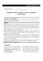
most common underlying lung pathology was idiopathic /<br />
primary bullous lung disease (19 out <strong>of</strong> 54 cases, 35.19 %)<br />
(Figure 1). COPD was <strong>the</strong> second most common<br />
underlying lung pathology (17 out <strong>of</strong> 54 cases, 31.48 %).<br />
Those with underlying idiopathic/primary bullous lung<br />
disease presented more commonly with spontaneous<br />
pneumothorax (13 out <strong>of</strong> 19 patients, 68.42%). Those with<br />
underlying COPD presented more commonly with<br />
breathlessness in <strong>the</strong> absence <strong>of</strong> spontaneous<br />
pneumothorax (9 out <strong>of</strong> 17 patients, 52.94%). The mean<br />
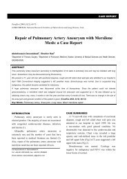
overall age <strong>of</strong> patients was 41.07 ± 14.92 years. Those with<br />
idiopathic / primary bullous lung disease were found to<br />
develop <strong>the</strong> disease at a younger age (mean age <strong>of</strong> 31 yrs)<br />
(Figure 2). The mean age <strong>of</strong> those with underlying collagen<br />
vascular disease was also younger (mean age <strong>of</strong> 26 yrs).<br />
Those with underlying COPD developed <strong>the</strong> disease at an<br />
older age (mean age <strong>of</strong> 56.53 years). The mean overall<br />
height was 67.91 ± 4.19 inches. Chest CT-scan confirmed<br />
<strong>the</strong> diagnosis in all patients. Bullae were more common in<br />
<strong>the</strong> upper lobes (40 out <strong>of</strong> 54 cases, 74.07 %). Open<br />
thoracotomy and bullectomy were done in 54% <strong>of</strong> cases<br />
while 46% needed bullectomy along with decortications.<br />
Mean chest tube drainage in <strong>the</strong> 1st 24 hours post<br />
empyema in 2 patients. All three patients who expired had<br />
diffuse disease in <strong>the</strong> surrounding lung parenchyma.<br />
Overall mortality rate at <strong>the</strong> end <strong>of</strong> study period was<br />
5.56%. The change in ABG parameters was statistically<br />
insignificant while <strong>the</strong> improvement in PFT parameters<br />
and subjective dyspnea score was statistically significant<br />
(Table 1). FEV1 is <strong>the</strong> most reliable indicator <strong>of</strong><br />
postoperative progress (correlation = 0.727) but<br />
improvement tends to decline over time (Figure 3).<br />
Patients with underlying diffuse lung disease had poorer<br />
baseline values and post operative outcome compared to<br />
those without underlying lung disease (Table 2). Two<br />
patients died post operatively without <strong>the</strong> opportunity for<br />
follow up PFT, thus <strong>the</strong> above values were computed for<br />
17 patients instead <strong>of</strong> 19 who had diffuse disease; 25<br />
patients did not have diffuse disease. All 3 patients who<br />
died post operatively also had underlying diffuse disease.<br />
20<br />
18<br />
16<br />
14<br />
No. <strong>of</strong> Case<br />
12<br />
10<br />
8<br />
6<br />
4<br />
2<br />
0<br />
Idiopathic<br />
COPD<br />
Tuberculosis<br />
Collagen Vascular <strong>Disease</strong><br />
Bronchiectasis<br />
IV Drug Abuse<br />
Figure 1. Underlying lung Pathology<br />
60<br />
Mean Age in years<br />
50<br />
40<br />
30<br />
20<br />
operatively was 450 ± 220 ml with a range <strong>of</strong> 250 – 900 ml.<br />
10<br />
Chest tubes were removed 9 – 14 days postoperatively<br />
(average day 5) excluding <strong>the</strong> 2 patients who developed<br />
empyema. Minor complications were seen distributed<br />
among 15 patients. Major complications were seen in only<br />
5 patients in <strong>the</strong> form <strong>of</strong> air leak > 1 week in 3 and<br />
0<br />
Idiopathic<br />
COPD<br />
Tuberculosis<br />
Collagen Vascular <strong>Disease</strong><br />
Figure 2. Mean age (in years) <strong>of</strong> various groups<br />
Bronchiectasis<br />
Drug Abuse<br />
<strong>Tanaffos</strong> 2012; 11(2): 27-33
30 <strong>Surgical</strong> <strong>Treatment</strong> <strong>of</strong> <strong>Bullous</strong> <strong>Lung</strong> <strong>Disease</strong><br />
Table 1. Depicts mean values (with standard deviation) <strong>of</strong> ABG and PFT parameters at different time periods. P values <strong>of</strong> intraoperative and postoperative parameters are<br />
given in comparison with respective preoperative values. The change in ABG parameters was statistically insignificant while <strong>the</strong> improvement in PFT parameters and<br />
subjective dyspnea score was statistically significant. FEV1 is <strong>the</strong> most reliable indicator <strong>of</strong> postoperative progress (correlation = 0.727) but improvement tends to decline<br />
over time.<br />
PRE OP INTRA OP<br />
POST OP POST OP POST OP POST OP<br />
1-3 DAYS 4-7 DAYS 3-6 MONTHS 1 YEAR<br />
PaO2 (mmHg) 74.06±7.08 73.29 ± 7.86 71.89 ± 11.28 75.88 ± 11.35 74.55 ± 8.08 73.92 ± 9.08<br />
SaO2 (%) 92.45±4.44 91±6.55 90.69 ± 7.2 92.82 ± 6.18 93.23 ± 1.8 92.1 ± 7.5<br />
PaCO2 (mmHg) 38.33±3.45 38.38 ± 3.96 38.99 ± 3.72 37.42 ± 3.55 37.43 ± 3.1 37.9 ± 4.08<br />
FEV1 (Liters) 1.47±0.56 2.18 ± 0.49 * 2.2 ± 0.38*<br />
FVC (Liters) 2.42±0.71 3.15 ± 0.65 * 3.3 ± 0.31*<br />
FEV1/FVC (%) 60.56±14.9 69.47 ± 11.28* 66.67 ± 7.88*<br />
Dyspnea Score 2.25±0.59 1.36 ± 0.9* 1.3 ± 0.45*<br />
* P
Lone YA, et al. 31<br />
in our state may also account for <strong>the</strong> high number <strong>of</strong> cases<br />
(9 out <strong>of</strong> 54 cases in our study). Four out <strong>of</strong> 54 cases had an<br />
underlying collagen vascular disease / multisystem<br />
autoimmune disease. Two <strong>of</strong> <strong>the</strong>se cases had Marfan<br />
syndrome, 1 had Kartagener syndrome and 1 had a<br />
multisystem autoimmune disease <strong>of</strong> unknown etiology,<br />
suspected to be a mild variant <strong>of</strong> Sjogren’s syndrome.<br />
Underlying pulmonary bronchiectasis also accounted for 4<br />
out <strong>of</strong> 54 cases and only 1 case out <strong>of</strong> 54 had a history <strong>of</strong> IV<br />
and inhalation drug abuse. Boushy et al. (5), Adeyemo et<br />
al. (2) and Palla et al. (6) reported COPD as <strong>the</strong> dominant<br />
underlying lung pathology in contrast to primary bullous<br />
lung disease in our study.<br />
In this study, <strong>the</strong> mean age <strong>of</strong> patients was 41.07 ± 14.92<br />
years with a range <strong>of</strong> 11 to 65 years. A total <strong>of</strong> 51.9% (28<br />
out <strong>of</strong> 54 cases) <strong>of</strong> patients in our study presented with a<br />
complication <strong>of</strong> bullous lung disease in <strong>the</strong> form <strong>of</strong><br />
spontaneous pneumothorax; 42.6% (23 out <strong>of</strong> 54 cases)<br />
presented with progressively worsening dyspnea. Two<br />
patients presented with post traumatic hemo / pneumo<br />
thorax with bullous lung disease as an incidental finding.<br />
One patient with a history <strong>of</strong> IV and inhalation drug abuse<br />
was <strong>the</strong> only one who presented with spontaneous<br />
hemothorax. Spontaneous pneumothorax was <strong>the</strong> more<br />
common presentation in those with primary bullous lung<br />
disease (13 out <strong>of</strong> 19 patients, 68.4%) while those with<br />
underlying COPD presented more commonly with<br />
progressively worsening dyspnea in <strong>the</strong> absence <strong>of</strong><br />
spontaneous pneumothorax (9 out <strong>of</strong> 17 patients, 53%).<br />
Those with underlying parenchymal lung disease like<br />
tuberculosis or bronchiectasis also presented more<br />
commonly with dyspnea without pneumothorax (66.7%<br />
and 75%, respectively). 3 out <strong>of</strong> 4 patients (75%) with<br />
underlying collagen vascular disease/multisystem<br />
autoimmune disease presented with spontaneous<br />
pneumothorax.<br />
Potgieter et al, (7) and Adeyemo et al. (2) mentioned<br />
progressively incapacitating dyspnea as <strong>the</strong> dominating<br />
presenting complaint in <strong>the</strong>ir respective series <strong>of</strong> bullous<br />
lung disease patients with spontaneous pneumothorax<br />
affecting a much smaller number <strong>of</strong> patients. Many o<strong>the</strong>r<br />
studies tried to confirm this finding. The much more<br />
common late presentation <strong>of</strong> bullous lung disease with <strong>the</strong><br />
complication <strong>of</strong> spontaneous pneumothorax in our study<br />
can be attributed to a number <strong>of</strong> factors. Due to <strong>the</strong> lack <strong>of</strong><br />
awareness regarding this disease in <strong>the</strong> general population,<br />
people do not seek proper medical treatment for<br />
progressive dyspnea until it becomes incapacitating. Even<br />
if <strong>the</strong>y do seek proper medical treatment, primary bullous<br />
lung disease is probably not thought <strong>of</strong> as commonly as it<br />
should be. Dyspnea in <strong>the</strong> elderly is taken more seriously<br />
and is promptly evaluated. Consequently a much higher<br />
percentage <strong>of</strong> older patients with underlying COPD were<br />
detected with bullous lung disease only with dyspnea<br />
before spontaneous pneumothorax complicated it (53%),<br />
even though this dyspnea may be attributed to diffuse<br />
disease in <strong>the</strong> surrounding lung parenchyma.<br />
The mean preoperative PaO 2 , SaO 2 and PaCO 2 in our<br />
study was 74.06 ± 7.08 mm Hg, 92.45 ± 4.44 % and 38.33 ±<br />
3.45 mm Hg respectively. The minimum SaO 2 was 89% and<br />
no patient with a saturation rate below this was operated.<br />
The mean rate <strong>of</strong> preoperative PaO 2 , SaO 2 and PaCO 2<br />
in our study among those who had diffuse disease (19<br />
patients) was 69.2 ± 10.6 mm Hg, 90.0 ± 4.8 % and 39.9 ±<br />
5.14 mm Hg, respectively. These parameters among those<br />
without diffuse disease (25 patients) were 79.83 ± 8.02 mm<br />
Hg, 93.1 ± 3.2 % and 37.5 ± 3.9 mm Hg, respectively.<br />
The mean PaO 2, SaO 2 and PaCO 2 3-6 months<br />
postoperatively in our study was 74.55 ± 8.08 mm Hg,<br />
93.23 ± 1.8 % and 37.43 ± 3.1 mm Hg, respectively. Even<br />
though <strong>the</strong> PaO 2 usually improved, <strong>the</strong> changes in <strong>the</strong><br />
mean PaO 2 , SaO 2 and PaCO 2 at 3-6 months postoperatively<br />
compared to <strong>the</strong> preoperative values were found to be<br />
statistically insignificant.<br />
The mean preoperative FEV 1 , FVC and FEV 1 / FVC in<br />
our study was 1.47 ± 0.56 L, 2.42 ± 0.71 L and 60.56 ± 14.88<br />
% respectively.<br />
The mean preoperative FEV 1 , FVC and FEV 1 / FVC in<br />
our study among those who had diffuse disease (19<br />
patients) was 1.3 ± 0.45 L, 2.3 ± 0.63 L and 56.5 ± 11.8 %,<br />
<strong>Tanaffos</strong> 2012; 11(2): 27-33
32 <strong>Surgical</strong> <strong>Treatment</strong> <strong>of</strong> <strong>Bullous</strong> <strong>Lung</strong> <strong>Disease</strong><br />
respectively. The same parameters among those without<br />
diffuse disease (25 patients) were 1.8 ± 0.6 L, 2.5 ± 0.49 L<br />
and 72.0 ± 13.4 %, respectively.<br />
The mean FEV 1, FVC and FEV 1 / FVC 3-6 months<br />
postoperatively in our study was 2.18 ± 0.49 L, 3.15 ± 0.65 L<br />
and 69.47 ± 11.28 %, respectively. The improvement in<br />
mean FEV 1, FVC and FEV 1 / FVC 3-6 months<br />
postoperatively compared to <strong>the</strong> preoperative values was<br />
found to be statistically significant with <strong>the</strong> strongest<br />
correlation for FEV 1 values.<br />
The mean FEV 1 , FVC and FEV 1 / FVC values 3-6<br />
months postoperatively in a study by Pearson et al. (8)<br />
was 1.77 ± 0.33 L, 2.84 ± 0.37L and 62.3%, respectively. The<br />
improvement in mean FEV 1 , FVC and FEV 1 / FVC values<br />
3-6 months postoperatively compared to <strong>the</strong> preoperative<br />
values was found to be statistically significant with <strong>the</strong><br />
strongest correlation for FEV 1 values. Palla et al. (6) also<br />
observed a statistically significant improvement in <strong>the</strong><br />
FEV 1, FVC and FEV 1 / FVC values 3-6 months<br />
postoperatively.<br />
The mean postoperative dyspnea score significantly<br />
improved from a preoperative value <strong>of</strong> 2.25 ± 0.59 to 1.36 ±<br />
0.9 and showed an inverse correlation with <strong>the</strong> change in<br />
FEV 1 . Palla et al. (6) in his study on 41 patients observed<br />
that <strong>the</strong> dyspnea score improved from 1.8 ± 0.9 to 1.4 ± 0.8<br />
and showed a significant inverse correlation with <strong>the</strong> FEV 1<br />
trend. Our observations are consistent with this study to a<br />
fair extent.<br />
The mean postoperative dyspnea score in those with<br />
underlying diffuse disease (19 patients) improved from a<br />
preoperative value <strong>of</strong> 2.45 ± 0.62 to 1.66 ± 0.72. The mean<br />
postoperative dyspnea score in those without underlying<br />
diffuse disease (25 patients) improved from a preoperative<br />
value <strong>of</strong> 2.05 ± 0.5 to 1.05 ± 0.61.<br />
The most common postoperative complication was air<br />
leak which occurred in a total <strong>of</strong> 13 patients (25.9%) but<br />
persisted for more than one week in only 3 patients,<br />
excluding <strong>the</strong> 2 patients with empyema. It sealed<br />
spontaneously with conservative management alone in all<br />
patients. Empyema with persistent drainage occurred in 2<br />
patients. It healed with IV antibiotics and <strong>the</strong> chest tube<br />
was removed after 2 months in one patient but <strong>the</strong> o<strong>the</strong>r<br />
one never recovered and succumbed to infection after 4<br />
months. Both air leak and empyema occurred more<br />
commonly in those with underlying COPD and diffuse<br />
disease (9 out <strong>of</strong> 16 patients had COPD and 10 out <strong>of</strong> 16<br />
had diffuse disease).<br />
Within two and a half years after <strong>the</strong> first case, 3 <strong>of</strong> our<br />
understudy patients died out <strong>of</strong> 54 with a mortality rate <strong>of</strong><br />
about 5.56% (at <strong>the</strong> end <strong>of</strong> 2 ½ years). One patient with<br />
Kartagener syndrome died within one week<br />
postoperatively and was <strong>the</strong> only one who needed<br />
ventilatory support during <strong>the</strong> immediate postoperative<br />
period. The o<strong>the</strong>r one had empyema with persistent<br />
drainage and air leak from which he never recovered and<br />
succumbed to infection after 4 months. The third patient<br />
died one year after surgery due to superimposed<br />
pneumonia on a lung that had not shown significant<br />
postoperative improvement. All three patients had diffuse<br />
emphysematous changes in <strong>the</strong>ir lung parenchyma, and 2<br />
had bronchiectatic changes.<br />
The mortality rate was reported by Gunstensen et al, to<br />
be 9.5%. This rate was reported by Fitzgerald as 2.1% and<br />
by Potgeiter et al, as 9.5%. (7) Antonio Palla et al (6)<br />
reported a mortality rate <strong>of</strong> zero after 3-6 months, 7.3%<br />
after 1 year, 4.9% after 2 years with an overall mortality<br />
rate <strong>of</strong> 12.2% after 5 years (5 <strong>of</strong> 41 patients). All 5 patients<br />
had underlying diffuse emphysematous changes. The<br />
mortality rate in our study was quite comparable with that<br />
observed in <strong>the</strong> above studies and consistent with <strong>the</strong> fact<br />
that deaths occur almost exclusively in those with<br />
underlying diffuse disease in <strong>the</strong>ir surrounding lung<br />
parenchyma.<br />
We draw <strong>the</strong> following conclusions from our study:<br />
- The most common underlying lung pathology was<br />
primary bullous lung disease followed by COPD.<br />
- The most common presenting complaint was<br />
spontaneous pneumothorax.<br />
- The presented patients were usually tall young adults in<br />
<strong>the</strong>ir fourth decade <strong>of</strong> life (31 – 40 years) with a history <strong>of</strong><br />
smoking.<br />
<strong>Tanaffos</strong> 2012; 11(2): 27-33
Lone YA, et al. 33<br />
- Improvement in mean PaO 2 , SaO 2 and PaCO 2 values was<br />
seen in most cases after surgery but was statistically<br />
insignificant.<br />
- Improvement in mean FEV 1, FVC and FEV 1 / FVC was<br />
statistically significant, with FEV 1 being <strong>the</strong> most reliable<br />
indicator <strong>of</strong> postoperative progress.<br />
- Improvement in subjective dyspnea score was statistically<br />
significant and showed an inverse correlation with FEV 1 .<br />
- The above ABG and PFT parameters as well as <strong>the</strong><br />
dyspnea score showed similar improvement when<br />
considered separately among those with diffuse disease<br />
and those without diffuse disease, though <strong>the</strong> former had<br />
poorer baseline values and postoperative improvement.<br />
- Complications occurred more commonly in those with<br />
diffuse disease.<br />
- Mortality was seen exclusively in those with diffuse<br />
disease.<br />
We conclude that surgery for bullous lung disease is<br />
needed more frequently in our community since we have a<br />
high number <strong>of</strong> patients with primary bullous lung disease<br />
and localized disease and <strong>the</strong>se patients usually have a<br />
good surgical outcome. Therefore, late diagnosis and<br />
potentially fatal complications like pneumothorax and<br />
recurrent infections would be prevented in <strong>the</strong>m. Those<br />
with underlying diffuse disease and severely decreased<br />
FEV 1 (especially below 1 L) also benefit from surgery but<br />
careful patient selection is necessary.<br />
4. Baldi S, Palla A, Mussi A, Falaschi F, Carrozzi L, Giuntini C, et<br />
al. Influence <strong>of</strong> bulla volume on postbullectomy outcome. Can<br />
Respir J 2001; 8 (4): 233- 8.<br />
5. Boushy SF, Kohen R, Billig DM, Heiman MJ. <strong>Bullous</strong><br />
emphysema: clinical, roentgenologic and physiologic study <strong>of</strong><br />
49 patients. Dis Chest 1968; 54 (4): 327- 34.<br />
6. Palla A, Desideri M, Rossi G, Bardi G, Mazzantini D, Mussi A,<br />
et al. Elective surgery for giant bullous emphysema: a 5-year<br />
clinical and functional follow-up. Chest 2005; 128 (4): 2043- 50.<br />
7. Potgieter PD, Benatar SR, Hewitson RP, Ferguson AD.<br />
<strong>Surgical</strong> treatment <strong>of</strong> bullous lung disease. Thorax 1981; 36<br />
(12): 885- 90.<br />
8. Pearson MG, Ogilvie C. <strong>Surgical</strong> treatment <strong>of</strong> emphysematous<br />
bullae: late outcome. Thorax 1983; 38 (2): 134- 7.<br />
REFERENCES<br />
1. Greenberg JA, Singhal S, Kaiser LR. Giant bullous lung<br />
disease: evaluation, selection, techniques, and outcomes. Chest<br />
Surg Clin N Am 2003; 13 (4): 631- 49.<br />
2. Adeyemo AO, Andy JJ. <strong>Surgical</strong> considerations in <strong>the</strong><br />
management <strong>of</strong> giant emphysematous bullae. J Natl Med<br />
Assoc 1987; 79 (9): 945- 9.<br />
3. Morgan MD, Denison DM, Strickland B. Value <strong>of</strong> computed<br />
tomography for selecting patients with bullous lung disease<br />
for surgery. Thorax 1986; 41 (11): 855- 62.<br />
<strong>Tanaffos</strong> 2012; 11(2): 27-33