LifeSign MI® Myoglobin/CK-MB/Troponin I Rapid ... - Drug Testing
LifeSign MI® Myoglobin/CK-MB/Troponin I Rapid ... - Drug Testing
LifeSign MI® Myoglobin/CK-MB/Troponin I Rapid ... - Drug Testing

Create successful ePaper yourself
Turn your PDF publications into a flip-book with our unique Google optimized e-Paper software.
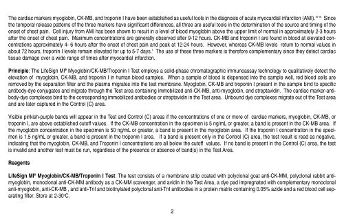
The cardiac markers myoglobin, <strong>CK</strong>-<strong>MB</strong>, and troponin I have been established as useful tools in the diagnosis of acute myocardial infarction (AMI). 10-16 Since<br />
the temporal release patterns of the three markers have significant differences, all three are useful tools in the determination of the source and timing of the<br />
onset of chest pain. Cell injury from AMI has been shown to result in a level of blood myoglobin above the upper limit of normal in approximately 2-3 hours<br />
after the onset of chest pain. Maximum concentrations are generally observed after 9-12 hours. <strong>CK</strong>-<strong>MB</strong> and troponin I are found in blood at elevated concentrations<br />
approximately 4- 6 hours after the onset of chest pain and peak at 12-24 hours. However, whereas <strong>CK</strong>-<strong>MB</strong> levels return to normal values in<br />
about 72 hours, troponin I levels remain elevated for up to 5-7 days. 1 The use of these three markers is therefore complementary since they detect cardiac<br />
tissue damage over a wide range of times after myocardial infarction.<br />
Principle: The <strong>LifeSign</strong> MI ® <strong>Myoglobin</strong>/<strong>CK</strong>-<strong>MB</strong>/<strong>Troponin</strong> I Test employs a solid-phase chromatographic immunoassay technology to qualitatively detect the<br />
elevation of myoglobin, <strong>CK</strong>-<strong>MB</strong>, and troponin I in human blood samples. When a sample of blood is dispensed into the sample well, red blood cells are<br />
removed by the separation filter and the plasma migrates into the test membrane. <strong>Myoglobin</strong>, <strong>CK</strong>-<strong>MB</strong> and troponin I present in the sample bind to specific<br />
antibody-dye conjugates and migrate through the Test area containing immobilized anti-<strong>CK</strong>-<strong>MB</strong>, anti-myoglobin, and streptavidin. The cardiac marker-antibody-dye<br />
complexes bind to the corresponding immobilized antibodies or streptavidin in the Test area. Unbound dye complexes migrate out of the Test area<br />
and are later captured in the Control (C) area.<br />
Visible pinkish-purple bands will appear in the Test and Control (C) areas if the concentrations of one or more of cardiac markers, myoglobin, <strong>CK</strong>-<strong>MB</strong>, or<br />
troponin I, are above established cutoff values. If the <strong>CK</strong>-<strong>MB</strong> concentration in the specimen is 5 ng/mL or greater, a band is present in the <strong>CK</strong>-<strong>MB</strong> area. If<br />
the myoglobin concentration in the specimen is 50 ng/mL or greater, a band is present in the myoglobin area. If the troponin I concentration in the specimen<br />
is 1.5 ng/mL or greater, a band is present in the troponin I area. If a band is present only in the Control (C) area, the test result is read as negative,<br />
indicating that the myoglobin, <strong>CK</strong>-<strong>MB</strong>, and <strong>Troponin</strong> I concentrations are all below the cutoff values. If no band is present in the Control (C) area, the test<br />
is invalid and another test must be run, regardless of the presence or absence of band(s) in the Test Area.<br />
Reagents<br />
<strong>LifeSign</strong> MI ® <strong>Myoglobin</strong>/<strong>CK</strong>-<strong>MB</strong>/<strong>Troponin</strong> I Test: The test consists of a membrane strip coated with polyclonal goat anti-<strong>CK</strong>-MM, polyclonal rabbit antimyoglobin,<br />
monoclonal anti-<strong>CK</strong>-MM antibody as a <strong>CK</strong>-MM scavenger, and avidin in the Test Area, a dye pad impregnated with complementary monoclonal<br />
anti-myoglobin, anti-<strong>CK</strong>-<strong>MB</strong> , and anti-TnI and biotinylated polyclonal anti-TnI antibodies in a protein matrix containing 0.05% azide and a red blood cell separating<br />
filter. Store at 2-30 o C.<br />
2