LifeSign MI® Myoglobin/CK-MB/Troponin I Rapid ... - Drug Testing
LifeSign MI® Myoglobin/CK-MB/Troponin I Rapid ... - Drug Testing
LifeSign MI® Myoglobin/CK-MB/Troponin I Rapid ... - Drug Testing

You also want an ePaper? Increase the reach of your titles
YUMPU automatically turns print PDFs into web optimized ePapers that Google loves.
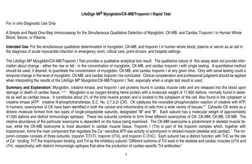
<strong>LifeSign</strong> MI ® <strong>Myoglobin</strong>/<strong>CK</strong>-<strong>MB</strong>/<strong>Troponin</strong> I <strong>Rapid</strong> Test<br />
For in vitro Diagnostic Use Only<br />
A Simple and <strong>Rapid</strong> One-Step Immunoassay for the Simultaneous Qualitative Detection of <strong>Myoglobin</strong>, <strong>CK</strong>-<strong>MB</strong>, and Cardiac <strong>Troponin</strong> I in Human Whole<br />
Blood, Serum, or Plasma.<br />
Intended Use: For the simultaneous qualitative determination of myoglobin, <strong>CK</strong>-<strong>MB</strong>, and troponin I in human whole blood, plasma or serum as an aid in<br />
the diagnosis of acute myocardial infarction in emergency room, critical care, point-of-care, and hospital settings.<br />
The <strong>LifeSign</strong> MI ® <strong>Myoglobin</strong>/<strong>CK</strong>-<strong>MB</strong>/<strong>Troponin</strong> I Test provides a qualitative analytical test result. The qualitative nature of this assay does not provide information<br />
about change - either the rise or fall - in the concentration of myoglobin, <strong>CK</strong>-<strong>MB</strong>, and cardiac troponin I with single testing. A quantitative method<br />
should be used, if desired, to quantitate the concentration of myoglobin, <strong>CK</strong>-<strong>MB</strong>, and cardiac troponin I at any given time. Only with serial testing could a<br />
temporal change in the level of myoglobin, <strong>CK</strong>-<strong>MB</strong>, and cardiac troponin I be concluded. Clinical consideration and professional judgment should be applied<br />
when interpreting the results of the <strong>LifeSign</strong> MI ® <strong>Myoglobin</strong>/<strong>CK</strong>-<strong>MB</strong>/<strong>Troponin</strong> I Test, especially when a single test result is used.<br />
Summary and Explanation: <strong>Myoglobin</strong>, creatine kinase, and troponin I are proteins found in cardiac muscle cells and are released into the blood upon<br />
damage or death of cardiac tissue. 1,2,3,4 <strong>Myoglobin</strong> is an oxygen-binding heme protein with a molecular weight of 17,800 daltons, normally found in skeletal<br />
as well as cardiac tissue. It constitutes about 2% of the total muscle protein and is located in the cytoplasm of the cell. Also found in the cytoplasm is<br />
creatine kinase (ATP: creatine N-phosphotransferase, E.C. No. 2.7.3.2) (<strong>CK</strong>). <strong>CK</strong> catalyzes the reversible phosphorylation reaction of creatine with ATP.<br />
In humans, isoenzymes of <strong>CK</strong> have been identified in both the cytosol and mitochondria of cells from a wide variety of tissues. 5,6 Cytosolic <strong>CK</strong> exists as a<br />
dimeric molecule formed from two types of single-polypeptide subunits, designated “M” and “B”. Each subunit has a molecular weight of approximately<br />
41,000 daltons and distinct immunologic epitopes. These two subunits combine to form three different isoenzymes of <strong>CK</strong>: <strong>CK</strong>-MM, <strong>CK</strong>-BB, <strong>CK</strong>-<strong>MB</strong>. The<br />
relative abundance of the particular isoenzyme is dependent on the tissue being examined. The <strong>CK</strong>-MM isoenzyme is predominant in skeletal muscle tissue,<br />
while the <strong>CK</strong>-<strong>MB</strong> isoenzyme is most abundant in cardiac muscle tissue. <strong>Troponin</strong> I (TnI) is part of the troponin complex which, together with<br />
tropomyosin, forms the main component that regulates the Ca ++ -sensitive ATP-ase activity of actomyosin in striated muscle (skeletal and cardiac). 7 The troponin<br />
complex consists of three subunits, troponin T(TnT), troponin I(TnI), and troponin C (TnC). Each subunit has a distinct function with TnC as the site<br />
of Ca ++ binding, TnT the tropomyosin binding, and TnI as the inhibitory subunit. 8 Different isoforms of TnI exist in the skeletal and cardiac muscles (sTnI and<br />
cTnI, respectively) with distinct immunologic epitopes that allow the production of cardiac-specific TnI antibodies. 9<br />
1
The cardiac markers myoglobin, <strong>CK</strong>-<strong>MB</strong>, and troponin I have been established as useful tools in the diagnosis of acute myocardial infarction (AMI). 10-16 Since<br />
the temporal release patterns of the three markers have significant differences, all three are useful tools in the determination of the source and timing of the<br />
onset of chest pain. Cell injury from AMI has been shown to result in a level of blood myoglobin above the upper limit of normal in approximately 2-3 hours<br />
after the onset of chest pain. Maximum concentrations are generally observed after 9-12 hours. <strong>CK</strong>-<strong>MB</strong> and troponin I are found in blood at elevated concentrations<br />
approximately 4- 6 hours after the onset of chest pain and peak at 12-24 hours. However, whereas <strong>CK</strong>-<strong>MB</strong> levels return to normal values in<br />
about 72 hours, troponin I levels remain elevated for up to 5-7 days. 1 The use of these three markers is therefore complementary since they detect cardiac<br />
tissue damage over a wide range of times after myocardial infarction.<br />
Principle: The <strong>LifeSign</strong> MI ® <strong>Myoglobin</strong>/<strong>CK</strong>-<strong>MB</strong>/<strong>Troponin</strong> I Test employs a solid-phase chromatographic immunoassay technology to qualitatively detect the<br />
elevation of myoglobin, <strong>CK</strong>-<strong>MB</strong>, and troponin I in human blood samples. When a sample of blood is dispensed into the sample well, red blood cells are<br />
removed by the separation filter and the plasma migrates into the test membrane. <strong>Myoglobin</strong>, <strong>CK</strong>-<strong>MB</strong> and troponin I present in the sample bind to specific<br />
antibody-dye conjugates and migrate through the Test area containing immobilized anti-<strong>CK</strong>-<strong>MB</strong>, anti-myoglobin, and streptavidin. The cardiac marker-antibody-dye<br />
complexes bind to the corresponding immobilized antibodies or streptavidin in the Test area. Unbound dye complexes migrate out of the Test area<br />
and are later captured in the Control (C) area.<br />
Visible pinkish-purple bands will appear in the Test and Control (C) areas if the concentrations of one or more of cardiac markers, myoglobin, <strong>CK</strong>-<strong>MB</strong>, or<br />
troponin I, are above established cutoff values. If the <strong>CK</strong>-<strong>MB</strong> concentration in the specimen is 5 ng/mL or greater, a band is present in the <strong>CK</strong>-<strong>MB</strong> area. If<br />
the myoglobin concentration in the specimen is 50 ng/mL or greater, a band is present in the myoglobin area. If the troponin I concentration in the specimen<br />
is 1.5 ng/mL or greater, a band is present in the troponin I area. If a band is present only in the Control (C) area, the test result is read as negative,<br />
indicating that the myoglobin, <strong>CK</strong>-<strong>MB</strong>, and <strong>Troponin</strong> I concentrations are all below the cutoff values. If no band is present in the Control (C) area, the test<br />
is invalid and another test must be run, regardless of the presence or absence of band(s) in the Test Area.<br />
Reagents<br />
<strong>LifeSign</strong> MI ® <strong>Myoglobin</strong>/<strong>CK</strong>-<strong>MB</strong>/<strong>Troponin</strong> I Test: The test consists of a membrane strip coated with polyclonal goat anti-<strong>CK</strong>-MM, polyclonal rabbit antimyoglobin,<br />
monoclonal anti-<strong>CK</strong>-MM antibody as a <strong>CK</strong>-MM scavenger, and avidin in the Test Area, a dye pad impregnated with complementary monoclonal<br />
anti-myoglobin, anti-<strong>CK</strong>-<strong>MB</strong> , and anti-TnI and biotinylated polyclonal anti-TnI antibodies in a protein matrix containing 0.05% azide and a red blood cell separating<br />
filter. Store at 2-30 o C.<br />
2
Specimen Collection and Preparation<br />
Whole blood, plasma or serum may be used as samples for this procedure. For whole blood or plasma, collect blood in a tube containing heparin as the<br />
anticoagulant. If serum samples are to be used, collect the blood in a tube without anticoagulant and allow to clot. Since cardiac proteins are relatively<br />
unstable, it is recommended that fresh samples be used as soon as possible. Blood samples should be tested within 4 hours of collection. If specimens<br />
must be stored, the blood cells should be removed. Plasma or serum samples may be refrigerated for 24 hours at 2-8°C. If plasma or serum samples must<br />
be stored for more than 24 hours, they should be frozen at -20°C or below.<br />
Materials Provided:<br />
Each box contains the following:<br />
<strong>LifeSign</strong> MI ® <strong>Myoglobin</strong>/<strong>CK</strong>-<strong>MB</strong>/<strong>Troponin</strong> I Test sealed in a foil pouch with desiccant and dropper<br />
Result sticker<br />
Directions for use<br />
Materials Required But Not Provided:<br />
1. Vacutainer ® (Becton Dickinson) tube, or equivalent, containing heparin as the anticoagulant or a tube designed for collection of serum.<br />
2. Timer<br />
3. Micropipettor and disposable pipet tips which are necessary only if the dropper provided is not used.<br />
Procedure<br />
1. Open the foil pouch, remove the <strong>LifeSign</strong> MI ® <strong>Myoglobin</strong>/<strong>CK</strong>-<strong>MB</strong>/<strong>Troponin</strong> I Test and lay the test on a<br />
level surface.<br />
2. Label the test with the patient’s identification.<br />
3. Using the dropper provided, add 3 drops (120 µl) of whole blood or plasma or serum into the sample well.<br />
4. Read the test results at 15 minutes.<br />
Procedural Notes<br />
• Do not use this product beyond the expiration date.<br />
• All patient samples should be handled as if they are potentially infectious. Observe established procedures for proper disposal of specimens and the<br />
used test device.<br />
3
<strong>LifeSign</strong> <strong>MI®</strong> <strong>Myoglobin</strong>/<strong>CK</strong>-<strong>MB</strong>/<strong>Troponin</strong> I Test Procedure and Results<br />
1. Add 3 drops (120 µL) of<br />
whole blood, serum or<br />
plasma sample.<br />
2. Read at 15 minutes.<br />
CONTROL (VALIDATION) BAND (C)<br />
The Control/Validation band serves two purposes:<br />
1. Functional test of the dye conjugates: and<br />
2. Proof of sample migration.<br />
If no control band appears, the test is NOT VALID.<br />
Repeat the test using a new <strong>LifeSign</strong> MI ® <strong>Myoglobin</strong>/<strong>CK</strong>-<strong>MB</strong>/<br />
<strong>Troponin</strong> I Test, and follow the procedure carefully.<br />
C<br />
Myo<br />
<strong>CK</strong>-<strong>MB</strong><br />
TnI<br />
C<br />
Myo<br />
<strong>CK</strong>-<strong>MB</strong><br />
TnI<br />
C<br />
Myo<br />
<strong>CK</strong>-<strong>MB</strong><br />
TnI<br />
C<br />
Myo<br />
<strong>CK</strong>-<strong>MB</strong><br />
TnI<br />
<strong>Myoglobin</strong> (+)<br />
<strong>CK</strong>-<strong>MB</strong> (+)<br />
<strong>Troponin</strong> I (+)<br />
Myo (–)<br />
<strong>CK</strong>-<strong>MB</strong> (–)<br />
TnI (+)<br />
Myo (+)<br />
<strong>CK</strong>-<strong>MB</strong> (+)<br />
TnI (–)<br />
Myo (–)<br />
<strong>CK</strong>-<strong>MB</strong> (–)<br />
TnI (–)<br />
Invalid<br />
4
Interpretation of Results<br />
1. Negative (-)<br />
A single pinkish purple colored band in the Control (C) area, with the absence of a distinct colored band in the Test area, indicates that the concentration<br />
of myoglobin is below 50 ng/ml, the concentrations of <strong>CK</strong>-<strong>MB</strong> is below 5 ng/ml, and the concentration of troponin I is below 1.5 ng/ml and the test<br />
result is negative.<br />
2. Positive (+)<br />
The presence of a pinkish purple colored band in the C area and the presence of one or more distinct bands in the Test area indicates a positive test<br />
result. If a band is present in the Myo area, the myoglobin concentration is 50 ng/ml or greater. If a band is present in the <strong>CK</strong>-<strong>MB</strong> area, the <strong>CK</strong>-<strong>MB</strong> concentration<br />
is 5 ng/ml or greater. If a band is present in the TnI area, the troponin I concentration is 1.5 ng/ml or greater.<br />
Notes:<br />
• A positive test result for myoglobin, <strong>CK</strong>-<strong>MB</strong>, or troponin I can be read as soon as a distinct colored band appears in both the C area and in the Test area<br />
for that cardiac marker.<br />
• Positive test results from strong positive samples may appear within 5 minutes.<br />
• The myoglobin, <strong>CK</strong>-<strong>MB</strong>, and TnI bands may appear sooner and darker than the Control band in samples that are very strongly positive.<br />
• The myoglobin, <strong>CK</strong>-<strong>MB</strong>, and TnI bands may appear after the appearance of the Control band and be fainter in samples that are weakly positive.<br />
• The <strong>LifeSign</strong> MI ® <strong>Myoglobin</strong>/<strong>CK</strong>-<strong>MB</strong>/<strong>Troponin</strong> I Test has been optimized to ensure that high concentrations of the cardiac<br />
markers will not result in false negative test results which are commonly referred to as a “high dose hook” or “prozone effect” for<br />
quantitative immunoassays. Concentrations of myoglobin, <strong>CK</strong>-<strong>MB</strong>, and troponin I of 50,000, 50,000, and 1,100 ng/mL, respectively were<br />
demonstrated to produce the expected positive test results in the <strong>LifeSign</strong> MI ® <strong>Myoglobin</strong>/<strong>CK</strong>-<strong>MB</strong>/<strong>Troponin</strong> I Test.<br />
3. Invalid<br />
A distinct colored band in the C area should always appear. If no pinkish purple band is present in the C area at the end of the 15 minute test period,<br />
the test is invalid, and the sample must be retested using a new test.<br />
Limitations:<br />
• The results of the <strong>LifeSign</strong> MI ® <strong>Myoglobin</strong>/<strong>CK</strong>-<strong>MB</strong>/<strong>Troponin</strong> I Test are to be used in conjunction with other clinical information such as clinical signs and<br />
5
symptoms and electrocardiographic test results to diagnose cardiac ischemia. A positive test result from a patient suspected of AMI may be used as an<br />
indicator of myocardial damage and requires further confirmation. Sampling of patients suspected of AMI at multiple time points is recommended due to<br />
the delay between onset of symptoms and the release of cardiac protein markers into the bloodstream.<br />
• Samples containing unusually high titers of certain antibodies, such as human anti-mouse or human anti-rabbit antibodies, may affect the performance<br />
of the test.<br />
• Hematocrit values in the range of 20 - 60% did not significantly affect the <strong>LifeSign</strong> MI ® Test results.<br />
User Quality Control: A quality control test using positive and negative controls such as the Princeton BioMeditech <strong>LifeSign</strong> MI ® controls (<strong>Myoglobin</strong>/<strong>CK</strong>-<br />
<strong>MB</strong>/<strong>Troponin</strong> I) should be performed before using a new lot of <strong>LifeSign</strong> MI ® <strong>Myoglobin</strong>/<strong>CK</strong>-<strong>MB</strong>/<strong>Troponin</strong> I Test and at regular intervals as part of a good quality<br />
control (QC) practice. The positive control should be selected to produce a moderate positive result in the myoglobin, <strong>CK</strong>-<strong>MB</strong>, and TnI areas as well as<br />
the C area. The negative control should produce a negative result (only control band present). Upon confirmation of the expected results, the lot is ready<br />
to use with patient specimens. Controls should also be used whenever the validity of test results is questioned and in accordance to local Quality Assurance<br />
policy. For information about obtaining controls contact the <strong>LifeSign</strong> MI ® local distributor for assistance.<br />
The presence of a band in the C area acts as an internal procedural control to ensure the valid performance of the test. In the absence of a band in the C<br />
area, the test is invalid and must be repeated. If a problem persists, contact the <strong>LifeSign</strong> MI ® Technical Support for assistance, 800-526-2125 or 732-246-<br />
3366.<br />
Expected Values: Specimens from 21 healthy adults were tested in the <strong>LifeSign</strong> MI ® <strong>Myoglobin</strong>/<strong>CK</strong>-<strong>MB</strong>/<strong>Troponin</strong> I Test and all specimens were determined<br />
to be negative for all three cardiac markers.<br />
The <strong>LifeSign</strong> MI ® <strong>Myoglobin</strong>/<strong>CK</strong>-<strong>MB</strong>/<strong>Troponin</strong> I Test has been calibrated against the <strong>LifeSign</strong> MI ® <strong>Myoglobin</strong>/<strong>CK</strong>-<strong>MB</strong> Test and the <strong>LifeSign</strong> MI ® <strong>Troponin</strong> I Test.<br />
The <strong>LifeSign</strong> MI ® <strong>Myoglobin</strong>/<strong>CK</strong>-<strong>MB</strong> Test uses a diagnostic cutoff of 5 ng/mL for <strong>CK</strong>-<strong>MB</strong> and 50 ng/mL for myoglobin. The <strong>LifeSign</strong> MI ® <strong>Troponin</strong> I Test uses<br />
a diagnostic cutoff of 1.5 ng/mL . The <strong>LifeSign</strong> MI ® <strong>Myoglobin</strong>/<strong>CK</strong>-<strong>MB</strong>/<strong>Troponin</strong> I Test is designed to yield a positive result when the concentration of one<br />
or more of the cardiac markers, myoglobin, <strong>CK</strong>-<strong>MB</strong>, or troponin I, is above these established cut-off values.<br />
6
Performance Characteristics<br />
Interfering Substances: Substances at the following levels do not interfere with the <strong>LifeSign</strong> MI ® <strong>Myoglobin</strong>/<strong>CK</strong>-<strong>MB</strong>/<strong>Troponin</strong> I Test :<br />
Human albumin<br />
Bilirubin (unconjugated)<br />
Free hemoglobin<br />
Triglycerides<br />
16 g/dL<br />
60 mg/dL<br />
4 g/dL<br />
1,300 mg/dL<br />
The following drugs were evaluated for potential positive and negative interference by the addition of these materials to (1) a serum sample containing elevated<br />
levels of myoglobin, <strong>CK</strong>-<strong>MB</strong>, and TnI and (2) a serum sample negative for the 3 cardiac markers. These drugs were tested at approximately twice<br />
the recommended therapeutic level. No interference was observed for any of these medications:<br />
Acetaminophen Digoxin Nystatine<br />
Acetylsalicylic acid Dipyridamole Oxazepam<br />
Allopurinol Erythromycin Oxytetracycline<br />
Ambroxol Furosemide Phenobarbital<br />
Ampicillin Glibenclamide Phenytoin<br />
Ascrobic acid Hydrochlorothiazide Probenecid<br />
Atenolol Indomethacin Procainamide<br />
Caffeine Isosorbide dinitrate D,L - Propanolol<br />
Captopril L-thyroxine Quinidine<br />
Chloramphenicol Methaqualone Sulfamethoxazol<br />
Chlordiazepoxide D,L-alpha-Methyldopa Theophylline<br />
Cinnarizine Nicotinic acid Trimethoprim<br />
Cyclosporine Nifedipine Verapamil<br />
Diclofenac<br />
Nitrofurantoin<br />
7
Cross-Reactivity Studies: Related human proteins were purified and added to normal human plasma to test for their potential reactivity in the <strong>LifeSign</strong> MI ®<br />
<strong>Myoglobin</strong>/<strong>CK</strong>-<strong>MB</strong>/TnI Test. A negative result was obtained with the proteins at the following concentrations:<br />
Protein<br />
Concentration<br />
(ng/mL)<br />
<strong>CK</strong>-BB 1000<br />
cardiac myosin light chain 200<br />
cardiac troponin T 5000<br />
cardiac troponin C 1000<br />
fast-twitch skeletal TnI 314<br />
Recovery Study: Normal human blood was supplemented with human myoglobin at concentrations of 125, 200, and 400 ng/mL, <strong>CK</strong>-<strong>MB</strong> at concentrations<br />
of 6, 10, and 20 ng/mL, and troponin I at concentrations of 2, 4, and 8 ng/mL. The samples were tested using the <strong>LifeSign</strong> MI ® <strong>Myoglobin</strong>/<strong>CK</strong>-<strong>MB</strong>/TnI Test<br />
in 6 replicates. There was a 100% agreement between the expected and the observed results at each concentration of cardiac marker.<br />
Proficiency <strong>Testing</strong>: Four different hospitals were provided with blinded whole blood samples. One group of samples had been supplemented with myoglobin,<br />
<strong>CK</strong>-<strong>MB</strong>, and TnI at concentrations of 100,10 and 3 ng/mL , respectively (Level 1). A second set of samples had been supplemented with myoglobin,<br />
<strong>CK</strong>-<strong>MB</strong>, and TnI at concentrations of 200, 20, and 6 ng/mL , respectively (Level 2). A third set of samples was not supplemented with any of the cardiac<br />
markers and thereby served as a negative control group. Each site received five replicates of each sample for a total of 15 samples per site. As shown<br />
in the data table below, there was a mean within run agreement of 98.8% and a mean between sites agreement of 98.9% in this study.<br />
8
<strong>LifeSign</strong> MI ® <strong>Myoglobin</strong>/<strong>CK</strong>-<strong>MB</strong>/<strong>Troponin</strong> I Test<br />
(Number of Positive Results/ Total)<br />
Sites<br />
Cardiac Marker<br />
Band<br />
No Additions<br />
(negative)<br />
Supplemented<br />
with Level 1<br />
Supplemented<br />
with Level 2<br />
% Agreement<br />
of Within Run<br />
Site 1<br />
myoglobin<br />
<strong>CK</strong>-<strong>MB</strong><br />
troponin I<br />
0/5<br />
0/5<br />
0/5<br />
5/5<br />
5/5<br />
5/5<br />
5/5<br />
5/5<br />
5/5<br />
100<br />
100<br />
100<br />
Site 2<br />
Site 3<br />
Site 4<br />
myoglobin<br />
<strong>CK</strong>-<strong>MB</strong><br />
troponin I<br />
myoglobin<br />
<strong>CK</strong>-<strong>MB</strong><br />
troponin I<br />
myoglobin<br />
<strong>CK</strong>-<strong>MB</strong><br />
troponin I<br />
0/5<br />
0/5<br />
0/5<br />
0/5<br />
0/5<br />
0/5<br />
0/5<br />
0/5<br />
0/5<br />
5/5<br />
4/5<br />
4/5<br />
5/5<br />
5/5<br />
5/5<br />
5/5<br />
5/5<br />
5/5<br />
5/5<br />
5/5<br />
5/5<br />
5/5<br />
5/5<br />
5/5<br />
5/5<br />
5/5<br />
5/5<br />
100<br />
93.3<br />
93.3<br />
100<br />
100<br />
100<br />
100<br />
100<br />
100<br />
% Agreement<br />
Between Sites<br />
100 96.7 100<br />
9
Correlation of Assay Results Between Whole Blood and Plasma: Heparinized whole blood from 20 individuals with or without elevated cardiac markers<br />
were tested prior to removal of the red cells and after red cell removal. The agreement between the use of whole blood and plasma was 100% as shown<br />
in the table below.<br />
Cardiac Result Whole blood(+) Whole blood(-) Whole blood(+) Whole blood(-)<br />
Marker and plasma (+) and plasma (-) and plasma (-) and plasma (+)<br />
<strong>Myoglobin</strong> 11 9 0 0<br />
<strong>CK</strong>-<strong>MB</strong> 11 9 0 0<br />
<strong>Troponin</strong> I 17 3 0 0<br />
Correlation of Assay Results Between Whole Blood and Serum: Whole blood (collected without anti-coagulant) from 20 individuals with or without elevated<br />
cardiac markers were tested prior to removal of the red cells and after red cell removal. The agreement between the use of whole blood and serum<br />
was 100% as shown in the table below.<br />
Cardiac Result Whole blood(+) Whole blood(-) Whole blood(+) Whole blood(-)<br />
Marker and serum (+) and serum (-) and serum (-) and serum (+)<br />
<strong>Myoglobin</strong> 11 9 0 0<br />
<strong>CK</strong>-<strong>MB</strong> 11 9 0 0<br />
<strong>Troponin</strong> I 17 3 0 0<br />
10
Method Comparison:<br />
Serum Samples (n = 251) were collected from 183 individuals at different time intervals after being admitted to a hospital emergency department with chest<br />
pain. The samples were tested with the <strong>LifeSign</strong> MI ® <strong>Myoglobin</strong>/<strong>CK</strong>-<strong>MB</strong>/<strong>Troponin</strong> I Test and with the <strong>LifeSign</strong> MI ® <strong>Myoglobin</strong>/<strong>CK</strong>-<strong>MB</strong> Test and the <strong>LifeSign</strong><br />
MI ® TnI Test. The correlation between the tests is shown below:<br />
<strong>LifeSign</strong> MI ® <strong>LifeSign</strong> MI ® <strong>LifeSign</strong> MI ®<br />
<strong>Myoglobin</strong>/<strong>CK</strong>-<strong>MB</strong> <strong>Myoglobin</strong>/<strong>CK</strong>-<strong>MB</strong> <strong>Troponin</strong> I<br />
Test Test Test Total<br />
myoglobin results <strong>CK</strong>-<strong>MB</strong> results troponin I results<br />
Pos Neg Pos Neg Pos Neg<br />
<strong>LifeSign</strong> MI ® Pos 69 2 89 5 65 2<br />
<strong>Myoglobin</strong>/<strong>CK</strong>-<strong>MB</strong>/<br />
<strong>Troponin</strong> I Test Neg 3 177 3 154 2 182<br />
Total 72 179 92 159 67 184 251<br />
<strong>Myoglobin</strong> <strong>CK</strong>-<strong>MB</strong> <strong>Troponin</strong> I<br />
Comparative 95.8% (69/72) 96.7% (89/92) 97% (65/67)<br />
Sensitivity<br />
Comparative 98.9% (177/179) 96.8% (154/159) 98.9% (182/184)<br />
Specificity<br />
Overall Agreement 98% (246/251) 96.8% (243/251) 98.4% (247/251)<br />
11
References<br />
1. Adams, J.E., et al, Biochemical markers of myocardial injury. Is <strong>MB</strong> creatine kinase the choice for the 1990’s. Circulation 88:750 (1993)<br />
2. Cairns, J. et al., usefulness of serial determinations of myoglobin and creatine kinase in serum compared for assessment of acute myocardial infarction.<br />
Clin.Chem. 29:469 (1983)<br />
3. Ohman, E.M. et al., Early detection of acute myocardial infarction: Additional diagnostic information from serum concentrations of myoglobin in patients<br />
without ST elevation. Br. Heart J. 63:335 (1990)<br />
4. McComb, J.M. et al., <strong>Myoglobin</strong> in the very early phase acute myocardial infarction. Ann. Clin. Biochem. 22:162 (1985)<br />
5. Neumeier, D., Tissue specific and subcellular distribution of creatine kinase isoenzymes. In Lang, H. (ed):Creatine Kinase Isoenzymes, Springer Verlag<br />
p.85 (1981)<br />
6. Lott, J.A. and Abbott, L.B., Creatine kinase isoenzymes Clin.Lab.Med. 6:547 (1986)<br />
7. Mehegan, J.P. And Tobacman, L.S. Cooperative interaction between troponin molecules bound to the cardiac thin filament. J.Biol.Chem. 266:966<br />
(1991)<br />
8. Ebashi,S., Ca2+ and the contractile proteins. J.Mol.Cell.Cardiol. 16:129 (1984)<br />
9. Bodor, S.G., et al., Development of monoclonal antibody for an assay of cardiac troponin I and preliminary results in suspected cases of myocardial<br />
infarction. Clin.Chem. 38:2203 (1992)<br />
10.Hamm,C.W., Cardiac-specific <strong>Troponin</strong>s in Acute Coronary Syndromes, in Heart Disease, a textbook of Cardiovascular Medicine, ed. Braunwald, E.<br />
,W.B. Saunders Co. Update 3 (1977)<br />
11. Panteghini, M., et al., Comparison of the Diagnostic Performance of Two <strong>Rapid</strong> Bedside Biochemical Assays in the Early Detection of Acute<br />
Myocardial Infarction, Clin.Cardiol. 21:394 (1998)<br />
12. Tucker, J.F., et al., Value of serial myoglobin levels in the early diagnosis of patients admitted for acute myocardial infarction, Ann.Emerg.Med. 24:704<br />
(1994)<br />
13. Hamm, C.W., et al., Emergency room triage of patients with acute chest pain by means of rapid testing for cardiac troponin T or troponin I. New<br />
Eng.J.Med. 337:1648 (1997)<br />
14. Antman, E.M., et al., Cardiac-specific troponin I levels to predict the risk of mortality inpatients with acute coronary syndromes. New Eng.J.Med.<br />
335:1342 (1996)<br />
15. Brogan, G.X. Jr., et al., Evaluation of Cardiac STATus <strong>CK</strong>-<strong>MB</strong>/<strong>Myoglobin</strong> device for rapidly ruling out acute myocardial infarction, Clin.Lab Med.<br />
17(4):655 (1997)<br />
16. Sylven,C. et al., Excellent reliability of nurse-based bedside diagnosis of acute myocardial infarction by rapid dry-strip creatine kinase, myoglobin, and<br />
troponin T. Amer.Heart J. 135(4):677 (1998)<br />
12
FOR IN VITRO DIAGNOSTIC USE ONLY<br />
Manufactured by:<br />
PBM<br />
Princeton BioMeditech Corp.<br />
Princeton, NJ 08543-7139<br />
USA<br />
Tel: 732-274-1000<br />
Fax: 732-274-1010<br />
111302BL<br />
13