

Eμ-BRD2 transgenic mice develop B-cell lymphoma and leukemia
Eμ-BRD2 transgenic mice develop B-cell lymphoma and leukemia
Eμ-BRD2 transgenic mice develop B-cell lymphoma and leukemia

Create successful ePaper yourself
Turn your PDF publications into a flip-book with our unique Google optimized e-Paper software.
NEOPLASIA<br />
E-<strong>BRD2</strong> <strong>transgenic</strong> <strong>mice</strong> <strong>develop</strong> B-<strong>cell</strong> <strong>lymphoma</strong> <strong>and</strong> <strong>leukemia</strong><br />
Rebecca J. Greenwald, Joseph R. Tumang, Anupama Sinha, Nicolas Currier, Robert D. Cardiff,<br />
Thomas L. Rothstein, Douglas V. Faller, <strong>and</strong> Gerald V. Denis<br />
Transgenic <strong>mice</strong> with lymphoid-restricted<br />
overexpression of the double bromodomain<br />
protein bromodomain-containing 2<br />
(Brd2) <strong>develop</strong> splenic B-<strong>cell</strong> <strong>lymphoma</strong> <strong>and</strong>,<br />
upon transplantation, B-<strong>cell</strong> <strong>leukemia</strong> with<br />
leukemic infiltrates in liver <strong>and</strong> lung. Brd2 is<br />
a nuclear-localized transcription factor kinase<br />
that is most closely related to TATA<br />
box binding protein–associated factor, 250<br />
kDa (TAF II 250) <strong>and</strong> the Drosophila <strong>develop</strong>mental<br />
protein female sterile homeotic. Constitutive<br />
expression of <strong>BRD2</strong> in the lymphoid<br />
compartment increases cyclin A<br />
Introduction<br />
transcription, “priming” <strong>transgenic</strong> B <strong>cell</strong>s<br />
for proliferation. Mice stochastically <strong>develop</strong><br />
an aggressive B-<strong>cell</strong> <strong>lymphoma</strong> with<br />
the features of B-1 <strong>cell</strong>s, including CD5 <strong>and</strong><br />
surface IgM expression. The B-<strong>cell</strong> <strong>lymphoma</strong><br />
is monoclonal for immunoglobulin<br />
gene rearrangement <strong>and</strong> is phenotypically<br />
stable. The lymphoblasts are very large <strong>and</strong><br />
express a transcriptome that is similar to<br />
human non-Hodgkin <strong>lymphoma</strong>s. Both a<br />
wild-type <strong>BRD2</strong> transgene <strong>and</strong> a kinase-null<br />
point mutant drive <strong>lymphoma</strong>genesis; therefore<br />
we propose that, rather than kinase<br />
activity, Brd2-mediated recruitment of E2<br />
promoter binding factors (E2Fs) <strong>and</strong> a specific<br />
histone acetyltransferase to the cyclin<br />
A promoter by both types of transgene is a<br />
mechanistic basis for neoplasia. This report<br />
is the first to describe a <strong>transgenic</strong> mouse<br />
model for constitutive expression of a protein<br />
with more than one bromodomain.<br />
(Blood. 2004;103:1475-1484)<br />
© 2004 by The American Society of Hematology<br />
Proper transcriptional control of the <strong>cell</strong> cycle is necessary to avoid<br />
errors that can lead to neoplastic transformation. Bromodomaincontaining<br />
proteins 1-3 recently garnered attention for their roles as<br />
transcriptional regulators <strong>and</strong> their links to chromatin-modifying<br />
activities. The bromodomain motif 4-6 is found in proteins with<br />
intrinsic histone acetyltransferase (HAT) activity, 7-9 such as CREB<br />
(cyclic adenosine monophosphate [cAMP]–response element binding<br />
protein) binding protein (CBP), Gcn5, <strong>and</strong> TAF II 250; in protein<br />
adapters that associate with HAT-containing multiprotein complexes,<br />
such as polybromo 10 ; in certain transcription factors, 11 such<br />
as suppressor of Ty 7 (Spt7); or in chromatin remodeling machines,<br />
such as switch 2/sucrose nonfermenting 2 (Swi2/Snf2) <strong>and</strong> brahma. 12<br />
Bromodomain proteins bind -aminoacetyl-lysines of core histones,<br />
at least in the case of p300/CBP-associated factor (p/CAF),<br />
facilitating changes in nucleosome structure. 13 The bromodomain/<br />
extraterminal domain (BET) protein family, defined by aminoterminal<br />
bromodomains <strong>and</strong> an “extraterminal domain,” 14 includes<br />
(formerly 15 RING3 [really interesting new gene 3]) <strong>and</strong> female<br />
sterile homeotic. 16 These transcriptional regulators contain 2 mutually<br />
related bromodomains that are most similar in primary<br />
sequence to the bromodomain of CBP, yet in overall structural<br />
organization most similar to TATA box binding protein–associated<br />
factor, 250 kDa (TAF II 250), which has 2 bromodomains. 14 The<br />
yeast BET protein bromodomain factor 1 (Bdf1) binds acetylated<br />
nucleosomal lysines 17,18 <strong>and</strong> combines with yeast TATA box<br />
binding protein-associated factor, 145 kDa (yTaf145) to perform a<br />
function similar to TAF II 250 in mammals. 19 Recent studies implicated<br />
bromodomain proteins in multiple transcriptional regulatory<br />
systems, including those that control the <strong>cell</strong> cycle, 20-22 generating<br />
interest in their role in carcinogenesis. 23,24<br />
Human leukemic <strong>cell</strong> lines or primary leukemic blasts have<br />
high levels of bromodomain-containing 2 (Brd2) autophosphorylation<br />
<strong>and</strong> substrate-directed phosphorylation activity, unlike normal<br />
lymphocytes. 25 We hypothesized a functional link between the<br />
increased kinase activity <strong>and</strong> oncogenic transformation, particularly<br />
because mitogenic signals increase autophosphorylation 25-27<br />
<strong>and</strong> induce nuclear translocation. 28 Nuclear-localized Brd2 or<br />
Brd2-like proteins participate in multiprotein transcription complexes,<br />
such as murine mediator, where Brd2 associates with<br />
proteins homologous to yeast transcriptional repressors suppressor<br />
of RNA polymerase B 7 (Srb7) <strong>and</strong> required for glucose repression<br />
in yeast 1 (Rgr1), <strong>and</strong> coactivator RNA polymerase transcriptional<br />
regulation mediator subunit 7 (Med7), consistent with the contention<br />
that Brd2 provides a transcriptional end point for mitogenic<br />
signaling. 29 Synergistically with oncogenic ras 30 or ras effectors,<br />
overexpressed Brd2 forms foci in monolayers of NIH/3T3 <strong>cell</strong>s <strong>and</strong><br />
transactivates reporter constructs of the promoters of the E2<br />
promoter binding factor (E2F)–regulated <strong>cell</strong> cycle genes cyclin<br />
D1, cyclin E, cyclin A, <strong>and</strong> dihydrofolate reductase. 30 Endogenous<br />
Brd2 participates in E2F-containing nuclear complexes. 30 A kinaseinactive<br />
point mutant of Brd2 does not transactivate synergistically<br />
with constitutively active mitogen-activated kinase kinase kinase 1<br />
From the Department of Pathology, Immunology Research Division, Brigham<br />
<strong>and</strong> Women’s Hospital, Harvard Medical School, Boston, MA; Immunobiology<br />
Unit, Departments of Medicine <strong>and</strong> Microbiology, Evans Biomedical Research<br />
Center, Boston Medical Center, Boston, MA; Cancer Research Center, Boston<br />
University School of Medicine, Boston, MA; <strong>and</strong> Center for Comparative<br />
Medicine, University of California at Davis, Davis, CA.<br />
Submitted June 26, 2003; accepted October 8, 2003. Prepublished online as<br />
Blood First Edition Paper, October 23, 2003; DOI 10.1182/blood-2003-06-2116.<br />
Supported by United States Public Health Service grants RR14905 from<br />
National Institutes of Health (NIH; National Regional Resource Centers;<br />
R.D.C.); <strong>and</strong> CA84294 (R.D.C.), CA84193 (D.V.F.) <strong>and</strong> CA75107 (G.V.D.) from<br />
the National Cancer Institute.<br />
Reprints: Gerald V. Denis, Cancer Research Center, K521, Boston University<br />
School of Medicine, 80 East Concord St, Boston, MA 02118; e-mail:<br />
gdenis@bu.edu.<br />
The publication costs of this article were defrayed in part by page charge<br />
payment. Therefore, <strong>and</strong> solely to indicate this fact, this article is hereby<br />
marked ‘‘advertisement’’ in accordance with 18 U.S.C. section 1734.<br />
© 2004 by The American Society of Hematology<br />
BLOOD, 15 FEBRUARY 2004 VOLUME 103, NUMBER 4<br />
1475
1476 GREENWALD et al BLOOD, 15 FEBRUARY 2004 VOLUME 103, NUMBER 4<br />
(MEKK), a ras effector, but wild-type does 30 ; however, both forms<br />
transactivate synergistically with oncogenic ras (G.V.D., unpublished<br />
data, December 2002). These results likely mean that both<br />
kinase-dependent <strong>and</strong> independent ras pathways are involved in<br />
Brd2 transactivation. Ectopically expressed Brd2 transactivates<br />
endogenous cyclin A <strong>and</strong> accelerates the G 1 3 S transition (A.S.,<br />
G.V.D., unpublished data, May 2003), providing a mechanism for<br />
its observed transforming properties.<br />
A logical next step was <strong>develop</strong>ing a <strong>transgenic</strong> mouse model,<br />
wherein constitutive overexpression of recombinant Brd2 would be<br />
predicted to deregulate the <strong>cell</strong> cycle <strong>and</strong> lead to transformation.<br />
We used a murine immunoglobulin (Ig) heavy-chain promoterenhancer<br />
construct pIgTE/N to express <strong>BRD2</strong> cDNA in the<br />
lymphoid compartment (E-<strong>BRD2</strong>; <strong>transgenic</strong> [Tg]), or the same<br />
vector incorporating a kinase-inactive point mutant (E-<strong>BRD2</strong> K574A ;<br />
Tg mut ), to test the hypothesis that kinase activity is linked to<br />
oncogenesis. The E system has been used successfully to express<br />
myc in the B lineage to produce B-<strong>cell</strong> 31 <strong>and</strong> pre–B-<strong>cell</strong> 32<br />
<strong>lymphoma</strong>s <strong>and</strong> the subunit of casein kinase II in the T lineage to<br />
produce thymomas. 33 Malignancies arise stochastically in these<br />
models <strong>and</strong> in E-<strong>BRD2</strong> <strong>mice</strong>: transgene expression is elevated in<br />
B <strong>cell</strong>s but not T <strong>cell</strong>s or other tissues; they sporadically <strong>develop</strong> a<br />
B-<strong>cell</strong> <strong>lymphoma</strong>. The malignancy is monoclonal, transplantable,<br />
<strong>and</strong> phenotypically uniform, bearing many of the hallmarks of B-1<br />
<strong>cell</strong>s. 34 Both Tg <strong>and</strong> Tg mut <strong>mice</strong> <strong>develop</strong> the same disease but<br />
littermate controls do not. This result suggests that intrinsic kinase<br />
activity of Brd2 is not required for oncogenesis in this system <strong>and</strong><br />
evinces that <strong>BRD2</strong> has oncogenic properties in vivo.<br />
Materials <strong>and</strong> methods<br />
Molecular cloning<br />
A consensus Kozak sequence was engineered into the 2.28-kilobase (kb)<br />
human Brd2 cDNA with Vent DNA Polymerase (New Engl<strong>and</strong> Biolabs,<br />
Beverly, MA). The cDNA was cloned into the XhoI site of pIgTE/N <strong>and</strong><br />
propagated in Escherichia coli STBL2 (Invitrogen Life Technologies,<br />
Carlsbad, CA). Restriction analysis <strong>and</strong> DNA sequencing verified the Tg<br />
construct. A Tg construct harboring a point mutation of catalytic lysine 574<br />
to alanine 25 was generated simultaneously (Tg mut ). A 5.22-kb SalI/MluI<br />
fragment containing the E enhancer <strong>and</strong> simian virus 40 (SV40) poly A<br />
was purified <strong>and</strong> injected into FVB oocytes.<br />
Construction of <strong>transgenic</strong> <strong>mice</strong> <strong>and</strong> genotyping<br />
Southern blotting <strong>and</strong> polymerase chain reaction (PCR) analysis of tail<br />
DNA identified founders. A primer set distinguished between endogenous<br />
<strong>and</strong> <strong>transgenic</strong> DNA (5-CAAGAGCTGGTAGTGACCATCCCT-3 <strong>and</strong><br />
5AGGAGGGCTAGCTGGAGAACCAGGAGC-3). Taq DNA Polymerase<br />
(Invitrogen Life Technologies) amplified the templates in 2.0 mM Mg 2<br />
for 35 cycles at a melting temperature of 94°C (t m ; 30 seconds), an<br />
annealing temperature of 68°C (t a ; 30 s), <strong>and</strong> an extension temperature of<br />
72°C (t e ; 1 minute). Three Tg founders <strong>and</strong> 10 Tg mut founders were<br />
obtained; breeding lines were culled to 2 Tg <strong>and</strong> 2 Tg mut lines, based on<br />
transgene passage <strong>and</strong> mRNA expression.<br />
Expression analysis<br />
Anti-CD45R (B220) or anti-CD3 magnetic beads (Miltenyi Biotec, Auburn,<br />
CA) positively selected B or T <strong>cell</strong>s, respectively, from spleens, in<br />
phosphate-buffered saline (PBS) supplemented with 2 mM ethylenediaminetetraacetic<br />
acid (EDTA) <strong>and</strong> 0.5% bovine serum albumin. RNA was<br />
isolated from purified B <strong>and</strong> T <strong>cell</strong>s or other tissues by guanidine<br />
denaturation, resolved, blotted, <strong>and</strong> detected with a <strong>BRD2</strong> probe in<br />
ExpressHyb (BD Biosciences, Palo Alto, CA). To detect cyclin A, transgene,<br />
or Ig gene expression, RNA was reverse-transcribed with r<strong>and</strong>om<br />
hexamer primers using ImProm-II reverse transcriptase (RT; Promega,<br />
Madison, WI) <strong>and</strong> amplified with PCR. The cyclin A gene was amplified<br />
with Taq DNA Polymerase (Invitrogen Life Technologies) using primers<br />
5-TGAAGGCCGGGAACGTGCGTGGACCCGCGC-3, 5-CTTCTC-<br />
CCACCTCAACCAGCCAGTCCACAA-3; <strong>and</strong> 5-CTCTGGGATTA-<br />
AAGGTATGTACCACAATGCACT-3, 5-TGGTCGCTGGGTGGCGC-<br />
CTAGGCAGGAGCG-3 in 2.5 mM Mg 2 for 40 cycles at 95°C (t m ;1<br />
min), 55°C (t a ; 1 min), <strong>and</strong> 72°C (t e ; 1.5 min). The <strong>BRD2</strong> transgene was<br />
amplified with forward primer 5-CTCGAGGCCGCCGCCATGGCTTCG-<br />
GTG-3 <strong>and</strong> reverse primer 5-AGTACCCATGTCCATAGGCTG-3 in 2.0<br />
mM Mg 2 for 35 cycles at 94°C (t m ; 30 s), 68°C (t a ; 30 s), <strong>and</strong> 72°C (t e ;1<br />
min). Ig gene cDNA templates were amplified with 3 primers to constant<br />
regions of expressed Ig genes <strong>and</strong> degenerate 5 primers to variable regions. 35<br />
Engineered EcoRI sites permitted cloning <strong>and</strong> sequencing. Primers were 3C ,<br />
5-CCCGAATTCGCTCTCGCAGGAGAC-3;3C ,5-CCCGAATTCTA-<br />
ACTGCTCACTGGA-3;5V H 1, 5-CCCGAATTCGAGGTGAAGCTGGT-<br />
GGAG A / T C A / T GG-3; 5V H 2, 5-CCCGAATTCCAGGTCCAGTTGCAG-<br />
CAG A / T C A / T GG-3; 5V 1, 5-CCCGAATTCGATATTGTGATGAC A /T /<br />
CCAG-3;5V 2, 5-GCGGAATTCCATATTGTTCTGACCCAGTCTCC-3<br />
<strong>and</strong> were used in pairs to amplify expressed Ig gene chains in 2.5 mM<br />
Mg 2 for 30 cycles at 95°C (t m ; 1 min), 55°C (t a ; 1 min), <strong>and</strong> 72°C (t e ;2<br />
min). PCR products were resolved in ethidium bromide <strong>and</strong> DNA<br />
fluorescence was measured. If the gene product was derived from a<br />
polyclonal Ig gene, a smear was expected after gel separation, but if the Ig<br />
gene was monoclonal, a single b<strong>and</strong> was expected. Single b<strong>and</strong>s were<br />
isolated <strong>and</strong> cloned into a plasmid for cDNA expression pcDNA(I) (Amp)<br />
(Invitrogen Life Technologies) for sequencing.<br />
To assess B-<strong>cell</strong> chronic lymphocytic <strong>leukemia</strong> <strong>lymphoma</strong> 6 (Bcl6),<br />
B-lymphocyte–induced maturation protein-1 (Blimp-1), or2-microglobulin<br />
expression, cDNA was amplified with a real-time PCR detection system<br />
(BioRad, Richmond, CA) using primers 5-GCAAC ATCTACTCGC-<br />
CCAAG-3,5-CTTCTTCTTTGCTGGCTTTGTC-3; <strong>and</strong> 5-AAGAGGT-<br />
TATTGGCGTGGTAAG-3, 5-ACTTCCTGTTGGCATTCTTGG-3; <strong>and</strong><br />
5-CCCGCCTCACATTGAAATCC-3, 5-GCGTATGTATCAGTCTCA-<br />
GTGG-3 correspondingly for 40 cycles at 95°C (t m ; 0.5 min) <strong>and</strong> 62°C<br />
(t a/e ; 1 min).<br />
Proliferation assays<br />
B <strong>cell</strong>s were isolated with B220 magnetic beads (Miltenyi Biotec); cultured<br />
in a 96-well plate in RPMI1640 5% fetal bovine serum supplemented<br />
with glutamine, penicillin, <strong>and</strong> streptomycin; <strong>and</strong> stimulated for 48 hours<br />
with different concentrations of mitogens. Goat antimouse IgM F(ab’) 2<br />
antibody, -chain specific, was from Jackson ImmunoResearch (West<br />
Grove, PA) <strong>and</strong> rat antimouse CD40 was from BD Pharmingen (San Diego,<br />
CA). Proliferation was measured in quadruplicate by incorporation of [ 3 H]<br />
thymidine (New Engl<strong>and</strong> Nuclear, Boston, MA).<br />
Flow cytometry<br />
Single <strong>cell</strong> suspensions were prepared from spleen, thymus, or lymph nodes<br />
in Hanks buffered salt solution (HBSS) after erythrocyte lysis <strong>and</strong> stained in<br />
PBS supplemented with 0.1% bovine serum albumin. Peritoneal washes<br />
were prepared with 5 mL of HBSS. Fluorescein isothiocyanate (FITC)– <strong>and</strong><br />
phycoerythrin (PE)–coupled antibodies against mouse CD5, CD11b, CD19,<br />
CD49b, CD117, CD127, IgM, <strong>and</strong> Ly-6G for surface cytofluorimetric<br />
analyses were from eBioscience (San Diego, CA). Antibodies against CD4,<br />
CD5, CD8, CD9, CD11b, CD25, CD44, CD62L, CD69, B220, B7-1, B7-2,<br />
Syndecan-1, <strong>and</strong> rat isotype controls were from BD Pharmingen. Cell<br />
staining was performed in the presence of Fc receptor blocking antibody<br />
(clone 2.4G2; eBioscience), <strong>and</strong> <strong>cell</strong>s were detected by flow cytometry with<br />
a FACSCalibur system <strong>and</strong> Cell Quest software (Becton Dickinson, San<br />
Jose, CA). Viability ( 99%) was determined by trypan blue. Cells were<br />
stained with CD19-FITC or B220-FITC antibodies <strong>and</strong> compared with<br />
PE-coupled antibodies for other antigens.<br />
Histology<br />
Tissue sections were fixed overnight in 10% formalin in PBS, preserved in<br />
70% ethanol, mounted, <strong>and</strong> stained with hematoxylin <strong>and</strong> eosin (H&E) or<br />
anti-B220 antibody. Blood smears were stained with Wright-Giemsa<br />
(Fisher, Pittsburgh, PA).
BLOOD, 15 FEBRUARY 2004 VOLUME 103, NUMBER 4<br />
B-CELL LYMPHOMA IN E-<strong>BRD2</strong> TRANSGENIC MICE 1477<br />
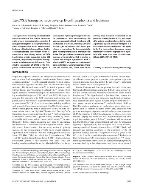
B-<strong>cell</strong> proliferation analysis<br />
Figure 1. Expression of transgene in lymphoid tissues. (A) RT-PCR analysis of<br />
E-<strong>BRD2</strong> expression in different Tg tissues. Lane 1, non-Tg spleen control; lane 2,<br />
Tg heart control; lane 3, Tg skeletal muscle control; lane 4, Tg spleen; lane 5, Tg<br />
thymus. Primers amplified both endogenous murine message <strong>and</strong> Tg message.<br />
Negative controls without RT verified the RT dependence of the signal (results not<br />
shown). (B) Tg expression in Tg B <strong>and</strong> T <strong>cell</strong>s. The 5 primer recognizes a Tg<br />
vector–specific sequence at the translation start site <strong>and</strong> the 3 primer a sequence<br />
within the first bromodomain, common to both endogenous <strong>and</strong> Tg sequences.<br />
Analysis was performed with purified B-<strong>cell</strong> RNA (B220) <strong>and</strong> T-<strong>cell</strong> RNA (CD3).<br />
Control reaction without reverse transcriptase is shown (RT control).<br />
Transplantation<br />
FVB female <strong>mice</strong> (6 to 8 weeks old; Taconic Farms, Germantown, NY)<br />
were sublethally irradiated with 6 or 8 Gy from a 137 Cs gamma source <strong>and</strong><br />
maintained on acidified water. For adoptive transfer experiments, irradiated<br />
<strong>mice</strong> were each injected subcutaneously with 1 10 7 B <strong>cell</strong>s positively<br />
selected from donor spleens by B220 magnetic beads. Mice were monitored<br />
for the <strong>develop</strong>ment of <strong>lymphoma</strong> or <strong>leukemia</strong> <strong>and</strong> were killed at the first<br />
appearance of clinical symptoms, without undergoing unnecessary pain <strong>and</strong><br />
suffering, in compliance with Federal <strong>and</strong> Institutional Animal Care <strong>and</strong><br />
Use Committees (IACUC) requirements.<br />
GeneChip analysis of transcriptomes<br />
In accordance with st<strong>and</strong>ard Affymetrix (Santa Clara, CA) protocols,<br />
splenic B-<strong>cell</strong> RNA from <strong>lymphoma</strong> or control spleen was labeled <strong>and</strong><br />
hybridized to a set of 3 arrays (Murine Genome MG-U74v2 arrays A, B,<br />
<strong>and</strong> C; Affymetrix). GeneChips were scanned; scans were quantified with<br />
Affymetrix Microarray Suite 5.0 software; normalization <strong>and</strong> analysis was<br />
performed in Microsoft Excel (Redmond, WA).<br />
Results<br />
Tg passage <strong>and</strong> expression<br />
PCR analysis established heterozygous passage in Tg <strong>and</strong> Tg mut<br />
<strong>mice</strong>, which Southern blot confirmed (results not shown). Heterozygous<br />
male <strong>mice</strong> were mated with FVB females to propagate 4<br />
independent lines; passage has been maintained through 6 generations.<br />
RT-PCR analysis of lymphoid <strong>and</strong> control tissues from Tg<br />
<strong>mice</strong> <strong>and</strong> littermate controls revealed greatly increased transcription<br />
of <strong>BRD2</strong> in Tg spleen <strong>and</strong> negligible expression in thymus<br />
(Figure 1A). The underexposed image emphasizes comparison of<br />
non-Tg <strong>and</strong> Tg spleen. All tissues express an endogenous Brd2<br />
message, apparent at longer exposures. This evidence shows that<br />
E drives transgene expression exceptionally well in B <strong>cell</strong>s. E<br />
expression in T lineages has been reported, 33,36,37 although malignancies<br />
arise more frequently in B lineages. 31 To confirm B-<br />
restriction, we performed RT-PCR with B220-selected <strong>and</strong> CD3-<br />
selected splenocytes (Figure 1B). A Tg-specific message was<br />
highly expressed in B220 Tg <strong>cell</strong>s <strong>and</strong> low but detectable in CD3 <br />
Tg <strong>cell</strong>s, about one tenth that of the B-<strong>cell</strong> level. However, Tg or<br />
Tg mut <strong>mice</strong> never <strong>develop</strong> T-<strong>cell</strong> <strong>lymphoma</strong>s.<br />
To explore whether Tg <strong>and</strong> Tg mut constructs confer the hypothesized<br />
proliferative advantage to B <strong>cell</strong>s, we measured in vitro<br />
proliferative responses to anti-CD40 <strong>and</strong> anti-IgM stimulation.<br />
Purified splenic B <strong>cell</strong>s from age-matched Tg, Tg mut , <strong>and</strong> littermate<br />
control <strong>mice</strong> (20 weeks; asymptomatic by visual inspection)<br />
responded to anti-CD40 <strong>and</strong> anti-IgM (10 g/mL). Control <strong>cell</strong>s<br />
showed a maximal burst of proliferation at 24 hours <strong>and</strong> no more<br />
through 48 hours (Figure 2). Tg <strong>and</strong> Tg mut B <strong>cell</strong>s proliferated at<br />
twice control levels at 24 hours <strong>and</strong> 3 times control through 48<br />
hours. Tg <strong>and</strong> Tg mut B <strong>cell</strong>s responded similarly <strong>and</strong> both underwent<br />
apoptosis in a few days. Without mitogens, Tg <strong>and</strong> Tg mut B<br />
<strong>cell</strong>s did not proliferate (results not shown).<br />
Incidence of <strong>lymphoma</strong><br />
All 4 founder lines of Tg <strong>and</strong> Tg mut <strong>mice</strong> sporadically <strong>develop</strong>ed<br />
clinical signs (ruffled fur, failure to nest, listlessness, hunched back,<br />
loss of appetite) after 28 weeks among PCR-positive littermates at<br />
approximately 10% annual incidence. Symptomatic <strong>mice</strong> were<br />
killed; spleen, thymus, liver, mesenteric lymph node, <strong>and</strong> bone<br />
marrow were examined. Splenomegaly was always apparent,<br />
accompanied by abdominal lymphadenopathy <strong>and</strong> leukemic infiltrations<br />
of liver <strong>and</strong> lung. To date, the numbers of Tg <strong>and</strong> Tg mut<br />
<strong>mice</strong> that sporadically <strong>develop</strong>ed <strong>lymphoma</strong> are 6 <strong>and</strong> 5, respectively,<br />
out of colonies of 38 <strong>and</strong> 59. The calculated 2 for this<br />
dataset is 0.61, below the 3.84 value for statistical significance<br />
( 0.05). Therefore, Tg is not different from Tg mut if “<strong>lymphoma</strong>”<br />
is the dependent variable. Thus, kinase function per se is<br />
not important for <strong>lymphoma</strong>genesis. Another 10% of <strong>mice</strong> die<br />
annually of causes unrelated to <strong>lymphoma</strong> or <strong>leukemia</strong>.<br />
Tg- <strong>and</strong> Tg mut -driven B-<strong>cell</strong> <strong>lymphoma</strong>s exhibit<br />
B-1 characteristics<br />
Flow cytometry was performed with unfractionated splenocytes<br />
from leukemic Tg <strong>and</strong> Tg mut <strong>mice</strong> <strong>and</strong> compared with littermate<br />
controls. Tg <strong>and</strong> Tg mut <strong>mice</strong> did not differ significantly. The vast<br />
majority of <strong>cell</strong>s were B220 CD19 , with very few CD3 , CD4 ,<br />
or CD8 <strong>cell</strong>s. Note that there are very few B220 <strong>cell</strong>s among<br />
unfractionated <strong>lymphoma</strong> <strong>cell</strong>s. B220 splenocytes expressed<br />
surface IgM, suggesting they are mature B <strong>cell</strong>s, <strong>and</strong> CD5 (Figure<br />
3A row 1). The proportion of CD5 B220 <strong>cell</strong>s in the control is<br />
higher than usually reported because of the particular isotype<br />
control antibody used to define flow cytometry parameters. B220<br />
was compared with CD19 to confirm B-<strong>cell</strong> lineage. Both were<br />
consistently expressed in independently arising <strong>lymphoma</strong>s. The<br />
pattern of CD5 or B7-1 versus B220 is very similar to versus CD19<br />
(Figure 3 rows 1-2). B-<strong>cell</strong> activation markers B7-1 <strong>and</strong> B7-2 were<br />
also elevated (Figure 3 row 2) <strong>and</strong> CD23 was low (Figure 3A row<br />
3). CD11b (which associates with CD18 to form macrophage<br />
Figure 2. Proliferation of Tg <strong>and</strong> Tg mut B <strong>cell</strong>s in response to anti-CD40 <strong>and</strong><br />
anti-IgM stimulation. B <strong>cell</strong>s from spleens of littermate control (f), Tg line 28 (u), Tg<br />
line 23 (), or Tg mut line 33 (o) or were stimulated in vitro for 24 or 48 hours with<br />
anti-CD40 <strong>and</strong> anti-IgM. Incorporation of [ 3 H] thymidine is shown with st<strong>and</strong>ard<br />
deviation (n 4) for each bar.
1478 GREENWALD et al BLOOD, 15 FEBRUARY 2004 VOLUME 103, NUMBER 4<br />
Figure 3. Flow cytometry of <strong>lymphoma</strong> <strong>cell</strong>s.<br />
(A) Unfractionated splenocytes from <strong>mice</strong> diagnosed with<br />
splenic <strong>lymphoma</strong> <strong>and</strong> peripheral <strong>leukemia</strong> (<strong>lymphoma</strong>)<br />
were stained for various markers of B-<strong>cell</strong> differentiation<br />
<strong>and</strong> activation <strong>and</strong> compared with asymptomatic non-Tg<br />
littermate splenocytes (control). Numbers in each square<br />
refer to percentages of gated events in a particular<br />
quadrant. (B) Peritoneal lymphocytes of premalignant,<br />
asymptomatic Tg <strong>mice</strong> (asymptomatic) were compared<br />
with age-matched littermate (control) to assess expansion<br />
of the B-1 <strong>cell</strong> population in the peritoneum.<br />
antigen-1 [Mac-1] <strong>and</strong> is expressed on granulocytes, natural killer<br />
[NK] <strong>cell</strong>s, <strong>and</strong> macrophages) <strong>and</strong> Ly-6G (a myeloid marker) were<br />
both low (Figure 3A row 3), as was CD49b (an NK marker, results<br />
not shown), supporting the interpretation of lymphoid lineage. In<br />
addition, a number of markers for immature B <strong>cell</strong>s were not<br />
detected, including CD117 (c-kit) <strong>and</strong> CD127 (interleukin-7 receptor<br />
-chain; results not shown). Lymphoma <strong>cell</strong>s were negative for<br />
CD25, syndecan-1, <strong>and</strong> CD69; positive for CD9; <strong>and</strong> showed a<br />
high IgM/low IgD ratio (Figure 3A rows 4-6). Collectively, these<br />
markers suggested that the <strong>lymphoma</strong> <strong>cell</strong>s bear B-1 hallmarks. 34<br />
Levels of B7-1 <strong>and</strong> B7-2 on splenic B <strong>cell</strong>s were comparable in<br />
asymptomatic Tg <strong>and</strong> Tg mut <strong>mice</strong> as they aged (8 through 60 weeks,<br />
not shown) <strong>and</strong> similar to those in littermate controls. Expression<br />
patterns also held constant with age for T-<strong>cell</strong> markers in asymptomatic<br />
Tg <strong>and</strong> Tg mut <strong>mice</strong> such as CD4 versus CD8 in spleen <strong>and</strong><br />
thymus <strong>and</strong> CD4 versus CD25, CD4 versus CD44, CD4 versus<br />
CD62L, <strong>and</strong> CD4 versus CD69 in spleen (results not shown).<br />
However, we observed increased CD5 versus B220, CD23 versus<br />
B220, CD11b versus B220, <strong>and</strong> IgM versus IgD markers among<br />
peritoneal <strong>cell</strong>s of premalignant <strong>mice</strong> with no clinical signs or<br />
splenomegaly, compared with age-matched control <strong>mice</strong>, suggesting<br />
that peritoneal B-1 <strong>cell</strong>s had exp<strong>and</strong>ed in the premalignant <strong>mice</strong><br />
<strong>and</strong> supporting the B-1 origin of the <strong>lymphoma</strong>.<br />
Histochemistry of splenic <strong>lymphoma</strong> <strong>and</strong> peripheral <strong>leukemia</strong><br />
Lymphoid organs of asymptomatic, heterozygous Tg, <strong>and</strong> Tg mut<br />
<strong>mice</strong> were compared with littermate controls with B220, CD3, <strong>and</strong><br />
H&E stains <strong>and</strong> were normal. However, spleens of leukemic <strong>mice</strong><br />
were enlarged between 5 <strong>and</strong> 50, splenic architecture was<br />
disrupted (Figure 4A; Tg, panel ii; Tg mut , panel iii), <strong>and</strong> invasive<br />
lymphoid populations showed numerous mitoses (Figure 4Av; Tg).<br />
Bone marrow showed lymphocytic <strong>lymphoma</strong> (Figure 4Avi; Tg).<br />
Elevated numbers of apoptotic <strong>cell</strong>s were not observed.<br />
Normal, syngeneic female <strong>mice</strong> were irradiated <strong>and</strong> received<br />
transplanted B <strong>cell</strong>s purified from splenic <strong>lymphoma</strong>s. Mice that<br />
received transplants <strong>develop</strong>ed <strong>leukemia</strong>s <strong>and</strong> blood-clotting problems;<br />
peripheral blood contained large numbers of morphologically
BLOOD, 15 FEBRUARY 2004 VOLUME 103, NUMBER 4<br />
B-CELL LYMPHOMA IN E-<strong>BRD2</strong> TRANSGENIC MICE 1479<br />
Figure 4. Tg-driven splenic <strong>lymphoma</strong> <strong>and</strong> peripheral <strong>leukemia</strong>. (A) A B220-stained section of asymptomatic Tg spleen (i; bar, 200 m) is compared with a B220-stained<br />
section of a Tg mouse (ii; bar, 100 m) or Tg mut mouse (iii; bar, 100 m) with lymphocytic <strong>lymphoma</strong>. An H&E-stained section of asymptomatic Tg spleen (iv; bar, 50 m) is<br />
compared with an H&E-stained section of a Tg mouse with lymphocytic <strong>lymphoma</strong> (v; bar, 50 m). An H&E-stained section of bone marrow of Tg mut mouse with lymphocytic<br />
<strong>lymphoma</strong> is also shown (vi; bar, 100 m). (B) A Wright-Giemsa–stained smear of peripheral blood of a leukemic mouse that received Tg B-<strong>cell</strong> <strong>lymphoma</strong> transplants shows<br />
overproliferation of lymphoblasts (i; bar, 50 m). Blasts exhibit prominent, multiple chromocenters <strong>and</strong> scanty dark blue cytoplasm (ii; bar, 20 m). Note the polymorphonuclear<br />
granulocyte for comparison. Additional field emphasizes homotypic adhesion <strong>and</strong> mixed chromatin states (iii; bar, 20 m).<br />
similar, large lymphoblasts (Figure 4Bi). The white blood count<br />
was 790 ( 40) 10 9 /L (7.9 [ 0.4] 10 5 /mm 3 ). The ratio of<br />
erythrocytes to lymphoblasts was 6.6 to 1. Leukemic blasts (Figure<br />
4Bii) exhibited scant cytoplasm, cytoplasmic vacuolation, homotypic<br />
adhesion, <strong>and</strong> abundant chromocenters that reflect mixed<br />
heterochromatin <strong>and</strong> euchromatin states, indicating transcriptional<br />
activity (Figure 4Biii). Together, these histologic features suggest<br />
greatest similarity to the centroblastic variant of diffuse large B-<strong>cell</strong><br />
<strong>lymphoma</strong>, following current convention for classifying murine<br />
lymphoid neoplasms. 38<br />
Monoclonal immunoglobulin gene rearrangement<br />
To determine whether the splenic B-<strong>cell</strong> neoplasm comprised a<br />
clonal or polyclonal population, total RNA from B220-selected<br />
<strong>cell</strong>s was isolated for cDNA synthesis. Whereas normal littermate<br />
control spleens weighed 0.2 g <strong>and</strong> yielded approximately 300 gof<br />
total RNA, spleens from <strong>lymphoma</strong> <strong>mice</strong> weighed up to 10 g <strong>and</strong><br />
yielded 30 mg total RNA. Expressed Ig sequences were amplified<br />
with 24-mer primers to the 3 constant region (3C <strong>and</strong> 3C ) <strong>and</strong><br />
degenerate primers to the variable regions (5V H <strong>and</strong> V ) to<br />
determine if a specific Ig gene rearrangement, or none, predominates<br />
in the exp<strong>and</strong>ed B-<strong>cell</strong> population. 35 B220 splenocytes of a<br />
non-Tg littermate were the control group. B-<strong>cell</strong> <strong>lymphoma</strong> RNA<br />
gave a single RT-PCR product (Figure 5A left), but polyclonal<br />
B-<strong>cell</strong> RNA gave smears with each primer pair (Figure 5A right). In<br />
this particular <strong>lymphoma</strong>, an approximately 0.5-kb product arose<br />
only from the C V H 1 primer pair. Cloning <strong>and</strong> sequencing identified<br />
a GenBank entry for Mus musculus V23-D-J-C mRNA,<br />
accession no. AB069913. Three independent, monoclonal IgM ,<br />
B-<strong>cell</strong> <strong>lymphoma</strong>s have been sequenced to date, but the number<br />
of clones remains too small to determine if the transgene promotes<br />
the emergence of a particular clonotype. Premalignant <strong>mice</strong><br />
without clinical signs or splenomegaly showed expansion of<br />
oligoclonal splenic B <strong>cell</strong>s in the weeks before clonal malignancy<br />
emerged (results not shown). We determined Bcl6 expression using<br />
real-time PCR with a st<strong>and</strong>ard set of primers for cDNAs synthesized<br />
from RNA of purified splenic B-<strong>cell</strong> <strong>lymphoma</strong> <strong>cell</strong>s of the<br />
first <strong>and</strong> sixth passage after transplantation (to assess stability of<br />
phenotype) <strong>and</strong> compared the result with Bcl6 expression in splenic<br />
B2 <strong>cell</strong>s (Figure 5B). Bcl6 expression was low <strong>and</strong> remained
1480 GREENWALD et al BLOOD, 15 FEBRUARY 2004 VOLUME 103, NUMBER 4<br />
Figure 6. Kaplan-Meier plots showing survival of <strong>mice</strong> that received Tg B-<strong>cell</strong><br />
<strong>lymphoma</strong> transplants. Non-Tg <strong>mice</strong> were irradiated with 6 Gy (dashed line <strong>and</strong><br />
dotted line) or 8 Gy (solid line) <strong>and</strong> inoculated subcutaneously with 10 7 purified B <strong>cell</strong>s<br />
from the spleen of a Tg donor with <strong>lymphoma</strong> (dotted line <strong>and</strong> solid line) or 10 7<br />
purified B <strong>cell</strong>s from the spleen of an asymptomatic Tg donor (dashed line). For<br />
dashed line, n 4; for dotted line, n 6; <strong>and</strong> for solid line, n 15. Control indicates<br />
B <strong>cell</strong>s from asymptomatic Tg donor; <strong>and</strong> DLCL, diffuse large B-<strong>cell</strong> <strong>lymphoma</strong>.<br />
Figure 5. Immunoglobulin gene rearrangements are monoclonal. (A) Ig RNAs<br />
were isolated from purified B <strong>cell</strong>s of a Tg splenic B-<strong>cell</strong> <strong>lymphoma</strong>, amplified by<br />
RT-PCR, <strong>and</strong> resolved. RT control is shown (left). Primers to the 3 constant region<br />
(3C <strong>and</strong> 3C ) Ig sequences <strong>and</strong> degenerate primers to the variable regions (5V H<br />
<strong>and</strong> V ) were used in pairs as indicated. RNA of purified, polyclonal B <strong>cell</strong>s from<br />
normal, non-Tg littermate control (right) is compared with Tg <strong>lymphoma</strong> (left). (B)<br />
Lymphoma RNA from first or sixth transplanted passage of malignancy was amplified<br />
with primers specific for Bcl6 (f) or Blimp-1 genes () by real-time PCR <strong>and</strong><br />
compared with splenic B2 control. Relative expression levels were determined by the<br />
2 –Ct method where C t C t, sample C t, reference with Bcl6 or Blimp-1 serving as<br />
samples <strong>and</strong> 2-microglobulin as a reference.<br />
unaltered over 6 months of serial transplantations, described in the<br />
next paragraph.<br />
Transplantation<br />
Malignant B <strong>cell</strong>s were purified from Tg <strong>lymphoma</strong> <strong>and</strong> transplanted<br />
(Figure 6). Radiation dose partly determined susceptibility<br />
to establishment of the <strong>lymphoma</strong> in the transplant recipient 39,40 ;<br />
median survival time for <strong>mice</strong> that received transplants of 10 7<br />
malignant B <strong>cell</strong>s (DLCL) after irradiation with 6 Gy was 28 days<br />
(dotted line), whereas median survival time after irradiation with 8<br />
Gy was 19 days (solid line). In all cases, autopsy confirmed that<br />
<strong>mice</strong> that received transplants died of complications from <strong>lymphoma</strong>,<br />
including massive splenomegaly, peripheral <strong>leukemia</strong>,<br />
internal hemorrhage, <strong>and</strong> leukemic infiltrates in liver <strong>and</strong> lung.<br />
Irradiated controls (6 Gy) that received B <strong>cell</strong>s purified from an<br />
asymptomatic Tg donor survived beyond 60 days (long dashed<br />
line). Splenocytes from Tg leukemic donors <strong>and</strong> transplants were<br />
indistinguishable by flow cytometry, Ig gene rearrangement, in<br />
vivo growth characteristics, organ sites of leukemic infiltration, or<br />
morphology of peripheral blood lymphoblasts (results not shown),<br />
suggesting the characteristics of the B-<strong>cell</strong> <strong>lymphoma</strong> were stable,<br />
even after 6 serial transplantations. The <strong>lymphoma</strong> cannot be<br />
maintained in ordinary tissue culture, as the <strong>cell</strong>s appear not to have<br />
undergone an immortalizing event that permits proliferation in<br />
media without B-<strong>cell</strong> mitogens (lipopolysaccharide, interleukin-4,<br />
or anti-CD40 anti-IgM). We have maintained individual <strong>lymphoma</strong><br />
clones by serial transplantation into irradiated recipients.<br />
Cyclin A expression<br />
We examined cyclin A transcription in the malignant <strong>cell</strong>s, based on<br />
our published model for Brd2-dependent transcriptional activation<br />
of the cyclin A locus, <strong>and</strong> on the <strong>cell</strong> cycle acceleration that we<br />
propose arises from E-<strong>BRD2</strong> Tg expression 30 (A.S., G.V.D.,<br />
unpublished data, May 2003). RT-PCR analysis of several independently<br />
arising splenic B-<strong>cell</strong> <strong>lymphoma</strong>s in both Tg <strong>and</strong> Tg mut <strong>mice</strong><br />
showed that, in all cases, cyclin A transcription was elevated in the<br />
primary malignant B <strong>cell</strong>s (Figure 7; <strong>lymphoma</strong> lanes 1-2) <strong>and</strong><br />
remained stably elevated in malignant splenic B <strong>cell</strong>s isolated from<br />
each passage of <strong>mice</strong> that received serial transplants that had<br />
<strong>develop</strong>ed B-<strong>cell</strong> <strong>lymphoma</strong> <strong>and</strong> in all individuals that received<br />
transplants, all of which <strong>develop</strong>ed the same <strong>lymphoma</strong> <strong>and</strong><br />
peripheral B-<strong>cell</strong> <strong>leukemia</strong> (Figure 7; <strong>lymphoma</strong> lanes 3-4, second<br />
passage, 2 representative recipients; <strong>lymphoma</strong> 5-6, fourth passage,<br />
2 representative recipients). Cyclin A message was slightly<br />
elevated in B <strong>cell</strong>s purified from asymptomatic Tg <strong>mice</strong> or Tg mut<br />
<strong>mice</strong> (Tg B <strong>cell</strong>s) but barely detectable in T <strong>cell</strong>s from Tg <strong>mice</strong> (Tg<br />
T <strong>cell</strong>s). Tg <strong>mice</strong> with B-<strong>cell</strong> <strong>lymphoma</strong> had very few splenic T<br />
<strong>cell</strong>s (Figure 3). This pattern of cyclin A expression may represent a<br />
premalignant marker.<br />
The annual rate of 10% of sporadic Tg <strong>lymphoma</strong> suggests that<br />
an additional “hit” is required for tumorigenesis to progress. That<br />
second hit could be engineered various ways. We chose an<br />
amphotropic retrovirus system that expresses the H-ras oncogene.<br />
Tg B <strong>cell</strong>s “primed” to proliferate are expected to be susceptible to<br />
cooperation with the activated ras gene. Lymphomagenesis was<br />
accelerated from several months to 4 weeks. Seven Tg <strong>and</strong> 7<br />
Figure 7. Cyclin A expression in Tg B-<strong>cell</strong> malignancy. RT-PCR was performed<br />
with RNA from purified B <strong>cell</strong>s (Tg B <strong>cell</strong>s) or purified T <strong>cell</strong>s (Tg T <strong>cell</strong>s) from spleens<br />
of asymptomatic Tg <strong>mice</strong>, using primers to the cyclin A gene. Results were compared<br />
with negative control (RT) <strong>and</strong> to RNA from B <strong>cell</strong>s obtained from independently<br />
arising B-<strong>cell</strong> <strong>lymphoma</strong>s in 6 different <strong>mice</strong>. Lymphoma lane 1, Tg leukemic mouse<br />
(transplant donor); lane 2, Tg mut leukemic mouse; lane 3, second serial transplantation;<br />
lane 4, second serial transplantation; lane 5, fourth serial transplantation; lane 6,<br />
fourth serial transplantation.
BLOOD, 15 FEBRUARY 2004 VOLUME 103, NUMBER 4<br />
B-CELL LYMPHOMA IN E-<strong>BRD2</strong> TRANSGENIC MICE 1481<br />
non-Tg <strong>mice</strong> were injected intraperitoneally with either H-ras–<br />
expressing amphotropic retrovirus or neo-expressing retrovirus<br />
alone. After 4 weeks, Tg <strong>mice</strong> injected with the H-ras retrovirus<br />
<strong>develop</strong>ed severe splenomegaly; non-Tg <strong>mice</strong> injected with H-ras<br />
did not <strong>develop</strong> illness. Analysis of histopathology, flow cytometry<br />
of <strong>cell</strong> surface antigens, transplantability, <strong>and</strong> monoclonality of Ig<br />
gene rearrangements indicated a diagnosis of B-<strong>cell</strong> <strong>lymphoma</strong><br />
indistinguishable from sporadic cases. We then performed Affymetrix<br />
GeneChip analysis with B-<strong>cell</strong> RNA purified from both<br />
sporadic <strong>and</strong> ras-accelerated splenic <strong>lymphoma</strong>s. Controls were,<br />
respectively, splenic B <strong>cell</strong>s purified from normal littermate<br />
controls <strong>and</strong> from apparently normal <strong>mice</strong> infected with neo<br />
retrovirus. The 3 mouse arrays A, B, <strong>and</strong> C permitted wholegenome<br />
analysis of the transcriptome of the Brd2-induced <strong>lymphoma</strong>s,<br />
resulting in molecular signatures for malignancy that we<br />
compared with published databases of human <strong>lymphoma</strong>s. Selected<br />
significant markers for named genes are shown in Table 1.<br />
In the Tg B-<strong>cell</strong> <strong>lymphoma</strong>, most transcriptional changes of<br />
greatest estimated significance were 50- to 100-fold decreases in<br />
mouse immune <strong>cell</strong> markers, mostly Ig gene -chain sequences<br />
(results not shown). Because the B-<strong>cell</strong> <strong>lymphoma</strong>s are monoclonal,<br />
the transcriptomes show a marked decrease in the abundance<br />
of diverse Ig gene transcripts when compared with the polyclonal<br />
Ig gene transcripts found in the normal littermate B-<strong>cell</strong> controls. In<br />
analyses of the most significant fold changes, we excluded Ig gene<br />
sequences because of this trivial explanation. To a first approximation,<br />
the transcriptome indicates similarity to diffuse large B-<strong>cell</strong><br />
<strong>lymphoma</strong> (DLCL) in humans, the most common type of non-<br />
Hodgkin <strong>lymphoma</strong>. 41 Histopathology supports this conclusion.<br />
The molecular signature of other B-<strong>cell</strong> phenotypes, such as resting<br />
or activated B <strong>cell</strong>s in the periphery, B-<strong>cell</strong> follicular <strong>lymphoma</strong>,<br />
transformed B-<strong>cell</strong> lines, or B-<strong>cell</strong> chronic lymphocytic <strong>leukemia</strong>,<br />
are not characteristic of this B-<strong>cell</strong> <strong>lymphoma</strong>. 41 The sporadically<br />
arising Tg B-<strong>cell</strong> <strong>lymphoma</strong>s (Table 1; sporadically arising Tg<br />
B-<strong>cell</strong> <strong>lymphoma</strong>s [SPORs]) <strong>and</strong> ras-accelerated <strong>lymphoma</strong>s (RASs)<br />
shared a close but not identical molecular signature. Most fold<br />
changes of named marker genes identified in the literature as<br />
lymphoid biomarkers were changed in the same direction <strong>and</strong> often<br />
by a similar magnitude in sporadic <strong>and</strong> ras-accelerated Tg <strong>lymphoma</strong>s.<br />
The Tg <strong>lymphoma</strong> transcriptional signature did not resemble<br />
signatures of human DLCL cases independently established through<br />
clinical measures to have a favorable or unfavorable prognosis 42<br />
(results not shown). We did not observe a good correlation between<br />
the Tg <strong>lymphoma</strong> transcriptional signature <strong>and</strong> signatures 41,42 of<br />
either germinal center–like or activated B-<strong>cell</strong>–like human DLCLs<br />
(Table 1).<br />
Discussion<br />
We designed <strong>transgenic</strong> <strong>mice</strong> to test our hypothesis that constitutive<br />
expression of <strong>BRD2</strong> in the lymphoid compartment would cause<br />
lymphoproliferative disease. We based this hypothesis on substantial<br />
in vitro evidence that ectopic overexpression of Brd2 transactivates<br />
E2F-regulated <strong>cell</strong> cycle genes such as cyclin D1, cyclin E,<br />
cyclin A, <strong>and</strong> dihydrofolate reductase in synergy with oncogenic<br />
ras 30 <strong>and</strong> that Brd2 participates in transcription complexes that<br />
contain a histone H4–specific HAT activity <strong>and</strong> E2F proteins.<br />
Furthermore, Brd2 responds to mitogenic signals; when overexpressed,<br />
it transforms NIH/3T3 fibroblasts in synergy with ras <strong>and</strong><br />
accelerates the G 1 3 S transition through direct action on the cyclin<br />
A promoter. Double bromodomain proteins such as Brd2 have been<br />
insufficiently studied. The occurrence of a B-<strong>cell</strong> <strong>lymphoma</strong> in<br />
Table 1. Molecular signature of Tg B-<strong>cell</strong> <strong>lymphoma</strong><br />
DLCL<br />
Gene SPOR RAS GC AB FL AT TC RPB CLL<br />
Bcl2 1.6 2.7 o <br />
Bcl6 4.4 13.4 o <br />
Casp1 5.0 3.2 o o o o<br />
Ccna1 2.9 1.6 o o <br />
Ccnb1 2.4 2.4 o o <br />
Ccne2 3.1 8.7 o o o o o o <br />
Cd3d 30.1 2.6 o <br />
Cd3e 40.8 2.6 o <br />
Cd38 1.6 3.2 o o o <br />
Cd44 10.6 5.9 o o<br />
Cdk5 1.6 2.6 o o o o o <br />
Ches1 3.1 1.4 o o o o<br />
Fos 52.2 20.0 o o <br />
Prkcb 2.4 2.0 o o o <br />
Rara 6.5 18.0 o o o o o<br />
Rb1 2.2 2.1 o o o o o <br />
Spib 1.5 2.8 o o o o <br />
Tcrb 5.7 3.9 o o o o<br />
Tubb1 6.1 1.9 o o <br />
Vegfa 9.5 6.6 o o o o o <br />
Most significant changes in gene expression profile of named genes in <strong>lymphoma</strong><br />
compared with control. About one third of the genome (12000 genes) was<br />
significantly expressed. Given the preliminary estimate of variance, about 35% of<br />
these were differentially expressed. Comparisons are made to entries in the online<br />
databases of Alizadeh et al 41 <strong>and</strong> Shipp et al. 42<br />
DLCL indicates diffuse large B-<strong>cell</strong> <strong>lymphoma</strong>; SPOR, sporadic Tg B-<strong>cell</strong><br />
<strong>lymphoma</strong>; RAS, ras-accelerated Tg B-<strong>cell</strong> <strong>lymphoma</strong>; GC, germinal center-like<br />
<strong>lymphoma</strong> 41,42 ; AB, activated peripheral B-<strong>cell</strong>-like <strong>lymphoma</strong> 41,42 ; FL, B-<strong>cell</strong> follicular<br />
<strong>lymphoma</strong> 41,42 ; AT, activated/resting T <strong>cell</strong> 41 ; TC, transformed <strong>cell</strong> lines 41 ; RPB,<br />
resting peripheral B <strong>cell</strong> 41 ; CLL, B-<strong>cell</strong> chronic lymphocytic <strong>leukemia</strong> 41 ; , small<br />
increase; , large increase; , small decrease; , large decrease; <strong>and</strong> o, no<br />
report, no significant change, or no consistent change within a subclass.<br />
Gene entries (mouse gene abbreviation/human homolog, name): Bcl2/BCL2<br />
indicates B-<strong>cell</strong> <strong>leukemia</strong>/<strong>lymphoma</strong> 2; Bcl6/BCL6, B-<strong>cell</strong> <strong>leukemia</strong>/<strong>lymphoma</strong> 6;<br />
Casp1/CASP1, caspase-1; Ccna1/CCNA1, cyclin A1; CCnb1/CCNB1, cyclin B1;<br />
Ccne2/CCNE2, cyclin E2; Cd3d/CD3D, CD3 antigen, polypeptide; Cd3e/CD3E,<br />
CD3 antigen, polypeptide; Cd38/CD38, CD38 antigen; Cd44/CD44, CD44 antigen;<br />
Cdk5/CDK5, cyclin-dependent kinase 5; Ches1/CHES1, checkpoint suppressor 1;<br />
Fos/FOS, FBJ osteosarcoma oncogene/v-fos FBJ murine osteosarcoma viral oncogene<br />
homolog; Prkcb/PRKCB1, protein kinase C; Rara/RARA, retinoic acid<br />
receptor , Rb1/RB1, retinoblastoma protein; Spib/SPIB, Spi-B transcription factor<br />
(Spi-1/PU.1 related); Tcrb/TCRB, T-<strong>cell</strong> receptor chain; Tubb1/TUBB1, tubulin 1;<br />
Vegfa/VEGF, vascular endothelial growth factor A.<br />
Most significant fold-changes are compared with Shipp et al. 42 Figure 5 (DLCL<br />
with GC-like vs AB-like signature) <strong>and</strong> Alizadeh et al. 41 Figure 1 (transcriptional<br />
profiles of various lymphoid <strong>cell</strong> types, including GC <strong>and</strong> AB signatures).<br />
these <strong>transgenic</strong> <strong>mice</strong> now establishes a link between this class of<br />
proteins <strong>and</strong> neoplasia.<br />
Considerable effort has been expended recently to explore the<br />
possible functions of the bromodomain motif in nuclear factors<br />
associated with transcription <strong>and</strong> chromatin restructuring. 4 The<br />
subject has been controversial because sometimes deletion of the<br />
motif has no phenotype. 6 Virtually all of the nuclear HATs contain<br />
bromodomains, but not all bromodomain proteins are HATs. 6 The<br />
solution of the bromodomain structure <strong>and</strong> interaction with nucleosomal<br />
histone acetyl-lysines 3,13 has clarified the matter. Recent<br />
studies suggest that bromodomain-containing proteins provide a<br />
“scaffolding” or platform function for transcription or chromatin<br />
remodeling complexes, anchoring them to nucleosomes. 1,2,23,24<br />
Furthermore, oncogenic fusion proteins that contain bromodomains<br />
participate in neoplasia, such as the BET protein 14 Brd4 (also<br />
called 22 mitotic chromosome-associated protein [MCAP]), which<br />
contains 1400 amino acids <strong>and</strong> is closely related to Brd2, sharing its<br />
dual bromodomain <strong>and</strong> extraterminal (ET) domain structure. 14<br />
BRD4 is rearranged in t(15;19) translocations associated with<br />
aggressive carcinomas of the respiratory tract <strong>and</strong> its fusion
1482 GREENWALD et al BLOOD, 15 FEBRUARY 2004 VOLUME 103, NUMBER 4<br />
partners likely encode oncoproteins. 43 Also, translocations that fuse<br />
the bromodomain <strong>and</strong> HAT domain of CBP to the mixed-lineage<br />
<strong>leukemia</strong> factor 44-48 (MLL; also called All-1) or to the monocytic<br />
<strong>leukemia</strong> zinc finger protein, 49 or that fuse p300 to MLL, create<br />
oncoproteins 50 that are associated with acute <strong>leukemia</strong>s (reviewed<br />
in Filetici <strong>and</strong> Ballario 23 ). The bromodomain is required for full<br />
transforming activity in at least one of these cases. 47 Deletion of the<br />
Brd2 bromodomains destroys Brd2-dependent transcriptional activation<br />
of cyclin A (G.V.D., unpublished data, May 2003) <strong>and</strong><br />
eliminates association of Brd2 with the multiprotein complex that<br />
contains a histone H4–specific HAT <strong>and</strong> E2F proteins.<br />
Ectopic expression of Brd2 leads both to <strong>cell</strong> cycle destabilization<br />
<strong>and</strong> transformation in synergy with oncogenic ras or its<br />
effectors. 30 We hypothesized that E-directed expression of <strong>BRD2</strong><br />
in the lymphoid compartment would lead to <strong>leukemia</strong> or <strong>lymphoma</strong>,<br />
<strong>and</strong> we obtained this result. Malignant <strong>cell</strong>s from enlarged<br />
spleens bear <strong>cell</strong> surface markers consistent with B-1 lineage. 34<br />
This apparently differentiated phenotype remains constant after 6<br />
serial transplantations, with respect to antigen expression in flow<br />
cytometry, Ig gene rearrangement, in vivo growth characteristics,<br />
elevated cyclin A transcription, organ sites of leukemic infiltration,<br />
<strong>and</strong> morphology. From these data we speculate that the malignant<br />
<strong>cell</strong>s do not have a high degree of genetic instability or flexibility in<br />
their transcriptional profile. Transcriptome analysis over time will<br />
be necessary to confirm this supposition.<br />
The Drosophila homolog of <strong>BRD2</strong> is female sterile homeotic,<br />
15,51,52 which encodes a homeotic protein <strong>and</strong> probable<br />
transcription regulator that is an upstream activator of the trithorax<br />
locus. 53 MLL, the human homolog of trithorax, is disrupted in<br />
mixed-lineage (myeloid <strong>and</strong> lymphoid) human <strong>leukemia</strong>s associated<br />
with 11q23 translocations. 44,45 The functional relationships<br />
established in Drosophila (between fsh <strong>and</strong> trithorax) may be<br />
conserved in humans (between Brd2 <strong>and</strong> MLL), as we hypothesized.<br />
25 We therefore looked for a mixed-lineage leukemic<br />
phenotype in the <strong>mice</strong> that received leukemic transplants, but flow<br />
cytometry suggests that myeloid components are not present<br />
among the leukemic B <strong>cell</strong>s.<br />
We have proposed a mechanism for Brd2-driven transcriptional<br />
activation of important <strong>cell</strong> cycle regulatory genes. 30 Brd2 recruits<br />
both E2F proteins <strong>and</strong> HAT activity to the cyclin A gene in<br />
particular (A.S., G.V.D., unpublished data, May 2003). These<br />
results agree with literature that has established a dual role for E2F<br />
recruitment <strong>and</strong> chromatin restructuring in cyclin A transcriptional<br />
control. 54-56 Coimmunoprecipitate complexes of Brd2 contain a<br />
histone H4–specific HAT (A.S., G.V.D., unpublished data, May<br />
2003). We speculate that a similar enzyme present in <strong>transgenic</strong> B<br />
<strong>cell</strong>s provides crucial nucleosomal modification functions to Brd2<br />
target genes. Several HATs capable of specific acetylation of<br />
histone H4 have been identified, including Tip60, implicated in<br />
<strong>leukemia</strong>s. 57,58 The composition of the multiprotein Brd2 complex<br />
29,30 may change across the <strong>cell</strong> cycle <strong>and</strong> contribute to cyclin A<br />
repression during G 0 or to transactivation during <strong>cell</strong> cycle<br />
progression. This model could explain why Tg <strong>and</strong> Tg mut <strong>mice</strong><br />
share a similar in vitro proliferative (Figure 2) <strong>and</strong> in vivo<br />
<strong>lymphoma</strong> phenotype (Figure 4A): apart from a point mutation in<br />
the kinase domain of the transgene of Tg mut <strong>mice</strong>, both lines<br />
overexpress the same Brd2 protein, probably involving the same<br />
HAT <strong>and</strong> E2F recruitment mechanism in both cases. This view<br />
implies that HAT <strong>and</strong> E2F recruitment is more important than<br />
kinase activity as an oncogenic mechanism in this model. The<br />
fine-structural characteristics of B-<strong>cell</strong> <strong>lymphoma</strong>s may nevertheless<br />
be different between Tg <strong>and</strong> Tg mut <strong>mice</strong>. Microarray technology<br />
will elucidate the transcriptomes of the <strong>lymphoma</strong>s in each<br />
breeding line. Reports of oncogenic bromodomain fusion proteins<br />
23,43,46-50 lead us to speculate that HAT recruitment to key <strong>cell</strong><br />
cycle regulatory genes, either via intrinsic HAT domains or via<br />
bromodomain protein–mediated “scaffolding,” is important for <strong>cell</strong><br />
cycle control. 1,23,24 Oncogenic fusion proteins or deregulated<br />
bromodomain protein function could upset the histone acetylation/<br />
deacetylation balance of these genes <strong>and</strong> lead to their improper<br />
transactivation, with possible neoplastic consequences.<br />
Ectopic overexpression of Brd2 transactivates a heterologous<br />
cyclin A promoter luciferase construct in synergy with mitogenic<br />
signals from ras or MEKK, an effector of ras, 59 <strong>and</strong> increases<br />
expression of endogenous cyclin A in synchronized fibroblasts that<br />
have been stimulated to enter the <strong>cell</strong> cycle; these <strong>cell</strong>s show<br />
Brd2-driven accelerated kinetics of S-phase entry <strong>and</strong> mobilization<br />
of cyclin A–associated cdk activity (A.S., G.V.D., unpublished<br />
data, May 2003). Accordingly, we examined cyclin A transcription<br />
in B220 <strong>cell</strong>s purified from several sporadically arising Tg B-<strong>cell</strong><br />
<strong>lymphoma</strong>s, compared with B220 <strong>cell</strong>s from asymptomatic Tg<br />
controls. Cyclin A is under negative transcriptional control until S<br />
phase 54-56<br />
<strong>and</strong> is frequently elevated in tumors 60 ; it is indeed<br />
transcriptionally activated in the Tg malignancy <strong>and</strong> slightly<br />
activated in B <strong>cell</strong>s from asymptomatic Tg <strong>mice</strong> but not T <strong>cell</strong>s.<br />
Therefore, the transgene destabilizes cyclin A in B <strong>cell</strong>s as predicted.<br />
The morphology, surface antigen phenotype, <strong>and</strong> transcriptome<br />
signature of E-<strong>BRD2</strong>–driven B-<strong>cell</strong> <strong>lymphoma</strong>s suggest they are<br />
most related to human non-Hodgkin <strong>lymphoma</strong>s of the DLCL<br />
type. 38,41 <strong>BRD2</strong> is not present in <strong>lymphoma</strong> databases we queried,<br />
41,42 so comparison of its level in lymphoid malignancies was<br />
not possible. We queried Cancer Genome Anatomy Project databases<br />
for evidence of transcriptional up-regulation of Brd2 message<br />
in human malignancies <strong>and</strong> found an increase of 4.2 fold<br />
(P .04) reported for 3 cancerous or precancerous lymph nodes<br />
compared with 4 normal lymph node controls. Furthermore,<br />
the Mitelman database (http://cgap.nci.nih.gov/chromosomes/<br />
mitelman) of recurrent chromosomal abnormalities associated<br />
with cancer identifies 386 references <strong>and</strong> 546 patient hematologic<br />
malignancies involving breakpoints at 6p21, which includes<br />
the class II major histocompatibility complex wherein<br />
<strong>BRD2</strong> is located, but only a small minority have received<br />
fine-structure mapping. Therefore, whereas some cases are<br />
likely to show breakpoints within the <strong>BRD2</strong> locus, insufficient<br />
data exist to make generalizations about the role of <strong>BRD2</strong> in the<br />
pathogenesis of human DLCL.<br />
Human DLCLs exhibit a variety of genetic abnormalities. The<br />
most common is structural alteration of the BCL6 locus, 61 often<br />
resulting in deregulated Bcl6 activity. 62,63 We determined that Bcl6<br />
transcript levels are low in Tg B-<strong>cell</strong> <strong>lymphoma</strong>, consistent with the<br />
view that Bcl6 activation is more commonly associated with<br />
lymphoblastic <strong>lymphoma</strong> in <strong>mice</strong> 64 ; furthermore the Bcl6 locus is<br />
not normally mutated in mouse B-<strong>cell</strong> lineage <strong>lymphoma</strong>s. 65<br />
Unaltered Bcl6 transcript levels in Tg <strong>lymphoma</strong> are also consistent<br />
with the posited function of the Tg as a transcriptional coactivator<br />
<strong>and</strong> <strong>cell</strong> cycle regulator 30 ; no current evidence shows a role for<br />
<strong>BRD2</strong> overexpression in genetic instability.<br />
Human DLCL is thought to arise from germinal center B <strong>cell</strong>s,<br />
or less commonly, from marginal zone B <strong>cell</strong>s. However, our<br />
expression profile data indicate that the Tg B-<strong>cell</strong> malignancy does<br />
not obviously share a signature with any subclass of human DLCL,<br />
<strong>and</strong> flow cytometry analysis unambiguously identifies the Tg<br />
malignancy as composed of B-1 <strong>cell</strong>s. A subclass of murine DLCL,<br />
which has neither follicular nor marginal zone origins, has been<br />
discussed 38 <strong>and</strong> seems appropriate to classify this Tg <strong>lymphoma</strong>.<br />
An ongoing theme in the <strong>lymphoma</strong> literature is the extreme
BLOOD, 15 FEBRUARY 2004 VOLUME 103, NUMBER 4<br />
B-CELL LYMPHOMA IN E-<strong>BRD2</strong> TRANSGENIC MICE 1483<br />
diversity of DLCL. Expression profile data 41,42 demonstrate that<br />
even among ostensible “biomarker” genes for specific subclasses<br />
of DLCL or clinical outcomes, remarkable variance exists across<br />
cases, as though each DLCL ought to be considered a completely<br />
unique neoplasm. Nevertheless, expression profiling with DNA<br />
microarrays remains the best method so far to link specific DLCLs<br />
with prognosis <strong>and</strong> most effective course of treatment. In addition,<br />
whereas splenic <strong>lymphoma</strong> <strong>cell</strong>s inoculated subcutaneously into<br />
irradiated transplant recipients (Figure 6) exhibit a stable splenic<br />
B-1 phenotype after several months of serial transplantations,<br />
intraperitoneal inoculation shifts the phenotype to peritoneal B-1<br />
after only a few days (J.R.T., T.L.R., G.V.D., unpublished observations,<br />
June 2003), suggesting that environment partially determines<br />
the transcriptome. Previously profiled B lymphocytes were obtained<br />
from tonsils, peripheral blood, cord blood or lymph nodes, 41<br />
or lymph nodes alone, 42 not from spleen as in our case, so both<br />
organ sites <strong>and</strong> species variation likely contributed to differences<br />
between databases <strong>and</strong> our profiles. Interestingly, cyclin A is<br />
elevated in several subclasses of DLCL but not in all cases 41 ;<br />
DLCL pathogenesis could conceivably follow multiple routes from<br />
initiation to malignancy. The early elevation of cyclin A transcription<br />
in Tg B <strong>cell</strong>s (Figure 7) may represent a premalignant change.<br />
Inroads are just being made into underst<strong>and</strong>ing the transcriptional<br />
<strong>and</strong> coactivator functions of double bromodomain proteins such as<br />
Brd2. The Tg-dependent transcriptional up-regulation of cyclin A<br />
represents a novel pathway for B-<strong>cell</strong> proliferation <strong>and</strong> may<br />
represent a useful model for <strong>lymphoma</strong>genesis.<br />
Like the E-<strong>BRD2</strong> system, the well-known E-myc system<br />
causes B-lineage–restricted malignancies that are monoclonal for<br />
Ig gene rearrangement, resemble non-Hodgkin <strong>lymphoma</strong>s, 66 <strong>and</strong><br />
may exhibit a lengthy premalignant phase. 31 This latent period,<br />
beginning prenatally, during which the myc transgene is active<br />
throughout the entire B-lymphoid compartment, suggests that an<br />
additional genetic change is required to effect full malignant<br />
transformation. 31,67 However, unlike E-myc <strong>mice</strong>, 31,66,67 the B-<strong>cell</strong><br />
<strong>lymphoma</strong>s of E-<strong>BRD2</strong> <strong>mice</strong> comprise only mature B <strong>cell</strong>s,<br />
despite E activity early in B-<strong>cell</strong> <strong>develop</strong>ment <strong>and</strong> despite the<br />
TAF-like properties of Brd2. We propose that E-<strong>BRD2</strong> acts at a<br />
specific, late stage of B-<strong>cell</strong> <strong>develop</strong>ment, probably on the B-1<br />
compartment, to promote the proliferation that gives rise to the<br />
DLCL presentation; we will investigate this system further. Weinberg<br />
<strong>and</strong> colleagues 68 originated a model of oncogenesis wherein<br />
cooperation between different oncogenes such as myc <strong>and</strong> ras<br />
greatly increases the frequency of neoplastic transformation; we<br />
observe analogous behavior here with ras acceleration of E-<strong>BRD2</strong>–<br />
driven <strong>lymphoma</strong>genesis. It would be interesting to determine<br />
References<br />
whether myc synergizes with Brd2 by mating E-myc <strong>mice</strong> with<br />
E-<strong>BRD2</strong> <strong>mice</strong>. Deregulated recruitment of HATs to susceptible<br />
promoters may provide a common mechanism for myc- <strong>and</strong><br />
Brd2-dependent tumorigenesis, as myc has been shown to recruit<br />
transformation transcription domain-associated protein (TRRAP)–<br />
<strong>and</strong> Gcn5-associated HAT activities. 69,70 TRRAP <strong>and</strong> Gcn5 also<br />
interact with E2F-1 <strong>and</strong> E2F-4 transactivation domains, 71 suggesting<br />
a model of transcriptional activation dependent on both specific<br />
promoter-bound transcriptional activators <strong>and</strong> recruited HATs to<br />
account for the oncogenic activity 72,73 of myc <strong>and</strong> E2Fs, recapitulating<br />
the link between HATs <strong>and</strong> E2Fs that Brd2 provides.<br />
Given the pleiotropic nature of similar bromodomain “scaffolds,”<br />
coactivators, <strong>and</strong> TAFs, such as TAF II 250, improper activation<br />
of cyclin A is unlikely to be the sole consequence of<br />
deregulated Brd2 signaling. Other transcriptional targets are likely<br />
to exist; Brd2 may not act solely in S phase. We speculate that some<br />
of these targets include virally transactivated genes associated with<br />
neoplasia, because Brd2 interacts with the latent nuclear antigen of<br />
Kaposi sarcoma–associated herpesvirus 74 (HHV-8), which also<br />
targets E2F-regulated genes. 75 We expect future studies will<br />
illuminate the role of bromodomain proteins as a scaffold that<br />
recruits chromatin restructuring activities <strong>and</strong> transcription factors<br />
to the cyclin A promoter, not unlike studies in TAF II 105 dominantnegative<br />
<strong>transgenic</strong> <strong>mice</strong>, which have revealed interactions between<br />
transcription activators <strong>and</strong> TAFs that have consequences for<br />
nuclear factor B (NFB) signaling <strong>and</strong> survival in B <strong>and</strong> T <strong>cell</strong>s. 76<br />
Underst<strong>and</strong>ing how Brd2 functions in normal <strong>cell</strong>s should provide<br />
insight into the origins <strong>and</strong> nature of B-<strong>cell</strong> <strong>lymphoma</strong>s that are<br />
promoted by improper Brd2 signaling in B <strong>cell</strong>s.<br />
Acknowledgments<br />
We thank Stephan Beck for cDNA that encodes Brd2; Philip Leder<br />
for pIgTE/N vector; Katya Ravid <strong>and</strong> the Transgenic Core Facility<br />
of Boston University School of Medicine (BUSM) for production<br />
of <strong>transgenic</strong> founders; Robin MacDonald of the BUSM Laboratory<br />
Animal Science Center for help with the mouse colony;<br />
Sherrie Sharp for help with flow cytometry; Bill Joyce for help with<br />
hematology; Robert J. Munn <strong>and</strong> Judith Walls for help with<br />
histology; Joan Press for help with determining immunoglobulin<br />
gene clonality; Norman Gerry, Marc Lenburg, <strong>and</strong> Garrett Frampton<br />
of the BUSM Microarray Resource for help with Affymetrix<br />
GeneChip technology; <strong>and</strong> Mary Ballestas, Janet Buhlmann, David<br />
Seldin, <strong>and</strong> Nancy Zeleznik-Le for critical review of the manuscript.<br />
1. Denis GV. Bromodomain motifs <strong>and</strong> “scaffolding”?<br />
Front Biosci. 2001;6:D1065-D1068.<br />
2. Horn PJ, Peterson CL. The bromodomain: a<br />
regulator of ATP-dependent chromatin remodeling?<br />
Front Biosci. 2001;6:D1019-1023.<br />
3. Zeng L, Zhou MM. Bromodomain: an acetyl-lysine<br />
binding domain. FEBS Lett. 2002;513:124-<br />
128.<br />
4. Haynes SR, Dollard C, Winston F, Beck S, Trowsdale<br />
J, Dawid IB. The bromodomain: a conserved<br />
sequence found in human, Drosophila <strong>and</strong> yeast<br />
proteins. Nucleic Acids Res. 1992;20:2603.<br />
5. Jeanmougin F, Wurtz J-M, Le Douarin B, Chambon<br />
P, Losson R. The bromodomain revisited.<br />
Trends Biochem Sci. 1997;22:151-153.<br />
6. Winston F, Allis CD. The bromodomain: a chromatin-targeting<br />
module? Nat Struct Biol. 1999;6:<br />
601-604.<br />
7. Bannister AJ, Kouzarides T. The CBP co-activator<br />
is a histone acetyltransferase. Nature. 1996;384:<br />
641-643.<br />
8. Struhl K. Histone acetylation <strong>and</strong> transcriptional<br />
regulatory mechanisms. Genes Dev. 1998;12:<br />
599-606.<br />
9. Dunphy EL, Johnson T, Auerbach SS, Wang EH.<br />
Requirement for TAF(II)250 acetyltransferase<br />
activity in <strong>cell</strong> cycle progression. Mol Cell Biol.<br />
2000;20:1134-1139.<br />
10. Nicolas RH, Goodwin GH. Molecular cloning of<br />
polybromo, a nuclear protein containing multiple<br />
domains including five bromodomains, a truncated<br />
HMG-box, <strong>and</strong> two repeats of a novel domain.<br />
Gene. 1996;175:233-240.<br />
11. Gansheroff LJ, Dollard C, Tan P, Winston F. The<br />
Saccharomyces cerevisiae SPT7 gene encodes<br />
a very acidic protein important for transcription in<br />
vivo. Genetics. 1995;139:523-536.<br />
12. Tamkun JW, Deuring R, Scott MP, et al. brahma –<br />
a regulator of Drosophila homeotic genes structurally<br />
related to the yeast transcriptional activator<br />
SWI2/SNF2. Cell. 1992;68:561-572.<br />
13. Dhalluin C, Carlson JE, Zeng L, He C, Aggarwal<br />
AK, Zhou M-M. Structure <strong>and</strong> lig<strong>and</strong> of a histone<br />
acetyltransferase bromodomain. Nature. 1999;<br />
399:491-496.<br />
14. Florence B, Faller DV. You bet-cha: a novel family<br />
of transcriptional regulators. Front Biosci. 2001;6:<br />
D1008-1018.<br />
15. Beck S, Hanson I, Kelly A, Pappin DJC, Trowsdale<br />
J. A homologue of the Drosophila female<br />
sterile homeotic (fsh) gene in the class II region of<br />
the human MHC. DNA Seq. 1992;2:203-210.<br />
16. Haynes SR, Mozer BA, Bhatia-Dey N, Dawid IB.<br />
The Drosophila fsh locus, a maternal effect gene,<br />
encodes apparent transmembrane proteins. Dev<br />
Biol. 1989;134:246-257.
1484 GREENWALD et al BLOOD, 15 FEBRUARY 2004 VOLUME 103, NUMBER 4<br />
17. Ladurner AG, Inouye C, Jain R, Tjian R. Bromodomains<br />
mediate an acetyl-histone encoded antisilencing<br />
function at heterochromatin boundaries.<br />
Mol Cell. 2003;11:365-376.<br />
18. Matangkasombut O, Buratowski S. Different sensitivities<br />
of bromodomain factors 1 <strong>and</strong> 2 to histone<br />
H4 acetylation. Mol Cell. 2003;11:353-363.<br />
19. Matangkasombut O, Buratowski RM, Swilling<br />
NW, Buratowski S. Bromodomain factor 1 corresponds<br />
to a missing piece of yeast TFIID. Genes<br />
Dev. 2000;14:951-962.<br />
20. Du J, Nasir I, Benton BK, Kladde MP, Laurent BC.<br />
Sth1p, a Saccharomyces cerevisiae Snf2p/Swi2p<br />
homolog, is an essential ATPase in RSC <strong>and</strong> differs<br />
from Snf/Swi in its interactions with histones<br />
<strong>and</strong> chromatin-associated proteins. Genetics.<br />
1998;150:987-1005.<br />
21. Trouche D, Le Chalony C, Muchardt C, Yaniv M,<br />
Kouzarides T. RB <strong>and</strong> hbrm cooperate to repress<br />
the activation functions of E2F1. Proc Natl Acad<br />
Sci U S A. 1997;94:11268-11273.<br />
22. Dey A, Ellenberg J, Farina A, et al. A bromodomain<br />
protein, MCAP, associates with mitotic chromosomes<br />
<strong>and</strong> affects G 2-to-M transition. Mol Cell<br />
Biol. 2000;20:6537-6549.<br />
23. Filetici P, Ballario P. The bromodomain: a chromatin<br />
browser? Front Biosci. 2001;6:D866-D876.<br />
24. Dyson MH, Rose S, Mahadevan LC. Acetyllysinebinding<br />
<strong>and</strong> function of bromodomain-containing<br />
proteins in chromatin. Front Biosci. 2001;6:D853-<br />
D865.<br />
25. Denis GV, Green MR. A novel, mitogen-activated<br />
nuclear kinase is related to a Drosophila <strong>develop</strong>mental<br />
regulator. Genes Dev. 1996;10:261-271.<br />
26. Rachie NA, Seger R, Valentine MA, Ostrowski J,<br />
Bomsztyk K. Identification of an inducible 85-kDa<br />
nuclear protein kinase. J Biol Chem. 1993;268:<br />
22143-22149.<br />
27. Ostrowski J, Florio SK, Denis GV, Suzuki H, Bomsztyk<br />
K. Stimulation of p85/RING3 kinase in multiple<br />
organs after systemic administration of mitogens<br />
into <strong>mice</strong>. Oncogene. 1998;16:1223-1227.<br />
28. Guo N, Faller DV, Denis GV. Activation-induced<br />
nuclear translocation of RING3. J Cell Sci. 2000;<br />
113:3085-3091.<br />
29. Jiang YW, Veschambre P, Erdjument-Bromage H.<br />
Mammalian mediator of transcriptional regulation<br />
<strong>and</strong> its possible role as an end-point of signal<br />
transduction pathways. Proc Natl Acad Sci U S A.<br />
1998;95:8538-8543.<br />
30. Denis GV, Vaziri C, Guo N, Faller DV. RING3 kinase<br />
transactivates promoters of <strong>cell</strong> cycle regulatory<br />
genes through E2F. Cell Growth Differ.<br />
2000;11:417-424.<br />
31. Adams JM, Harris AW, Pinkert CA, et al. The c-<br />
myc oncogene driven by immunoglobulin enhancers<br />
induces lymphoid malignancy in <strong>transgenic</strong><br />
<strong>mice</strong>. Nature. 1985;318:533-538.<br />
32. Schmidt EV, Pattengale PK, Weir L, Leder P.<br />
Transgenic <strong>mice</strong> bearing the human c-myc gene<br />
activated by an immunoglobulin enhancer: a<br />
pre-B-<strong>cell</strong> <strong>lymphoma</strong> model. Proc Natl Acad Sci<br />
U S A. 1988;85:6047-6051.<br />
33. Seldin DC, Leder P. Casein kinase II alpha transgene-induced<br />
murine <strong>lymphoma</strong>: relation to theileriosis<br />
in cattle. Science. 1995;267:894-897.<br />
34. Hardy RR, Hayakawa K. B <strong>cell</strong> <strong>develop</strong>ment<br />
pathways. Annu Rev Immunol. 2001;19:595-621.<br />
35. Kavaler J, Caton AJ, Staudt LM, Schwartz D,<br />
Gerhard W. A set of closely related antibodies<br />
dominates the primary antibody response to the<br />
antigenic site Cb of the A/PR/8/34 influenza virus<br />
hemagglutinin. J Immunol. 1990;145:2312-2321.<br />
36. Kemp DJ, Harris AW, Cory S, Adams JM. Expression<br />
of the immunoglobulin C gene in mouse T<br />
<strong>and</strong> B lymphoid <strong>and</strong> myeloid <strong>cell</strong> lines. Proc Natl<br />
Acad Sci U S A. 1980;77:2876-2880.<br />
37. Kemp DJ, Harris AW, Adams JM. Transcripts of<br />
the immunoglobulin C gene vary in structure<br />
<strong>and</strong> splicing during lymphoid <strong>develop</strong>ment. Proc<br />
Natl Acad Sci U S A. 1980;77:7400-7404.<br />
38. Morse HC, Anver MR, Fredrickson TN, et al. Hematopathology<br />
subcommittee of the Mouse Models<br />
of Human Cancers Consortium: Bethesda<br />
proposals for classification of lymphoid neoplasms<br />
in <strong>mice</strong>. Blood. 2002;100:246-258.<br />
39. Chiu KM, Knospe WH. Inhibitor of granulocytemacrophage<br />
colony formation in plasma of <strong>mice</strong><br />
rendered aplastic by allogeneic lymph node <strong>cell</strong>s.<br />
Exp Hematol. 1989;17:335-339.<br />
40. Hudson WA, Liu Q, Le C, Kersey JH. Xenotransplantation<br />
of human lymphoid malignancies is<br />
optimized in <strong>mice</strong> with multiple immunologic defects.<br />
Leukemia. 1998;12:2029-2033.<br />
41. Alizadeh AA, Eisen M, Davis RE, et al. Distinct<br />
types of diffuse large-B-<strong>cell</strong> <strong>lymphoma</strong> identified<br />
by gene expression profiling. Nature. 2000;403:<br />
503-511.<br />
42. Shipp MA, Ross KN, Tamayo P, et al. Diffuse<br />
large B-<strong>cell</strong> <strong>lymphoma</strong> outcome prediction by<br />
gene-expression profiling <strong>and</strong> supervised machine<br />
learning. Nat Med. 2002;8:68-74.<br />
43. French CA, Miyoshi I, Kubonishi I, Grier HE,<br />
Perez-Atayde AR, Fletcher JA. BRD4-NUT fusion<br />
oncogene: a novel mechanism in aggressive carcinoma.<br />
Cancer Res. 2003;63:304-307.<br />
44. Gu Y, Nakamura T, Alder H, et al. The t(4;11)<br />
chromosomal translocation of human acute <strong>leukemia</strong>s<br />
fuses the ALL-1 gene, related to Drosophila<br />
trithorax, to the AF-4 gene. Cell. 1992;71:701-708.<br />
45. Tkachuk DC, Kohler S, Cleary ML. Involvement<br />
of a homolog of Drosophila trithorax by 11q23<br />
chromosomal translocations in acute <strong>leukemia</strong>s.<br />
Cell. 1992;71:691-700.<br />
46. Sobulo OM, Borrow J, Tomek R, et al. MLL is<br />
fused to CBP, a histone acetyltransferase, in<br />
therapy-related acute myeloid <strong>leukemia</strong> with a<br />
t(11;16)(q23;p13.3). Proc Natl Acad Sci U S A.<br />
1997;94:8732-8737.<br />
47. Lavau C, Du C, Thirman M, Zeleznik-Le N. Chromatin-related<br />
properties of CBP fused to MLL<br />
generate a myelodysplastic-like syndrome that<br />
evolves into myeloid <strong>leukemia</strong>. EMBO J. 2000;19:<br />
4655-4664.<br />
48. Liedman D, Zeleznik-Le N. Retroviral transduction<br />
model of mixed lineage <strong>leukemia</strong> fused to<br />
CREB binding protein. Curr Opin Hematol. 2001;<br />
8:218-223.<br />
49. Borrow J, Stanton Jr VP, Andresen JM. The translocation<br />
t(8;16)(p11;p13) of acute myeloid leukaemia<br />
fuses a putative acetyltransferase to the CREB-binding<br />
protein. Nat Genet. 1996;14:33-41.<br />
50. Ida K, Kitabayashi I, Taki T, et al. Adenoviral E1Aassociated<br />
protein p300 is involved in acute myeloid<br />
<strong>leukemia</strong> with t(11;22)(q23;q13). Blood.<br />
1997;90:4699-4704.<br />
51. Forquignon F. A maternal effect mutation leading<br />
to deficiencies of organs <strong>and</strong> homeotic transformations<br />
in the adults of Drosophila. Wilhelm Roux<br />
Arch Dev Biol. 1981;190:132-138.<br />
52. Gans M, Forquignon F, Masson M. The role of<br />
dosage in the region 7D1-7D5-6 of the X chromosome<br />
in the production of homeotic transformations<br />
in Drosophila melanogaster. Genetics.<br />
1980;96:887-902.<br />
53. Mozer BA, Dawid IB. Cloning <strong>and</strong> molecular characterization<br />
of the trithorax locus of Drosophila<br />
melanogaster. Proc Nat’l Acad Sci U S A. 1989;<br />
86:3738-3742.<br />
54. Schulze A, Zerfass K, Spitkovsky D, et al. Cell<br />
cycle regulation of the cyclin A gene is mediated<br />
by a variant E2F site. Proc Natl Acad Sci U S A.<br />
1995;92:11264-11268.<br />
55. Stiegler P, De Luca A, Bagella L, Giordano A. The<br />
COOH-terminal region of pRb2/p130 binds to histone<br />
deacetylase 1 (HDAC1), enhancing transcriptional<br />
repression of the E2F-dependent cyclin<br />
A promoter. Cancer Res. 1998;58:5049-5052.<br />
56. Knudsen KE, Fribourg AF, Strobeck MW, Blanchard<br />
J-M, Knudsen ES. Cyclin A is a functional<br />
target of retinoblastoma tumor suppressor protein-mediated<br />
<strong>cell</strong> cycle arrest. J Biol Chem.<br />
1999;274:27632-27641.<br />
57. Clarke AS, Lowell JE, Jacobson SJ, Pillus L.<br />
Esa1p is an essential histone acetyltransferase<br />
required for <strong>cell</strong> cycle progression. Mol Cell Biol.<br />
1999;19:2515-2526.<br />
58. Bird AW, Yu DY, Pray-Grant MG, et al. Acetylation<br />
of histone H4 by Esa1 is required for DNA doublestr<strong>and</strong><br />
break repair. Nature. 2002;419:411-415.<br />
59. Lange-Carter CA, Johnson GL. Ras-dependent<br />
growth factor regulation of MEK kinase in PC12<br />
<strong>cell</strong>s. Science. 1994;265:1458-1461.<br />
60. Yam CH, Fung TK, Poon RY. Cyclin A in <strong>cell</strong> cycle<br />
control <strong>and</strong> cancer. Cell Mol Life Sci. 2002;59:<br />
1317-1326.<br />
61. Ye BH, Lista F, Lo Coco F, et al. Alterations of a<br />
zinc finger-encoding gene, BCL-6, in diffuse<br />
large-<strong>cell</strong> <strong>lymphoma</strong>. Science. 1993;262:747-<br />
750.<br />
62. Ye BH, Chaganti S, Chang CC, et al. Chromosomal<br />
translocations cause deregulated BCL6<br />
expression by promoter substitution in B <strong>cell</strong> <strong>lymphoma</strong>.<br />
EMBO J. 1995;14:6209-6217.<br />
63. Pasqualucci L, Migliazza A, Basso K, Houldsworth<br />
J, Chaganti RS, Dalla-Favera R. Mutations<br />
of the BCL6 proto-oncogene disrupt its<br />
negative autoregulation in diffuse large B-<strong>cell</strong><br />
<strong>lymphoma</strong>. Blood. 2003;101:2914-2923.<br />
64. Hori M, Xiang S, Qi CF, et al. Non-Hodgkin <strong>lymphoma</strong>s<br />
of <strong>mice</strong>. Blood Cells Mol Dis. 2001;27:<br />
217-222.<br />
65. Hori M, Qi CF, Torrey TA, Huppi K, Morse HC.<br />
The Bcl6 locus is not mutated in mouse B-<strong>cell</strong><br />
lineage <strong>lymphoma</strong>s. Leukemia Res.<br />
2002;26:739-743.<br />
66. Harris AW, Pinkert CA, Crawford M, Langdon WY,<br />
Brinster RL, Adams JM. The E-myc <strong>transgenic</strong><br />
mouse. A model for high-incidence spontaneous<br />
<strong>lymphoma</strong> <strong>and</strong> <strong>leukemia</strong> of early B <strong>cell</strong>s. J Exp<br />
Med. 1988;167:353-371.<br />
67. Langdon WY, Harris AW, Cory S, Adams JM. The<br />
c-myc oncogene perturbs B lymphocyte <strong>develop</strong>ment<br />
in E-myc <strong>transgenic</strong> <strong>mice</strong>. Cell. 1986;47:<br />
11-8.<br />
68. L<strong>and</strong> H, Parada LF, Weinberg RA. Cellular oncogenes<br />
<strong>and</strong> multistep carcinogenesis. Science.<br />
1983;222:771-778.<br />
69. Nikiforov MA, Ch<strong>and</strong>riani S, Park J, et al.<br />
TRRAP-dependent <strong>and</strong> TRRAP-independent<br />
transcriptional activation by Myc family oncoproteins.<br />
Mol Cell Biol. 2002;22:5054-5063.<br />
70. McMahon SB, Wood MA, Cole MD. The essential<br />
cofactor TRRAP recruits the histone acetyltransferase<br />
hGCN5 to c-Myc. Mol Cell Biol. 2000;20:<br />
556-562.<br />
71. Lang SE, McMahon SB, Cole MD, Hearing P.<br />
E2F transcriptional activation requires TRRAP<br />
<strong>and</strong> GCN5 cofactors. J Biol Chem. 2001;276:<br />
32627-32634.<br />
72. McMahon SB, Van Buskirk HA, Dugan KA, Copel<strong>and</strong><br />
TD, Cole MD. The novel ATM-related protein<br />
TRRAP is an essential cofactor for the c-Myc <strong>and</strong><br />
E2F oncoproteins. Cell. 1998;94:363-374.<br />
73. Frank SR, Schroeder M, Fern<strong>and</strong>ez P, Taubert S,<br />
Amati B. Binding of c-Myc to chromatin mediates<br />
mitogen-induced acetylation of histone H4 <strong>and</strong><br />
gene activation. Genes Dev. 2001;15:2069-2082.<br />
74. Platt GM, Simpson GR, Mittnacht S, Schulz TF.<br />
Latent nuclear antigen of Kaposi’s sarcoma-associated<br />
herpesvirus interacts with RING3, a homolog<br />
of the Drosophila female sterile homeotic<br />
(fsh) gene. J Virol. 1999;73:9789-9795.<br />
75. Radkov SA, Kellam P, Boshoff C. The latent<br />
nuclear antigen of Kaposi sarcoma-associated<br />
herpesvirus targets the retinoblastoma-E2F pathway<br />
<strong>and</strong> with the oncogene Hras transforms primary<br />
rat <strong>cell</strong>s. Nat Med. 2000;6:1121-1127.<br />
76. Silkov A, Wolstein O, Shachar I, Dikstein R. Enhanced<br />
apoptosis of B <strong>and</strong> T lymphocytes in<br />
TAF II105 dominant-negative <strong>transgenic</strong> <strong>mice</strong> is<br />
linked to nuclear factor-kappa B. J Biol Chem.<br />
2002;277:17821-17829.
