Lateropulsion
Lateropulsion
Lateropulsion

Create successful ePaper yourself
Turn your PDF publications into a flip-book with our unique Google optimized e-Paper software.
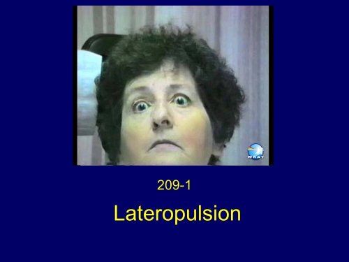
209-1<br />
<strong>Lateropulsion</strong>
<strong>Lateropulsion</strong> (deviation) of<br />
the eyes towards the side of<br />
the lesion, under closed<br />
lids.
Figure 1 shows a hypothetical scheme to account for lateropulsion of saccades.<br />
Interruption of climbing fibers originating from the inferior olivary nucleus may occur<br />
prior to their crossing in the medulla (1)or as they enter the inferior cerebellar<br />
peduncle in Wallenberg’s syndrome. (2) Loss of climbing fiber inputs to Purkinje<br />
cells in the dorsal vermis causes the latter to inhibit the fastigial nucleus (4), which<br />
causes ipsipulsion of saccades. Pharmacological inactivation of the dorsal vermis<br />
(3) causes contrapulsion (although clinical lesions produce bilateral hypometria).<br />
Interruption of crossed fastigial nucleus outputs in the superior cerebellar peduncle<br />
(uncinate fasciculus, (5) causes contrapulsion. Thus contrapulsion arises at sites 1,<br />
3 and 5 and ipsipulsion at sites 2-4.
Box 12-1. Leigh RJ, Zee DS. The Neurology of Eye Movements 4th Edition.<br />
Oxford University Press, New York 2006 with permission.
Lateral Medullary Infarct<br />
Figure 2 Axial T2WI in a patient with a classic Wallenberg syndrome shows a<br />
normal flow void in the left vertebral artery. The right vertebral artery is filled with<br />
thrombus which is isointense with brain. Note hyperintensity in the right olive as<br />
well as the lateral medulla. A small old infarct is also present in the left cerebellar<br />
hemisphere. Right PICA territory infarct. Courtesy of Anne Osborn, M.D.
Medial Medullary Infarct<br />
Figure 3 Axial T2WI shows a hyperintensity in the olive and medial medulla.
Medial Medullary Infarct<br />
Figure 4 Axial DWI shows restriction in the same territory.
Medial Medullary Infarct<br />
Figure 5 MRA shows an occluded left vertebral artery with sparing of the anterior<br />
inferior cerebellar artery which arises from the basilar artery above the vertebral<br />
artery confluence.
http://www.lib.med.utah.edu/NOVEL