THE ENITHARES (Hemiptera-Heteroptera: Notonectidae) OF THE ...
THE ENITHARES (Hemiptera-Heteroptera: Notonectidae) OF THE ...
THE ENITHARES (Hemiptera-Heteroptera: Notonectidae) OF THE ...

Create successful ePaper yourself
Turn your PDF publications into a flip-book with our unique Google optimized e-Paper software.
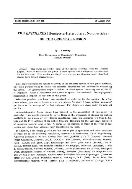
Pacific Insects 10(2): 353-442 20 August 1968<br />
<strong>THE</strong> <strong>ENITHARES</strong> (<strong>Hemiptera</strong>-<strong>Heteroptera</strong>: <strong>Notonectidae</strong>)<br />
<strong>OF</strong> <strong>THE</strong> ORIENTAL REGION<br />
By I. Lansbury<br />
HOPE DEPARTMENT <strong>OF</strong> ENTOMOLOGY, UNIVERSITY<br />
MUSEUM, OXFORD<br />
Abstract: This paper redescribes most of the species recorded from the Oriental<br />
Region. Keys to both sexes are given. Fifteen species and 1 subspecies are described<br />
for the first time. Five species are placed in synonymy and three previously described<br />
species have proved unrecognisable.<br />
This paper embodies the results of a study of the Oriental species of the genus Enithares.<br />
The main purpose being to collate the scattered descriptions and information concerning<br />
this genus. The geographical scope is limited to those species occurring east of the 60°<br />
of longitude. African, Mascarene and American species are excluded. No phylogenetic<br />
speculation is implied in any part of this paper.<br />
Wherever possible types have been examined in order to fix the species. In a few<br />
cases where types are no longer extant or available for study, I have utilized 'compared'<br />
specimens or the concept of the last reviewer. Full details are given under the relevant<br />
species.<br />
Acknowledgments: Many people have assisted in the preparation of this paper. In<br />
particular, I am deeply indebted to Dr G. Byers of the University of Kansas for making<br />
available to me a copy of G.T. Brooks unpublished thesis on Enithares. To Miss S. Nakata<br />
and Dr P. D. Ashlock of the Bishop Museum, Honolulu for the very large collection<br />
of un-named material sent to me. A glance at the location of many of the types of new<br />
species will show how valuable their contribution has been.<br />
In addition, I am deeply grateful for the loan or gift of specimens and other assistance<br />
afforded me by the following individuals, museums and institutions; Dr P. Wygodzinsky,<br />
American Museum of Natural History, New York (AMNH.) ; Dr T. Campbell, National<br />
Insect Collection, Canberra, A.C.T. (ANIC.) ; The Director, Naturhistorisches Museum,<br />
Basle (BASLE) ; Mrs Black, Dept. Entomology, Brit. Mus. (Nat. Hist.) (BMNH) ; Mr H.<br />
Synave, Institut Royal des Sciences Naturelles de Belgique, Bruxelles (BRUSSELS) ; Miss<br />
T. Gunawardane, National Museum, Colombo, Ceylon (COLOMBO) ; Dr A. Soos, Zoological<br />
Dept., Hungarian Museum of Natural History, Budapest (BUDAPEST) ; Dr Borge Petersen<br />
and Dr Nils Miller Andersen, Universitetets Zoologiske Museum, Copenhagen (COPENHA<br />
GEN) ; Mr R.G. Ordish, Dominion Museum, Wellington, N.Z. (DM) ; Dr M. Beier, Naturhistorisches<br />
Museum, Wien (VIENNA); Dr T. Jaczewski, Institute of Zoology, Polish
354 Pacific Insects Vol. 10, no. 2<br />
Academy of Sciences, Warsaw (WARSAW) ; Dr R. Froeschner, United States National<br />
Museum, Washington (USNM) ; Dr G.E. Hutchinson, Osborn Zoological Laboratory, Yale<br />
(YALE) ; Dr T.E. Woodward, University of Queensland, Brisbane (UQ) ; Dr I. M. Kerhzner,<br />
USSR Academy of Sciences, Leningrad (LENINGRAD) ; Dr F. S. Truxal, Los Angles<br />
County Museum, California (LA) ; Dr P.H. van Doesburg, Rijksmuseum van Naturlijke<br />
Historie, Leiden, The Netherlands (LEIDEN) ; Dr H. Andersson, Dept, of Entomology,<br />
Zoological Institute, University of Lund (LUND) ; Dr R. Benard, Museum d'histoire<br />
Naturelle, Paris (PARIS) ; Dr E. Kyellander, Naturhistoriska Riksmuseum, Stockholm (STOCK<br />
HOLM) ; Dr H. Weidner, Zoologisches Staatsinstitut und Zoologisches Museum, Hamburg<br />
(HAMBURG) ; Dr C. N. Smithers, The Australian Museum, Sydney (AMS) ; Dr K. S. Pradhan,<br />
Zoological Survey of India, Calcutta (Zool. Sur. India) ; Bernice P. Bishop Museum,<br />
Honolulu, Hawaii (BISHOP) ; Snow Entomological Collections, University of Kansas, Lawrence<br />
(SECK) ; Mr E. S. Brown, c/o Dept, of Entomology, Brit. Mus. (Nat. Hist.) (E.S.B.) ;<br />
Dr Marshall Laird, World Health Organisation, Geneva. Dr G. Gross, South Australian<br />
Museum, Adelaide (SAMA) ; Dr A. Neboiss, National Museum of Victoria, Melbourne<br />
(MELBOURNE) ; Mr A. W. Sweeney, Malaria Institute, Rabaul, T.P.N.G. Dr CH. Fernando,<br />
Dept, of Biology, University of Waterloo, Ontario, Canada.<br />
BIOLOGY<br />
Little is known about the biology of Enithares. Nowrojee (1911) iov E. ciliata (F.) ( =<br />
indica F. sensu Nowrojee) and Hoffmann (1931) for E. Sinica (Stal) give some data on<br />
life histories. Both report that the eggs are laid on submerged plant tissue. Nowrojee<br />
states that the eggs are glued on, not inserted in the plant. Enithares thereby closely resembles<br />
some species of Notonecta (Hungerford 1919). The number of eggs laid is variable.<br />
E. ciliata in an aquarium laid 8, E. Sinica "more than a dozen." I have a plant<br />
stem on which E. woodwardi Lansbury laid 19 eggs in about 90 minutes in an aquarium.<br />
Hungerford (1919) dissected a female N irrorata Uhler which was found to contain 252<br />
ova, several of which were nearly ready for laying. It is not known if Enithares is as<br />
prolific as this.<br />
The Qgg of E. woodwardi is about 1.45 mm long, width viewed from above .65 mm.<br />
Color in alcohol pale yellowish white. Shape, elongate rounded at both ends, anterior<br />
pole slightly depressed around the curved, cylindrical truncated, micropylar peg. Hatching<br />
line visible as a broken linear brown line extending from micropyle to caudal end.<br />
Chorion hard, covered with hexagonal reticulations. The Qgg of Enithares is very similar<br />
in general structure to that of Notonecta (Hungerford 1919).<br />
The incubation period is variable. The period for E. Sinica varies between 9-26 days<br />
(Hoffmann 1931). Nowrojee (1911) noted eye spots appearing just before hatching. The<br />
data given by Hoffmann who includes a precis of Nowrojee's figures show that development<br />
from hatch is dependant upon temperature - E. Sinica taking 38-49 days and E. ciliata<br />
33-38 days, the period of the 5th instar being the longest. Hoffmann using isolated specimens<br />
found that the highest mortality occurred in the first and second instars. E. Sinica<br />
took 2 or more days to become fully pigmented after the last moult. Hale (1923) stated<br />
that pale examples of E. woodwardi (=£*. bergrothi Montandon sensu Hale) confined in<br />
aquaria eventually became black, other than head, part of the pronotum and legs.
1968 Lansbury : Oriental Enithares (<strong>Notonectidae</strong>) 355<br />
There is no evidence that Enithares is attracted to light. Large collections of aquatic<br />
<strong>Heteroptera</strong> obtained in light traps from the Philippines and elsewhere have never included<br />
Enithares.<br />
Unlike Anisops, Enithares never seems to be found in large numbers. Miller (1964)<br />
comments on the small numbers of Enithares found as compared to Anisops in East<br />
Africa. Miller found that Enithares normally remained at the water surface and usually<br />
fed on prey caught in the surface film. When disturbed 3 distinct reactions were noticed.<br />
a) it swam horizontally without leaving the surface; b), it dived and anchored itself to a<br />
submerged object, or c), it dived and remained poised in mid-water 10-20 cm below the<br />
surface, keeping stationary by rapid strokes of the hind legs. The third reaction was<br />
the one most commonly observed in the laboratory. Hale (1923) also noticed the habit<br />
of floating at the surface in deep water or clinging to submerged objects. Kirkaldy<br />
(1904) quoting E. E. Green in litt, refers to Enithares preying on Metrocoris stali (Dohrn),<br />
Gerridae and Gyrnidae (Coleoptera).<br />
Laird (1947) found in New Britain that E. alexis lairdi Lansbury (=f£. hergrothi sensu<br />
Laird) fed on mosquito larvae, particularly culicines. DempwolfT (1904) in New Guinea<br />
found that habitats with many notonectids harbored no mosquito larvae.<br />
HISTORY <strong>OF</strong> <strong>ENITHARES</strong><br />
Spinola (1837) proposed the genus Enithares (an anagram of Theresind) for Notonecta<br />
indica F. 1803 and for a new species, braziliensis. The latter is no longer included in<br />
Enithares s.s. (Brooks 1953).<br />
Fieber (1851) renamed the genus Bothronotus because he did not apparently approve<br />
of anagrams.<br />
The only major revision has been by Kirkaldy (1904) who attempted to account for<br />
all the known species and gave a comprehensive key. In 1933, Lundblad gave a detailed<br />
resume of the species found in the Sunda Is. and Hutchinson (1933) similarly dealt with<br />
those from India, Burma and Ceylon. Finally Brooks (1948) described 17 new species,<br />
15 of which were Oriental; these are included in this paper.<br />
To date, within the geographical scope of this paper, 42 species and 1 subspecies have<br />
been described. Three were synonymised by earlier workers and 5 additional species are<br />
placed in synonymy in this paper. Two species described by Fieber (1851 & 1851a) and<br />
1 by Paiva (1918) have not proved recognisable.<br />
Fifteen species and 1 subspecies are described for the first time bringing the number<br />
to 46 species and 2 subspecies.<br />
TYPE SPECIES DESIGNATION<br />
Spinola (1837) proposed the genus Enithares and included 2 species Notonecta indica F.<br />
1803 "De Bombay, envoyee par M. Dupont" and E. braziliensis a new species "Du Bresil,<br />
enyoyee par M. Buquet." Kirkaldy (1897) designated by a footnote the type species of<br />
Enithares in the following phrase "I am not aware that any author has indicated a type
356 Pacific Insects Vol. 10, no. 2<br />
for this genus and therefore now set apart E. indica for that purpose." Kirkaldy on the<br />
same page in the main text made a lapsus calami in referring to Spinola's indica as N.<br />
indica L., whereas Spinola (1837) clearly referred to N. indica F. 1803.<br />
I have examined the holotype $ of N. indica F. 1803, described from Sumatra and<br />
preserved in the Zoological Museum, Copenhagen and find that it is conspecific with E.<br />
intricata Breddin 1905. The Fabrician name is however, preoccupied by Notonecta indica<br />
L. 1771 ; therefore, Breddin's name being the next available must continue to be used.<br />
In the course of this study, it has been found that E. intricata is distributionally limited<br />
to Sumatra and Java. It is, therefore, clear that Spinola's indica from Bombay must<br />
refer to another species.<br />
Fabricius (1798) described Notonecta ciliata from "Indiae aquis." Kirkaldy (1889)<br />
erroneously included N. ciliata in Anisops. Lundblad (1933) pointed out after seeing<br />
Fabricius' type that it was an Enithares. I have examined the holotype Sf. of ciliata preserved<br />
in the Zoological Museum, Copenhagen. The specimen is somewhat mutilated<br />
but there are sufficient diagnostic features left i.e. metaxyphus and nodal furrow to show<br />
that ciliata is conspecific with E. abbreviata (Kirby) 1891.<br />
Kirkaldy (1900) on discovering that JV. indica F. was preoccupied by N indica L. took<br />
the next available name which he thought was abbreviata. I have compared the type<br />
of ciliata with that of abbreviata in the British Museum (Nat. Hist.) and find them conspecific.<br />
Comparison of the type of indica F. with the types of ciliata and abbreviata<br />
shows that indica is clearly distinct.<br />
The distribution of Enithares in India is such that of the 5 species recorded, 3, E.<br />
triangularis (Guerin-Meneville), E. hungerfordi and E. fusca Brooks are all restricted to<br />
S. India; E. lineatipes Horvath is confined to the Punjab and Baluchistan leaving ciliata<br />
widespread over India and much of SE Asia. In view of the foregoing, it is therefore<br />
quite clear that Kirkaldy (1897) unwittingly based his type species designation on a<br />
misidentification of Spinola (1837).<br />
An application was therefore put before the International Commission on Zoological<br />
Nomenclature (Lansbury 1966) asking that Kirkaldy's type designation be set aside and<br />
nominating N. ciliata F. 1798 as the type species of Enithares.<br />
TECHNIQUE AND TERMINOLOGY<br />
Enithares are with few exceptions rather difficult to identify. The males of some species<br />
have prominent secondary sexual features i.e. spurs on legs, irregularly-shaped fore and<br />
mid-tibiae. There are however, many species without these 'spot' characters. In these<br />
cases, it is necessary to detach the male genital capsule and clear in the usual way. The<br />
aedeagus should be partially withdrawn from the capsule as it provides good diagnostic<br />
characters. Sometimes the sex of specimens is not immediately apparent from external<br />
features: If the 7th sternum is levered up slightly, the paired stylus-like gonoplacs of<br />
the female or the genital capsule of the male will become visible.<br />
Many species have at least 2 color forms. Lundblad (1933) was the first to comment<br />
on the baffling similarity between very pale or completely albino forms "leukokroismus."<br />
Where fairly large series of specimens have been available for study, i.e. E. woodwardi,
1968 Lansbury : Oriental Enithares (<strong>Notonectidae</strong>) 357<br />
every kind of intergrade between the pale and almost melanic form has been found.<br />
Therefore, no reliance should be placed on color in naming Enithares. The more dominant<br />
color forms are described in the text.<br />
The length of a specimen is measured along a median line from the tip of the vertex<br />
to the end of the elytra. To measure the anterior width of the vertex and synthlipsis<br />
etc., the specimen is placed in a horizontal position with the transverse and longitudinal<br />
axes horizontal. The synthlipsis is defined as the narrowest part of the vertex between<br />
the posterior margins of the eyes; the anterior width of vertex as the width of the vertex<br />
between the anterior margins of the eyes. To determine the position of the nodal<br />
furrow, one must view the specimen on its lateral surface with the head tilted downwards<br />
until the nodal furrow and membranal suture are at the same level. The distance<br />
between the nodal furrow and the membranal suture is measured from the inner tip of<br />
the furrow straight back to the suture along a line parallel to the lateral margins of<br />
the elytra.<br />
GENERIC KEY TO <strong>THE</strong> NOTONECTIDAE (Modified from Lansbury, 1966)<br />
1. Hemelytral commissure without a definite hair-lined pit at anterior end (Notonectinae)<br />
2<br />
Hemelytral commissure with a definite hair lined pit at anterior end (Anisopinae) 7<br />
2(1). Mid-femur with an anteapical pointed protuberance 3<br />
Mid-femur without an anteapical pointed protuberance 4<br />
3(2). Anterolateral margins of prothorax not foveate Notonecta<br />
Anterolateral margins of prothorax foveate Enithares<br />
4 (2). Eyes basally contiguous or forming an ocular commissure, c? parameres asymmetrical... 5<br />
Eyes basally widely spaced, & parameres symmetrical Aphelonecta<br />
5(4). Anterolateral margins of prothorax not foveate Neonychia<br />
Anterolateral margins of prothorax foveate 6<br />
6(5). Antennae 3-segmented Nychia<br />
Antennae 4-segmented Martarega<br />
7 (1). Ventral abdominal keel not extending onto last abdominal sternite.
358 Pacific Insects Vol. 10, no. 2<br />
Fig. 1-7. 1, E. stylata n. sp. lateral view of nodal furrow and membrane; 2, E. Ii neatipes<br />
Horvath, elytron; 3, Enithares sp., elytron; 4, E. hungerfordi Brooks, # ventral view<br />
of embolium and coxal plate; 5, E. stylata #, Ibid; 6, Enithares sp., lateral view of $<br />
genitalia; 7, E. simplex (Kirby) ibid.<br />
Nauk 7 : 206-7 ; 1851a, ibid: 470-72.<br />
Body from above convex. Head narrower than pronotum. Eyes large, reniform, 2x<br />
sinuate along outer margin and dorsally occupying about 2/3 of head. Inner lateral margins<br />
of eyes converging from anterior width of vertex. Ocelli absent. Antennae 4-segmented,<br />
arising from behind eyes and lying in trough of pronotal fovea. Rostrum 4-segmented.<br />
Pronotum broader than long, lateral margins diverging, anterolateral margins foveate. Scutellum<br />
triangular. Clavus and corium coriaceous. Corium with a cleft known as the nodal<br />
furrow directed inwards from lateral margins slightly anterior to membranal suture (fig.<br />
1-3). Membrane clearly divided into 2 zones and bilobed (fig. 1-3). Fore and median legs<br />
with 3 tarsi, middle tarsus vestigial. Hind leg with 2 tarsi. Two tarsal claws present on all<br />
legs. Mid-femur with a pointed anteapical protuberance. Infra-coxal plates bare and fringed
1968 Lansbury : Oriental Enithgres (<strong>Notonectidae</strong>) 359<br />
with hairs. Keel of abdominal venter bare and fringed with hairs along lateral margins.<br />
& genitalia : Terminology modified from Truxal (1952). Genital capsule 9th segment<br />
which is cleft medianly into an anterior and posterior lobe. Posterior lobe separated<br />
dorsally and along upper 1/2 of posterior margin. Parameres symmetrical, directed<br />
dorsad, basally inflected into genital chamber through median cleft. Basal plate large,<br />
fused ventrally into a ring, lateral arms of basal plate heavily sclerotised, dorsally ring<br />
very wide, ventrally narrow. Aedeagus clearly differentiated into a phallosoma and<br />
endosoma, the latter with prominent conjunctival appendages. Basal portion of phallosoma<br />
narrow and annulated, apically more membranous. Penis valves not visible. The<br />
9th tergum forms a broad bridge across anterior part of 9th segment. The lOth lightly<br />
sclerotised, protruding from dorsal margin<br />
of anterior lobe of 9th segment (fig. 8).<br />
£ genitalia : Terminology follows Scudder<br />
(1959). Sternum Vll produced posteriorly,<br />
partially covering genitalia. First<br />
gonocoxa very roughly triangular, anterior<br />
margin more sclerotised than remainder,<br />
broadest part covered with long fine hairs.<br />
First gonapophysis elongate, sclerotised<br />
with stout spines. Gonangulum elongate<br />
and sclerotised. Second gonocoxa slender,<br />
only very slightly sclerotised. Second ramus<br />
heavily sclerotised. Second gonapophysis<br />
sclerotised throughout, apically bent<br />
ventrad and set with spines. Gonoplacs<br />
separate, stylus like with very many long<br />
fine hairs. Single median Spermatheca<br />
present (fig. 6 & 7).<br />
%?<br />
KEY TO <strong>ENITHARES</strong> MALES<br />
Fig. 8. E. megalops n. sp. lateral view<br />
of cT genitalia showing aedeagus partially<br />
expanded.<br />
1. Embolium viewed ventrally greatly expanded towards head (fig. 4 & 27) 2<br />
Embolium viewed ventrally not as above (fig. 5) 5<br />
2(1). Apices of clavii clearly raised and roughened with many pits (fig. 12)<br />
. stridulata Brooks<br />
Apices of clavii not as above 3<br />
3 (2). Nodal furrow clearly more than its own length removed from membranal suture,<br />
pronotal humeral angles produced (fig. 15) producta n. sp.<br />
Nodal furrow clearly less than its own length removed from membranal suture,<br />
pronotal humeral angles not produced 4<br />
4(3). Mid-femoral hairs short (fig. 22); median length of metaxyphus 2x basal width<br />
(fig. 30) ; fore femur with triangular eminence ventrally (fig. 25) ...triangularis (Guerin)<br />
Mid-femoral hairs long and straggly (fig. 35) ; median length of metaxyphus equaling<br />
basal width (jig. 33); fore femur not as above hungerfordi Brooks<br />
5(1). Fore trochanter with a nodule ventrad (fig. 42) hackeri Hungerford<br />
Fore trochanter without a nodule ventrad 6
360 Pacific Insects Vol. 10, no. 2<br />
6 (5). Base of labrum with nodule-like projections on posterior corners (fig. 55)<br />
simplex (Kirby)<br />
Base of labrum not as above 7<br />
7(6). Head width less than or only 2x median head length 8<br />
Head width over 2>< median head length 13<br />
8 (7). Outer claw of mid-leg conspicuously thickened and bent inwards 9<br />
Outer claw of mid-leg not as above ll<br />
9(8). Hind femur with a large spur ventrad (fig. 66) Sinica (Stal)<br />
Hind femur not as above 10<br />
10(9). Lateral margins of metaxyphus convergent (fig. 75) mandalayensis Distant<br />
Lateral margins of metaxyphus more or less parallel (fig. 82) uncata Lundblad<br />
11 (8). Nodal furrow less than its own length removed from membranal suture<br />
hebridiensis n. sp.<br />
Nodal furrow more than its own length removed from membranal suture 12<br />
12 (ll). Anterior lobe with 2 dorsal projections, posterior lobe higher than anterior lobe<br />
(fig. 93) bakeri Brooks<br />
Dorsal margin of anterior lobe slightly convex distally, posterior lobe about as high<br />
as anterior lobe (fig. 99) genitalis Lundblad<br />
13(7). Head equaling or clearly longer than median pronotal length 14<br />
Head clearly shorter than median pronotal length 31<br />
14(13). Hind femur with a large spur ventrad (fig. 66) Sinica (Stal)<br />
Hind femur not as above 15<br />
15(14). Mid-femoral hairs very long, mesotrochanter spur-like at inner ventral angle 16<br />
Mid-femoral hairs not very long, mesotrochanter not spur-like at inner ventral<br />
angle 17<br />
16(15). Outer lobe of mid-tibia distally rounded (fig. 104) alexis alexis Lansbury<br />
Outer lobe of mid-tibia distally acuminate (fig. 110) alexis lairdi n. subsp.<br />
17(15). Outer claw of mid-leg conspicuously thickened and bent inwards (fig. 70); hind<br />
femur slightly expanded distally (fig. 72) mandalayensis Distant<br />
Outer claw of mid-leg and hind femur not as above 18<br />
18(17). Nodal furrow more than its own length removed from membranal suture 19<br />
Nodal furrow equal to or less than its own length removed from membranal suture ... 22<br />
19(18). More than ll mm long; lateral margins of 1st 3 and part of 4th visible ventral Connexival<br />
segments minutely transversely ridged chinensis Brooks<br />
Not more than 9.6 mm long; ventral Connexival segments not as above 20<br />
20(19). Over 9mm long; lateral margins of metaxyphus concave (fig. 132.) Dorsal margin<br />
of pronotal fovea directed caudad behind eyes (fig. 134) timorensis Brooks<br />
Not more than 8 mm long; lateral margins of metaxyphus straight or convex. Dorsal<br />
margin of pronotal fovea directed laterad from behind eyes 21<br />
21 (20). Pronotum with a transverse rugose band interrupted medianly (fig. 139). Posterior<br />
lobe finger-like (fig. 137) loria Brooks<br />
Pronotum smooth. Posterior lobe not finger-like (fig. 99) genitalis Lundblad<br />
22(18). Lateral margins of 1st 2 visible ventral abdominal segments with a series of transverse<br />
ridges (fig. 147) nigra n. sp.<br />
Lateral margins of 1st 2 visible ventral abdominal segments not as above 23<br />
23 (22). Dorsal margin of pronotal fovea curving laterad from behind eyes (fig. 149)<br />
bergrothi Montandon<br />
Dorsal margin of pronotal fovea directed caudad from behind eyes 24<br />
24(23). Head width slightly greater than 3X anterior width of vertex. Chaetotaxy of midtibia<br />
interrupted distally (fig. 322) rogersi Distant
1968 Lansbury : Oriental Enithares (<strong>Notonectidae</strong>) 361<br />
Head width slightly less than 3X anterior width of vertex. Chaetotaxy of mid-tibia<br />
not interrupted distally 25<br />
25(24). Over 10 mm long hippokleides Kirkaldy<br />
Less than 9.5 mm long 26<br />
26(25). Posterior lobe clearly bent caudad apically (fig. 176) ripleyana n. sp.<br />
Posterior lobe not bent caudad apically 27<br />
27(26). Over 9 mm long. Head width almost 2.5x median length timorensis Brooks<br />
Less than 9 mm long. Head width not more than 2.33x median length 28<br />
28(27). 8-9 mm long. Lateral arms of basal plate produced caudad (fig. 182) ... vulgaris n. sp.<br />
7-8 mm long. Lateral arms of basal plate rounded 29<br />
29 (28). Posterior margin of eyes directed caudad laterad, i.e. slanting from inner margin to<br />
anterolateral margins of pronotum. Outer lateral margins of eyes strongly divergent<br />
from anterolateral margins of pronotum (fig. 194 & 199) 30<br />
Posterior margin of eyes more or less straight. Outer lateral margins of eyes convergent<br />
almost from anterolateral margins of pronotum (fig. 189) ... intricata Breddin<br />
30 (29). Parameres basally constricted, apically expanded and rounded ; posterior lobe evenly<br />
rounded dorsally (fig. 8) megalops n. sp.<br />
Parameres crescentic; posterior lobe more acuminate dorsally (fig. 198)<br />
paramegalops n. sp.<br />
31 (13). Hind femur either greatly expanded or with a prominent spur or nodule 32<br />
Hind femur simple 38<br />
32(31). Over 11.5 mm long subparallel n. sp.<br />
Not more than 10 mm long 33<br />
33(32). Head width more than 3X anterior width of vertex. First tarsus of fore leg curved<br />
(fig. 213); hind femur greatly expanded distally (fig. 212) gibbera Brooks<br />
Head width less than 3X anterior width of vertex. First tarsus of fore leg not<br />
curved; hind femur with a spur or nodule 34<br />
34(33). Fore and mid-tibiae strongly produced (fig. 220-22). Metaxyphus evenly rounded<br />
(fig. 225) buhleri Brooks<br />
Fore and mid-tibiae not strongly produced. Metaxyphus not evenly rounded 35<br />
35 (34). Outer claw of mid-leg conspicuously thickened and bent inwards (fig. 62). Hind<br />
femur with a large spur ventrad (fig. 66^) Sinica (Stal)<br />
Outer claw of mid-leg not as above. Hind femur with a nodule, not a spur 36<br />
36 (35). Fore tibia clearly depressed distally. Mesotrochanter with a variable-sized patch of<br />
black spicules 37<br />
Fore tibia not depressed distally. Mesotrochanter without black spicules, but with<br />
fine sparse hairs (fig. 228) fusca Brooks<br />
37 (36). Mesotrochanter with a large patch of black spicules (fig. 235). First mid-tarsus roughly<br />
triangular. Apex of mid-tibia bluntly produced (fig. 236) ciliata (F.)<br />
Mesotrochanter with a small patch of black spicules (fig. 243). Apex of mid-tibia<br />
produced and acuminate with a large spine on inner surface distally (fig. 247). First<br />
mid-tarsus elongate (fig. 245) lombokensis n. sp.<br />
38(31). Median head length shorter than anterior width of vertex 39<br />
Median head length longer than anterior width of vertex 46<br />
39(38). Nodal furrow more than its own length removed from membranal suture (fig. 3) 40<br />
Nodal furrow less than its own length removed from membranal suture (fig. 1) 41<br />
40 (39). Lateral margins of 1st 3 and part of 4th visible ventral Connexival segments minutely<br />
transversely ridged chinensis Brooks<br />
Lateral margins of ventral Connexival segments not as above lineatipes H6rvath<br />
41 (39). Lateral margins of 1st 3 and part of 4th visible ventral Connexival segments minute-
362 Pacific Insects Vol. 10, no. 2<br />
ly transversely ridged biimpressa (Uhler)<br />
Lateral margins of ventral Connexival segments not as above 42<br />
42(41). More than 10 mm long metallica Brooks<br />
Less than 9.5 mm long 43<br />
43(42). Head width slightly more than 3X anterior width of vertex 44<br />
Head width less than 3X anterior width of vertex 45<br />
44(43). Inner surface of mid-tibia with a large nodule distally (fig. 264) ... thienemanni Lundblad<br />
Inner surface of mid-tibia distally without a large nodule (fig. 271) atra Brooks<br />
45 (43). Mid-tibia with many marginal spines (fig. 211}. Posterior lobe more or less upright<br />
(fig. 280) amboinensis n. sp.<br />
Mid-tibia with few marginal spines (fig. 174). Posterior lobe apically inclined<br />
caudad (fig. 116) ripleyana n. sp.<br />
46(38). More than 11.5 mm long 47<br />
Less than 10.5 mm long 48<br />
47(46). Posterior lobe apically finger-like set with spines, parameres short and rounded<br />
(fig. 332) horvathi Kirkaldy<br />
Posterior lobe broad and rounded, not set with spines apically, parameres very long<br />
(fig. 285) stylata n. sp.<br />
48(46). Mesotrochanter forming a distinct spur at inner ventral margin 49<br />
Mesotrochanter rounded or angulate 52<br />
49(48). Mid-femoral hairs very long, dorsal margin set with 5-6 stout spines (fig. 102) 50<br />
Mid-femoral hairs short, dorsal margin not set with stout spines (fig. 288) 51<br />
50(49). Outer lobe of mid-tibia distally rounded (fig. 103) alexis alexis Lansbury<br />
Outer lobe of mid-tibia distally acuminate (fig. 110) alexis lairdi n. subsp.<br />
51 (49). Fore tibia curved forward (fig. 293). Mid tibia distally swollen forming a convex<br />
arch (fig. 289) malayensis Brooks.<br />
Fore tibia not curved forward. Mid tibia not distally swollen (fig. 295) ... maai n. sp.<br />
52 (48). Lateral margins of 1st 2 visible ventral abdominal segments with a series of transverse<br />
ridges (fig. 147) nigra n. sp.<br />
Lateral margins of 1st 2 visible ventral abdominal segments not as above 53<br />
53(52). Lateral margins of pronotal fovea produced towards eyes as a spur or nodule 54<br />
Lateral margins of pronotal fovea not as above 55<br />
54 (53). Dorsal margin of pronotal fovea directed caudad behind eyes (fig. 307). Apex of<br />
posterior lobe inclined caudad (fig. 305) foveatus n. sp.<br />
Dorsal margin of pronotal fovea directed laterad behind eyes (fig. 157). Apex of<br />
posterior lobe not inclined caudad (fig. 158) woodwardi n. sp.<br />
55(53). Head width not more than 2.5X median head length 56<br />
Head width over 2.5X median head length 61<br />
56 (55). Dorsal margin of pronotal fovea directed laterad from behind eyes (fig. 149). Lateral<br />
margins of metaxyphus more or less straight (fig. 150) bergrothi Montandon<br />
Dorsal margin of pronotal fovea directed caudad from behind eyes. Lateral margins<br />
of metaxyphus not straight, usually shallowly concave distally 57<br />
57 (56). Parameres very long (fig. 336). First mid-tarsus with 4 stout spines set close together<br />
medianly (fig. 335) freyi quadrispinosus Lansbury<br />
Parameres very short. First mid-tarsus with a variable number of spines; not set<br />
close together but dispersed along lateral margins 58<br />
58(57). Head width more than 3X anterior width of vertex 59<br />
Head width less than 3X anterior width of vertex 60<br />
59 (58). Inner surface of mid-tibia with chaetotaxy interrupted distally (fig. 322); midtibia<br />
distally concave (fig. 324). First mid-tarsus with 4 stout spines (fig. 323).
1968 Lansbury ; Oriental Enithares (<strong>Notonectidae</strong>) 363<br />
rogersi Distant<br />
Chaetotaxy of mid-tibia not interrupted distally or concave (fig. 309). First midtarsus<br />
with 2 stout spines (fig. 310) vicintricata n. sp.<br />
60 (58). Not more than 8 mm long. Posterior lobe with dorsal margin evenly rounded (fig.<br />
187) intricata Breddin<br />
At least 8.75 mm long. Posterior lobe inclined caudad apically (fig. 177)<br />
ripleyana n. sp.<br />
61 (55). Parameres very long, almost or surpassing dorsal margin of posterior lobe 62<br />
Parameres short, not as above 64<br />
62(61). Mid-tibia with a large nodule on inner distal surface (fig. 264) ... thienemanni Lundblad<br />
Mid-tibia without a large nodule on inner distal surface 63<br />
63 (62). First mid-tarsus with 4 stout spines set close together medianly (fig. 335). Parameres<br />
acutely triangular (fig. 336) freyi quadrispinosus Lansbury<br />
First mid-tarsus with many very short stout setae (fig. 272). Parameres not as<br />
above (fig. 273) atra Brooks<br />
64(61). Length 8.75-9.75 mm. Posterior lobe inclined caudad apically (fig. 176)... ripleyana n. sp.<br />
Length 7.5-8 mm. Posterior lobe cleft on outer margin, not inclined caudad (fig.<br />
315) martini Kirkaldy<br />
KEY TO <strong>ENITHARES</strong> FEMALES<br />
1. Nodal furrow clearly more than its own length removed from membranal suture<br />
(fig. 128) 2<br />
Nodal furrow equal to or less than its own length removed from membranal suture<br />
(fig. 124) 8<br />
2(1). Lateral margins of 1st 3 and part of 4th visible ventral Connexival segments minutely<br />
transversely ridged chinensis Brooks<br />
Connexival segments not as above 3<br />
3(2). Pronotal humeral width more than 3 X median length 4<br />
Pronotal humeral width less than 3X median length 6<br />
4(3). Head longer than pronotum genitalis Lundblad<br />
Head shorter than pronotum 5<br />
5(4). Apex of metaxyphus spatulate stridulata Brooks<br />
Apex of metaxyphus acuminate producta n. sp.<br />
6(3). Less than 7 mm long loria Brooks<br />
Over 7.5 mm long 7<br />
7(6). Between 7.75-8.25 mm long bakeri Brooks<br />
Over 10 mm long lineatipes Horvath<br />
8(1). Lateral margins of 1st 3 and part of 4th visible ventral Connexival segments minutely<br />
transversely ridged biimpressa (Uhler)<br />
Lateral margins of ventral Connexival segments not as above 9<br />
9(8). Embolium viewed ventrally greatly expanded towards head (fig. 4) 10<br />
Embolium not greatly expanded towards head (fig. 5) ll<br />
10(9). Basal width of metaxyphus equaling median length hungerfordi Brooks<br />
Basal width of metaxyphus 1/2 median length triangularis (Guerin)<br />
ll (9). Not more than 7 mm long megalops n. sp.<br />
More than 7 mm long 12<br />
12(11). Pronotal humeral width 3x or more median length 13<br />
Pronotal humeral width less than 3X median length 40
364 Pacific Insects Vol. 10, no. 2<br />
13(12). Head length less than anterior width of vertex 14<br />
Head length greater than anterior width of vertex 22<br />
14(13). Metaxyphus with apex rounded buhleri Brooks<br />
Metaxyphus with apex acuminate 15<br />
15(14). Less than 10 mm long 16<br />
10 mm or more long 18<br />
16(15). Head width more than 3X median length atra Brooks & thienemanni Lundblad<br />
Head width less than 3X median length 17<br />
17(16). Not more than 9 mm long amboinensis n. sp.<br />
Over 9 mm long fusca Brooks & ripleyana n. sp.<br />
18 (15). Metaxyphus centrally depressed, lateral margins raised and thickened, infuscated<br />
dark brown to black (fig. 60 &61) simplex (Kirby)<br />
Metaxyphus not as above 19<br />
19(18). Synthlipsis 2/3 anterior width of vertex subparallel n. sp.<br />
Synthlipsis about 1/2 anterior width of vertex 20<br />
20(19). Clavus and corium metallic greenish bronze metallica Brooks<br />
Clavus and corium not metallic 21<br />
21(20). Not more than 10.25 mm long atra Brooks<br />
At least 11.5 mm long stylata n. sp.<br />
22(13). Over 12 mm long horvathi Kirkaldy<br />
Less than 12 mm long 23<br />
23 (22). Lateral margins of 1st 2 visible ventral abdominal segments with a series of transverse<br />
ridges (fig. 147) nigra n. sp.<br />
Lateral margins of 1st 2 visible ventral abdominal segments not as above 24<br />
24 (23). Lateral margins of pronotal fovea almost contiguous with posterior margin of eyes<br />
(fig. 306) foveatus n. sp.<br />
Lateral margins of pronotal fovea not as above 25<br />
25(24). Head shorter than pronotum 26<br />
Head longer than pronotum 32<br />
26(25). Viewed laterally, margins of pronotum carinate gibbera Brooks<br />
Viewed laterally, margins of pronotum rounded 27<br />
27(26). Head width more than 3X median length thienemanni Lundblad<br />
Head width less than 3X median length 28<br />
28(27). At least 10 mm long 29<br />
Not more than 9.75 mm long 30<br />
29(28). Clavus and corium metallic greenish bronze metallica Brooks<br />
Clavus and corium not metallic freyi quadrispinosus Lansbury<br />
30 (28). Viewed laterally, lower margin of pronotal fovea raised, appearing as a projection<br />
when viewed from above (fig. 320) martini Kirkaldy<br />
Viewed laterally, lower margin of pronotal fovea rounded, not appearing as a projection<br />
when viewed from above 31<br />
31(30). Not more than 8.5 mm long intricata Breddin<br />
Between 9-9.75 mm long ripleyana n. sp.<br />
32(25). More than 10 mm long simplex (Kirby)<br />
Less than 9.75 mm long 33<br />
33(32). Tomentose area of pronotal fovea hardly visible when viewed from above 34<br />
Tomentose area of pronotal fovea clearly visible from above 35<br />
34 (33). Not more than 7 mm long megalops n. sp.<br />
About 8 mm long paramegalops n. sp.<br />
35(33). Lateral margins of metaxyphus convergent from base 36
1968 Lansbury : Oriental Enithares (<strong>Notonectidae</strong>) 365<br />
Basal lateral margins of metaxyphus parallel, apically acuminate<br />
mandalayensis Distant & uncata Lundblad<br />
36(35). Hind femur indented adjacent to attachment with trochanter alexis Lansbury<br />
Hind femur not indented as above 37<br />
37(36). Not more than 9 mm long 38<br />
Over 9 mm long 39<br />
38 (37). I ateral margins of pronotum broadly rounded, dorsal margin of pronotal fovea<br />
directed caudad from posterior margin of eyes intricata Breddin<br />
Lateral margins of pronotum converging more or less straight from humeral angles,<br />
dorsal margin of pronotal fovea directed laterad from posterior margin of eyes<br />
vulgaris n. sp.<br />
39(37). Outer posterolateral margins of eyes straight timorensis Brooks<br />
Outer posterolateral margins of eyes rounded ripleyana n. sp.<br />
40(12). Greatest head width over 2X anterior width of vertex 41<br />
Greatest head width less than 2X anterior width of vertex mandalayensis Distant<br />
41 (40). Head longer than pronotum 42<br />
Head shorter than pronotum 50<br />
42(41). Lateral margins of 1st 2 visible ventral abdominal segments with a series of transverse<br />
ridges (fig. 141) nigra n. sp.<br />
Lateral margins of 1st 2 visible ventral abdominal segments not as above 43<br />
43 (42). Ventral margins of pronotal fovea slightly produced towards posterior margin of<br />
eyes hackeri Hungerford<br />
Ventral margins of pronotal fovea rounded 44<br />
44 (43). Dorsal margin of pronotal fovea curving laterad from posterior margin of eyes,<br />
tomentose area hardly visible 45<br />
Dorsal margin of pronotal fovea directed caudad from posterior margin of eyes,<br />
tomentose area clearly visible 46<br />
45(44). Not more than 9 mm long vulgaris n. sp.<br />
At least 9.75 mm long bergrothi Montandon & hebridiensis n. sp.<br />
46(44). At least 10 mm long hippokleides Kirkaldy<br />
Not more than 9.5 mm long 47<br />
47(46). Hind femur indented close to attachment with trochanter alexis Lansbury<br />
Hind femur not indented as above 48<br />
48(47). Basal lateral margins of metaxyphus divergent, raised and infuscated Sinica (Stal)<br />
Basal lateral margins of metaxyphus not as above 49<br />
49 (48). Anterior width of vertex clearly extending beyond anterior margin of eyes<br />
mandalayensis Distant & uncata Lundblad<br />
Anterior width of vertex not extending beyond anterior margin of eyes<br />
intricata Breddin<br />
50(41). Ventrolateral margins of pronotal fovea produced towards posterior margin of eyes... 51<br />
Ventrolateral margins of pronotal fovea rounded 53<br />
51 (50). Lateral margins of 1st 2 visible ventral abdominal segments with a series of<br />
transverse ridges (fig. 147) , nigra n. sp.<br />
Lateral margins of 1st 2 visible ventral abdominal segments not as above 52<br />
52(51). Not more than 9 mm long foveatus n. sp.<br />
Between 9.5-11 mm long woodwardi n. sp.<br />
53 (50). Mesotrochanter angulate malayensis Brooks<br />
Mesotrochanter rounded 54<br />
54(53). 10 mm or more long 55<br />
At most 9.6 mm long 58
366 Pacific Insects VoL 10, no. 2<br />
55 (54). Metaxyphus centrally depressed, lateral margins raised and thickened, infuscated<br />
dark brown-black (fig. 60 & 61) simplex (Kirby)<br />
Metaxyphus not as above 56<br />
56 (55). Dorsal margin of pronotal fovea curving laterad from posterior margin of eyes,<br />
tomentose area hardly visible bergrothi Montandon<br />
Dorsal margin of pronotal fovea directed caudad from posterior margin of eyes,<br />
tomentose area clearly visible 57<br />
57(56). Head narrower than anterior pronotal angles maai n. sp.<br />
Head wider than anterior pronotal angles freyi quadrispinosus Lansbury<br />
58(54). Basal lateral margins of metaxyphus divergent, raised and infuscated Sinica (Stal)<br />
Basal lateral margins of metaxyphus not as above 59<br />
59 (58). Dorsal margin of pronotal fovea curving laterad from posterior margin of eyes,<br />
tomentose area hardly visible bergrothi Montandon<br />
Dorsal margin of pronotal fovea directed caudad from posterior margin of eyes,<br />
tomentose area clearly visible 60<br />
60 (59). Viewed laterally, lower margin of pronotal fovea raised, appearing as a projection<br />
when viewed from above (fig. 320) martini Kirkaldy<br />
Viewed laterally, lower margin of pronotal fovea rounded, not appearing as a projection<br />
when viewed from above 61<br />
61(60). Not more than 8.5 mm long intricata Breddin<br />
Larger, at least 8.75 mm long 62<br />
62(61). Head almost 3X median length rogersi Distant<br />
Head never more than 2.75X median length 63<br />
63 (62). Basal lateral margins of metaxyphus parallel, apically convergent<br />
vicintricata n. sp. & ciliata (F.)<br />
Lateral margins of metaxyphus convergent from base to apex<br />
freyi quadrispinosus Lansbury<br />
Enithares stridulata Brooks Fig. 9-14.<br />
Enithares stridulata Brks., 1948, J. Kans. Ent. Soc. 21 : 37-38, pl. 1, fig. 1.- Lansbury, 1964, Ann.<br />
Zool. Warsz. 22: 210-11, fig. 4a-d.<br />
1968 Lansbury : Oriental Enithares (<strong>Notonectidae</strong>) 367<br />
Fig. 9-14. E. stridulata Brooks #. 9, side view of mid-tibia; 10,<br />
ibid tarsi and claws; ll, genital capsule; 12, apices of clavus; 13,<br />
head and pronotum from above; 14, metaxyphus.<br />
a slight depression on distal 1/2 of anterior margin. Mesotrochanter rounded (Brooks states<br />
that it is angulate). Male mid-tibia with many large spines (fig. 9), mid-tarsus with a series<br />
of transverse ridges (fig. 10). Apex of metaxyphus spatulate (fig. 14). Genital capsule as in<br />
fig. ll, posterior lobe with inner margin evenly rounded, parameres elongate almost reaching<br />
upper margin of posterior lobe, basally parameres transversely corrugated on inner margin.<br />
SPECIMENS EXAMINED: Holotype 6\ 4
368 Pacific Insects Vol. 10, no. 2<br />
Comparative notes : E. stridulata is very similar in general appearance and size to<br />
triangularis and hungerfordi, but can be separated by having the nodal furrow clearly<br />
more than its own length removed from the membranal suture and the apex of the<br />
metaxyphus spatulate.<br />
Enithares producta Lansbury, new species Fig. 15-21.<br />
&. Length 9 mm, maximum width 4.5 mm ; £, length 10 mm, maximum width 4.1 mm.<br />
Broad robust species, tapering abruptly from base of pronotum to end of abdomen, (fig. 15<br />
& 16).<br />
Color : Pale form, eyes dark brown flecked with black. Vertex, pronotum and scutellum<br />
yellowish brown. Elytra hyaline appearing brown due to underlying pigmentation showing<br />
through. Membrane hyaline, very pale yellow. Dark form, eyes and vertex yellowish brown.<br />
Anterior 1/2 of pronotum, scutellum, clavus, inner angle of corium and membrane dark brown<br />
to black. Pronotum with a median transverse pale brown band, posteriorly pronotum slightly<br />
darker. Pronotal humeral angles and outer margin of corium grayish. Legs and abdomen<br />
ventrally yellowish brown.<br />
Fig. 15 21. E. producta n. sp. 15, df dorsal aspect; 16, 9- ibid; 17, df genital capsule;<br />
18, c? mid-femur; 19,
1968 Lansbury : Oriental Enithares (<strong>Notonectidae</strong>) 369<br />
Structural characters : & with dorsal view of head rather acuminate, £ more rounded, anterior<br />
width of vertex extending beyond anterior margin of eyes. Greatest width of head 5/6<br />
pronotal humeral width, just over 3x anterior width of vertex and about 3X median head<br />
length. Median head length slightly greater than anterior width of vertex and about 3/4<br />
median pronotal length. Synthlipsis just over 1/2 anterior width of vertex. Pronotal humeral<br />
width about 3X median length, posterior margin concave, centrally emarginate. Male pronotal<br />
humeral angles clearly produced (fig. 15), # not so (fig. 16). Dorsal margin of pronotal<br />
fovea directed caudad before turning laterad behind eyes. Nodal furrow directed dorsad, tip<br />
inclined cephalad, furrow more than its own length removed from membranal suture. Embolium<br />
ventrally expanded towards head. Lateral margins of elytra sinuate. Mesotrochanter rounded<br />
(fig. 18). Male mid-tibia with a dense patch of long hairs on inner proximal surface (fig. 19).<br />
Chaetotaxy of $ mid-tarsus and claws as in fig. 20. Metaxyphus long, apex acuminate (fig.<br />
21). Male genital capsule as in fig. 17, posterior lobe apically bent over caudad and acuminate,<br />
parameres very short.<br />
Holotype
370 Pacific Insects Vol. 10, no. 2<br />
Fig. 22-28. E. triangularis (Guerin-Meneville)
1968 Lansbury : Oriental Enithares (<strong>Notonectidae</strong>) 371<br />
Fig. 29-31. E. triangularis &. 29, head and pronotum from above;<br />
30, metaxyphus; 31, ventral view of hind femur. Fig. 32-34. E. hunger-for'di<br />
Brooks
372 Pacific Insects Vol. 10, no. 2<br />
SPECIMENS EXAMINED: South India, Nilgiri Hills, Devala, 960m (3200 ft), X.1960, P.S.<br />
Nathan, 1£ ; West Bengal, Paresnath, 1200 rn (4000 ft), 9.IV.1909, A.N. Annandale, 1$ ;<br />
Madras, 1$, (SECK). India, Nilgiri Hills, H.L. Andrews, 1< anterior width of vertex and median head length.<br />
Synthlipsis 3/5 anterior width of vertex. Head length equaling anterior width of vertex and<br />
about 5/6 median pronotal length. Pronotal humeral width nearly 3>< median length, lateral<br />
margins diverging, 2/3 median length ; posterior margin almost straight. Dorsal margin of<br />
pronotal fovea curving laterad behind eyes. Nodal furrow curved cephalad and less than its<br />
own length removed from membranal suture. Anterior 1/2 of embolium greatly expanded (fig. 4$,<br />
40
1968 Lansbury : Oriental Enithares (<strong>Notonectidae</strong>) 373<br />
Fig. 35-41. E. hungerfordi &. 35, mid-femur; 36, mid-tibia; 37, mid-tarsus and claws;<br />
38, fore femur; 39, genital capsule; 40, embolium and adjacent structures ; 41, claws<br />
of hind leg.
374 Pacific Insects Vol. 10, no. 2<br />
(SECK). India, Shembagarrrur, St. Breuning, 1^ (LENINGRAD). India, Cinchona, Anamalai<br />
Hills, XI.1959, Nathan, 2
1968 Lansbury : Oriental Enithares (<strong>Notonectidae</strong>) 375<br />
Fig. 42-48. E. hackeri Hungerford
376 Pacific Insects Vol. 10, no. 2<br />
nudum).—Kirkaldy, 1900, Entomologist 33 : 10 (transferred to Enithares^).<br />
Notonecta templetonii Kirby, 1891, J. Linn. Soc. (zool.) 24 : 126. New Synonymy.<br />
Enithares templetonii : Kirkaldy, 1900, Entomologist 33 : 10; 1904, Wien. Ent. Ztg. 23 : 97, 102 &<br />
133.—Distant, 1906, Fauna Brit. India, Rhynchota 3 : 43-44 (ex parte). —Paiva, 1918, Rec.<br />
Indian Mus. 14 : 32 (ex parte).- In Annandale, 1919, Rec. Indian Mus. 16 : 156 (ex parte).<br />
—Hutchinson, 1933, Rec. Indian Mus. 35: 394-95, fig. 2-4a (ex parte). —Lundblad, 1933,<br />
Arch. Hydrobiol. Suppl. 12 : 146, listed only.<br />
Fig. 49-56. E. simplex (Kirby)
1968 Lansbury : Oriental Enithares (<strong>Notonectidae</strong>) 377<br />
Enithares triangularis var simplex Kirkaldy, 1904, Wien. Ent. Ztg. 23 : 97, 100 & 133.—Lundblad,<br />
1933, Arch. Hydrobiol. Suppl. 12 : 146; listed only.<br />
Enithares triangularis : Distant, 1906, Fauna Brit. India, Rhynchota 3 : 44 (ex parte).—Hutchinson,<br />
1933, Rec. Indian Mus. 35 : 395 (ex parte).<br />
#6\ Length 12.5-14.5 mm, maximum width 3.5-6 mm; #£, 10.5-12 mm, maximum width 4.5-5 mm.<br />
Color : Pale form : pale yellowish brown, sometimes with anterior 1/3 grayish. Dark form:<br />
Eyes dark brown to black. Vertex yellowish brown. Anterior 1/2 of pronotum pale brown to<br />
whitish yellow, sometimes with a median brown blotch. Pronotum posteriorly hyaline grayish<br />
appearing dark brown to black due to scutellar color showing through. Scutellum black with<br />
2 pear-shaped linear blotches. Clavus hyaline grayish, black along margins adjacent to scutellum<br />
and hemelytral commissure. Corium grayish yellow, hyaline, black along margins of membrane.<br />
Opaque zone of membrane black, anterior and posterior lobes dark smoky brown. Abdomen<br />
ventrally black with legs, keel and segmental margins dark brown.<br />
Structural characters : Head in dorsal view rounded (fig. 57 & 59). Greatest width of head<br />
3/4 pronotal humeral width; just less than 3X anterior width of vertex and varying between<br />
—3X to slightly more than 3X median head length. Synthlipsis about 2/3 anterior width of<br />
vertex. Median head length less than median pronotal length. Head length to anterior width<br />
of vertex variable, can be less, equal to or longer than anterior width of vertex. Pronotal<br />
humeral width just over 3X median length; lateral margins diverging, just over 1/2 median<br />
length; posterior margin of # almost straight, £ centrally emarginate. Dorsal margin of pronotal<br />
fovea directed caudad before turning laterad behind eyes. Nodal furrow basally straight,<br />
apical 1/3 inclined cephalad, less than its own<br />
length removed from membranal suture. Labrum<br />
of
378 Pacific Insects Vol. 10, no. 2<br />
and £# of templetonii could be found. Brooks (Th. unpubl.) listed only S& of simplex and W<br />
of templetonii. Despite their disimilar appearance, the relative measurements of head and pronotum<br />
are the same. The most striking and seemingly unique differences in Enithares is between<br />
the metaxyphii. The base of the metaxyphus of the $ is flat (fig. 61), whereas that of the tf<br />
is strongly convex (fig. 58).<br />
SPECIMENS EXAMINED : Holotype $ of "TV. simplex" Ceylon and lectotype £ & paralectotype<br />
of "AT. templetonii Ceylon (BMNH). Ceylon, Uinidoma, S. P., 26.1.1958, K.<br />
L. A. Perera, 2$$ & 2 o.o. ; Ceylon, Pundaloya, IIL1897, E. E. Green, 5££ ; Ceylon, Mai,<br />
1889, H. Fruhstrofer, 2£$ (SECK) ; Ceylon, Haputale, IX. 1907, 1$ (LENINGRAD) ; Ceylon,<br />
Adam's Peak, S. L. Hora, 1 $ ; Ceylon, Diyatalawa, IX. 1925, Hora, 1 £ ; Ceylon,<br />
Haputale, IX. 1907, Distant Coll., 1 £ (AMNH) ; Ceylon, Thwaites, 1872, det. E. triangularis<br />
Kirk. 1897, 4
1968 Lansbury : Oriental Enithares (<strong>Notonectidae</strong>) 379<br />
1899, Termeszetr. Fuz. 12 : 40.— Kirkaldy, 1904, Wien. Ent. Ztg. 23 : 98, 108, 133.— Oshanin,<br />
1909, Verzeichnis der Palaearktischen Hemipteren 3 : 974.— Oshanin, 1912, Verzeichnis der<br />
Palaearktischen Hemipteren 4 : 91.— Esaki, 1926, Ann. Hist. Nat. Mus. Nat. Hung. 24 : 187<br />
(E. formosanus Matsumura synonymised with Sinica).—Hoffmann, 1931, Lingnan Sci. J. 9(4) :<br />
432-33.— Esaki, 1932, Bull. Biol. Soc. Japan 2 : 210 ; 1932a, Iconogr. Ins. Japan : 1692 : fig. 3343<br />
(tf).— Lundblad, 1933, Arch. Hydobiol. Suppl. 12: 146.—Wu, 1933, Lingnan Sci. J. 12:214.—<br />
Hoffmann, 1933, Lingnan Sci. J. 12 : 255.— Wu, 1935, Cat. Insect. Sinen. 2 : 576.— Esaki, 1940,<br />
Notes Ent. Chin. 7(5) : 129-30.— Hoffmann, 1941, Lingnan Sci. J. 29: 61.— Brooks, 1948, J.<br />
Kans. Ent. Soc. 21: 40.— Lansbury, 1964, Ann. Zool. Warsz. 22 : 208-10, fig. 3.— Asahina et<br />
al, 1965, Icon. Insects Japon Col. Nat. Edit. 3 : 107, pl. 54, fig.7.<br />
Enithares formosana Matsumura, 1913, Thousands Insects Japan, Addit. 1: 97, pl. xi, fig. 7.— Esaki,<br />
1915, Ent. Mag. Kyoto 1: 32.— Matsumura, 1915, Ent. Mag. Kyoto 1: 110; 1931, 6000 Illust.<br />
Ins. Jap. Emp. Tokyo : 1227.<br />
tftf & W. Length 8.5-9.5 mm, maximum width 2.9-3.5 mm.<br />
Boat-shaped species, broadest across abdomen closest to hind margin of pronotum.<br />
Color : Somewhat variable, eyes gray to reddish brown. Pale form; yellowish brown. Dark<br />
form; vertex and anterior 1/2 of pronotum yellowish brown or creamy yellow tinged with<br />
green. Posterior 1/2 of pronotum hyaline, appearing black due to scutellar color showing<br />
through. Scutellum either entirely yellowish brown or with a triangular black area basally.<br />
Clavus and corium hyaline appearing either dark gray or black due to dorsal pigmentation<br />
showing through. Opaque zone of membrane hyaline yellowish, remainder hyaline. Both forms<br />
with legs and abdomen ventrally yellowish brown.<br />
Structural characters : Head in dorsal view rounded (fig. 68). Greatest width of head 5/7<br />
pronotal humeral width, between 2.33-2.5 X anterior width of vertex and usually just over 2X<br />
median head length, occasionally just less than 2X. Synthlipsis just less than 1/2 anterior width<br />
of vertex. Head length variable, either shorter, equaling or longer than pronotum and always<br />
greater than anterior width of vertex. Pronotal humeral width nearly 3X median length,<br />
lateral margins diverging, about 1/2 median length; posterior margin almost straight. Dorsal<br />
margin of pronotal fovea directed caudad before turning laterad behind eyes (fig. 68). Nodal<br />
furrow straight, directed cephalad, less than its own length removed from membranal suture.<br />
First fore tarsus of tf (fig. 65) with a dense patch of black spines. Mesotrochanter short and<br />
angulate (fig. 62). Male 1st mid-leg tarsus with 5 stout spines (fig. 64), outer claw of mid-leg<br />
thickened and bent inwards (fig. 64). Chaetotaxy of tf mid-leg (fig. 62 & 64). Hind femur of<br />
tf with a stout pointed projection on ventral surface (fig. 66). Lateroventral margins of abdomen<br />
with very stout spines (fig. 67). Metaxyphus (fig. 69) basal 3/4 clearly divergent, apically<br />
rounded. Male genital capsule as in fig. 63, parameres very short and rounded, posterior<br />
lobe inclined cephalad, parallel-sided.<br />
Lectotype
380 Pacific Insects Vol. 10, no. 2<br />
China (Kinb.) 1&; Sud. China, Pingshiang, Dr Kreyenberg, Coll. Breddin, det. O. Lundbland,<br />
35\J & 1 Sf. (STOCKHOLM) ; China, Hainan, Chenfu Wu, 1 £ ; China, Kusu, C. W. 2o.o. ;<br />
Hong Kong, 2££ ; Da-laen-Saen, Walker Coll, l
1968 Lansbury : Oriental Enithares (<strong>Notonectidae</strong>) 381<br />
or black due to scutellar color showing through. Scutellum yellowish brown, basally darker,<br />
sometimes black. Hemelytra hyaline, appearing gray or brown depending on dorsal abdominal<br />
coloration. Legs and abdomen ventrally brown.<br />
Structural characters : Head in dorsal view rounded, anterior width of vertex slightly produced<br />
(fig. 74). Greatest width of head variable, 60% of specimens examined less than 2X,<br />
remainder equal to or more than 2X anterior width of vertex ; likewise, head width to head<br />
length variable, can be less than 2X, equaling or slightly more than 2X head length. Synthlipsis<br />
just less than 1/2 anterior width of vertex. Head length always greater than pronotal<br />
length and anterior width of vertex. Pronotal humeral width usually less than 3 X median<br />
length, occasionally more than 3X median length. Lateral margins of pronotum diverging, 1/2<br />
median length ; posterior margin almost straight. Dorsal margin of pronotal fovea directed<br />
caudad before turning laterad behind eyes (fig. 74). Nodal furrow curved cephalad, less than<br />
its own length removed from membranal suture. Mesotrochanter rounded (fig. 70), that of
382 Pacific Insects Vol. 10, no. 2<br />
Fig. 76-82. E. uncata Lundblad
1968 Lansbury : Oriental Enithares (<strong>Notonectidae</strong>) 383<br />
Enithares hebridiensis Lansbury, new species Fig. 83-90.<br />
Enithares bergrothi : Laird, 1956, Bull. R. Soc. N. Z. 6 : 74.<br />
6*#. Length 10.2 mm, maximum width 3.6 mm; $$, length 9.75 mm, maximum width 3.5 mm.<br />
Color : Pale form; eyes black, frons yellowish tinged with green. Outer margins of vertex<br />
dark brown. Vertex medianly and anterior 1/2 of pronotum yellowish brown. Posterior 1/2 of<br />
pronotum hyaline, appearing brown due to scutellar color showing through. Scutellum yellowish<br />
Fig. 83-90. E. hebridiensis n. sp. &. 83, mid-femur; 84, mid-tibia; 85,<br />
mid-tarsus and claws; 86, fore tarsus; 87, genital capsule; 88, metaxyphus;<br />
89, lateral view of pronotal fovea; 90, dorsal aspect.<br />
brown. Elytra hyaline, appearing yellow due to abdominal coloration showing through. Dark<br />
form; eyes reddish brown flecked with black. Frons dark green often suffused with dark brown<br />
blotches. Anterior 1/2 of pronotum dark brown, posterior 1/2 hyaline, separated from anterior<br />
brown area by a transverse whitish line. Scutellum black, sometimes with yellowish pear-shaped<br />
blotches along lateral margins. Clavus black. Corium black with outer margin broadly<br />
grayish hyaline. Opaque zone of membrane black, remainder smoky hyaline. Both forms with<br />
legs pale brown, abdomen ventrally black with keel and segmental margins of the connexivum<br />
very dark brown.<br />
Structural characters : Head in dorsal view rounded (fig. 90). Greatest width of head just<br />
over 5/6 pronotal humeral width, $ head width 3X anterior width of vertex, $ just under 3X<br />
anterior width of vertex. Male head width slightly less than 2X median head length, #<br />
head width 2X or more median head length. Synthlipsis 1/2 anterior width of vertex. Head<br />
length at least 1/3 greater than anterior width of vertex and slightly longer than pronotum.<br />
Pronotal humeral width almost 3X median length, lateral margins diverging nearly 2/3 median<br />
length, posterior margin convex, centrally straight. Dorsal margin of pronotal fovea curving
384 Pacific Insects Vol. 10, no. 2<br />
laterad from behind eyes (fig. 90), viewed laterally (fig. 198). Nodal furrow slanting dorsad,<br />
less than its own length removed from membranal suture. Chaetotaxy of # mid-leg (fig. 83-<br />
85). First tarsus of 6* fore leg much wider than 2nd (fig. 86). Male genital capsule as in fig.<br />
87, parameres triangular. Metaxyphus laterally convergent (fig. 88).<br />
Holotype ^ SL 4 QQ paratypes, New Hebrides, Aneityum, Red Crest, 360 rn (1200 ft),<br />
5 km (3 mi) NE of Anelgauht, IV-V. 1955, L. E. Cheesman; 7 3\J & 18 QQ paratypes,<br />
New Hebrides, Erromanga, VII-VIII. 1930, L. E. C ; $ paratype, New Hebrides, Tanna,<br />
16.X.1930, E.E.C; 1# & 1$ paratype, New Hebrides, Banks Is., Central lake near lake<br />
shore, Gaua, 300 m (1000 ft), 2.VI.1927. J. R. Baker (Percy Sladen Mem. Fd.) 1927 (BMNH).<br />
6 $$ & 3 QQ paratypes, New Hebrides, Efate Is. (NW) Limestone Plateau N. of Maat,<br />
1000 m, 22.VIII.1957, J. L. Gressitt (BISHOP). \$ paratype, New Hebrides, Espiritu Santo,<br />
near Nevaka headwaters, 3.IX.1952, M. Laird (OXFORD).<br />
Comparative notes : E. hebridiensis is most closely allied to bergrothi and woodwardi.<br />
The very broad 1st tarsal segment and head width being less than 2x the median head<br />
length of the $$ distinguish hebridiensis from bergrothi and woodwardi.<br />
Enithares bakeri Brooks Fig. 91-95.<br />
Enithares bakeri Brks., 1948, J. Kans. Ent. Soc. 21 : 40, pl. 1, fig. 3.<br />
&&. Length 7.9 mm, maximum width 2.75 mm ; $#, length 7.75-8 mm, maximum width 2.8 mm.<br />
Fig. 91-95. E. bakeri Brooks &. 91, mid-femur; 92, mid-tibia, tarsus<br />
and claws; 93, genital capsule; 94, metaxyphus ; 95, head and pronotum<br />
from above.
1968 Lansbury : Oriental Enithares (<strong>Notonectidae</strong>) 385<br />
Short robust oval species, greatest width midway of body length.<br />
Color : Eyes light to dark reddish brown, flecked with darker spots. Vertex, pronotum, scutellum<br />
and elytra light to dark brown. Legs and abdomen ventrally yellowish brown.<br />
Structural characters : Head in dorsal view rounded (fig. 95). Greatest width of head 7/9<br />
pronotal humeral width, just over 2X anterior width of vertex and slightly less than 2X median<br />
head length. Synthlipsis just over 1/2 anterior width of vertex. Head length 1/6 greater than<br />
anterior width of vertex and median pronotal length. Pronotal humeral width just under 3X<br />
median length, lateral margins diverging, about 2/3 median length, posterior margin almost<br />
straight. Dorsal margin of pronotal fovea curving laterad behind eyes (fig. 95). Nodal furrow<br />
sinuate, directed dorsad, more than its own length removed from membranal suture. Mesotrochanter<br />
bluntly acuminate, chaetotaxy of
386 Pacific Insects Vol. 10, no. 2<br />
Enithares marginata Kirkaldy, 1904, Wien. Ent. Ztg. 23 : 108-109, ex parte, nee. E. marginata<br />
(Fieber) 1851.<br />
6*6* • Length 7 mm, maximum width 2.75 mm; $$, length 7-8 mm, maximum width 3 mm.<br />
Male boat-shaped, £ rather more parallel sided.<br />
Color : Pale form, shining opaque yellowish brown. Dark form, vertex, anterior 1/2 of pronotum<br />
and scutellum yellowish brown, posterior 1/2 of pronotum dark brown. Inner areas of<br />
clavus and corium smudged with black, lateral margins yellowish brown. Both forms with eyes<br />
reddish brown; inner lobe of membrane hyaline ; opaque zone, legs and abdomen ventrally<br />
yellowish brown.<br />
Structural characters : Head in dorsal view rounded (fig. 101). Greatest width of head of #<br />
6/7 pronotal humeral width, of $ 4/5. Head width between 2.25 and 2.50 anterior width of vertex<br />
and about 2>< median head length. Synthlipsis 1/2 anterior width of vertex. Head length<br />
slightly greater than anterior width of vertex and longer than pronotum. Pronotal humeral<br />
width just over 3X median length, lateral margins rounded, posterior margin almost straight.<br />
Dorsal margin of pronotal fovea curving laterad behind eyes. Nodal furrow curved cephalad,<br />
more than its own length removed from membranal suture. Mesotrochanter rounded (fig. 96)<br />
chaetotaxy of tf mid-leg (fig. 96-98). First tarsal segment of the tf mid-leg with a variable<br />
number of black spicules (fig. 97-98). Male genital capsule (fig. 99) very large and ovate;<br />
posterior lobe with a row of stout spines along dorsal margin ; anterior lobe almost as deep<br />
dorso-ventrally as posterior lobe. Parameres broad, almost diamond shaped. Metaxyphus (fig.<br />
100) short, lateral margins convergent.<br />
SPECIMENS EXAMINED : Lectotype $ & paralectotype $. Ostjava, Lamongan-See (Klakah-<br />
See), 240 m, 18.XI.1928 (STOCKHOLM). Java : Bogor Botanical Gardens, small pond with<br />
vegetation, bottom muddy, 23.IV. 1959, B. Pisarksi & J. Proszynski, 26*6* ; Java : Tjibulan,<br />
small pool in rice field, bottom mud, overgrown with grass, 16.IV.1959, Pisarksi & Proszynski,<br />
1
1968 Lansbury : Oriental Enithares (<strong>Notonectidae</strong>) 387<br />
Fig. 102-108. E. alexis Lansbury
388 Pacific Insects Vol. 10, no. 2<br />
behind eyes. Nodal furrow curved cephalad, less than its own length removed from membranal<br />
suture. Male fore tibia (fig. 105) with inner margin depressed. Mesotrochanter elongate with<br />
tip produced, mid-femur with a dense mat of long hairs and proximo-ventrally depressed,<br />
dorsal margin with several stout setae (fig. 102). Male mid-tibia with distal outer margins<br />
produced and rounded (fig. 103). Chaetotaxy of
1968 Lansbury : Oriental Enithares (<strong>Notonectidae</strong>) 389<br />
Outer lobe of mid-tibia distally rounded<br />
(fig. 103)<br />
First mid-tarsus with the stout setae<br />
widely spaced (fig. 104)<br />
Outer lobe of mid-tibia distally acuminate<br />
(fig. HO)<br />
First mid-tarsus with the stout setae all<br />
distal (fig. 110)<br />
Holotype 3 (BISHOP 7709), 1$ & 3££ paratypes, New Britain, Jaquinot Bay, Palmalmal,<br />
1945, M. Laird, (paratypes DM).<br />
Enithares chinensis Brooks Fig. 111-116, 127-128.<br />
Enithares chinensis Brks., 1948, J. Kans. Ent. Soc. 21 : 43, pl. 2, fig. 1.<br />
Fig. 111-116. E. chinensis Brooks #. 111, mid-femur; 112, mid-tibia; 113, mid-tarsus<br />
and claws; 114, genital capsule; 115, metaxyphus; 116, head and pronotum from above.
390 Pacific Insects Vol. 10, no. 2<br />
6*6*. Length ll mm, maximum width 4 mm; ##, length 11.25 mm, maximum width 3.75 mm.<br />
Color : Pale form, eyes reddish brown flecked with black. Vertex, pronotum and elytra including<br />
membrane yellowish brown and semi-hyaline. Abdomen ventrally dark brown, keel,<br />
lateral margins and legs yellowish brown. Dark form, eyes pale reddish brown. Vertex yellowish<br />
brown. Pronotum, scutellum, elytra and opaque zone of membrane very dark brown.<br />
Lateral margins of pronotum, scutellum laterally, clavus and corium with paler brown linear<br />
areas. Anterior and posterior lobes of membrane, legs, keel and lateral margins of sternites<br />
pale brown. Abdomen ventrally black.<br />
Structural characters : Head in dorsal view rounded. Greatest width of head about 5/6 pronotal<br />
humeral width, nearly 2.25 X anterior width of vertex and 2.5 X median head length. Synthopsis<br />
just over 1/2 anterior width of vertex. Median head length slightly shorter than anterior width<br />
of vertex and equaling median pronotal length in & ; $• slightly longer. Pronotal humeral<br />
width over 3X median length in & ', R- Just less than 3X median length. Lateral margins convex,<br />
about 1/2 median length, posterior margin almost straight. Dorsal margin of pronotal<br />
fovea directed obliquely caudad before turning laterad (fig. 116). Nodal furrow short and<br />
straight, directed dorsad, more than its own length removed from membranal suture (fig. 128).<br />
Both sexes with a row of small but distinct transverse ridges along 1st 3 and part of 4th visible<br />
ventral Connexival segments. These ridges are on extreme outer margin of connexivum. Mesotrochanter<br />
rounded (fig. 111) chaetotaxy of & mid-leg (fig. 111-113). Male genital capsule as<br />
in fig. 114, parameres elongate with hairs at apex, posterior lobe with a distinct 'step' dorsally.<br />
Metaxyphus as in fig. 115.<br />
Holotype &, China, Datchulan, 1939, T. H. Cheng (USNM), Brooks, (1948 : 43) also<br />
referred to a $ and £ paratype from China, Foochow, Chenfu F. Wu in the 'Imperial<br />
Museum in Japan' These specimens are in fact in the SECK. Through the courtesy of<br />
Dr Byers, I have been permitted to examine them. Also, I have seen the following material<br />
: Macao, J. C. K. Coll. 1 $ & 1 $ (BISHOP) The $ from Macao was sent to Dr<br />
Froeschner, Washington for comparison with the type, he reported as follows, lO.xii. 1965<br />
"First, my comparison with the type of chinensis. I can report with confidence that your<br />
specimen and the type are conspecific." China, Foochow, Chenfu F. Wu, Det China 1931<br />
as E. biimpressa (Uhler) 1# & 1$ (BMNH). China, Yen-Ping, 21.VII.1917, Ac. 5148,<br />
1$ (AMNH). Laos, R. V. de Salvaza, 1$ (LEIDEN).<br />
Comparative notes : Very closely allied to biimpressa by the minutely serrated ventral<br />
Connexival segments and the male genitalia. The nodal furrow of chinensis is more than<br />
its own length removed from the membranal suture (fig. 128) whereas in biimpressa it is<br />
equal to or less than its own length removed from the membranal suture (fig. 124). Viewed<br />
laterally, the posterior margin of the pronotal fovea of biimpressa (fig. 123 £) is produced<br />
towards the posterior margin of eye, but not so in chinensis (fig. 127 £).<br />
Enithares biimpressa (Uhler) Fig. 117-126.<br />
Bothronotus biimpressus Uni., 1860, Proc. Acad. Nat. Sci. Philad. 1860: 231.<br />
Enithares biimpressa : Hoffmann, 1933, Lingnan Sci. J. 12 : 256.—Wu, 1935, Cat. Ins. Sin. 2 : 576,<br />
ex parte.— Hoffmann, 1941, Lingnan Sci. J. 20 : 61.<br />
##. Length 10 mm, maximum width 4.5 mm; #£, length 10-10.5 mm, maximum width 4 mm.<br />
Color : Pale form, yellowish brown, hyaline and shining. Dark form with eyes reddish<br />
brown. Vertex and pronotum anteriorly yellowish brown, medianly almost hyaline yellowish,
1968 Lansbury : Oriental Enithares (<strong>Notonectidae</strong>) 391<br />
Fig. 117-122. E. biimpressa (Uhler) tf. 117, mid-femur; 118, mid-tibia; M9, mid-tarsus<br />
and claws; 120, genital capsule; 121, metaxyphus; 122, head and pronotum from above.<br />
posteriorly with lateral brown suffusions. Scutellum laterally dark yellowish brown, remainder<br />
almost black. Clavus appearing black, claval suture suffused with gray. Corium anteriorly<br />
grayish with yellowish brown vermiculate blotches along lateral margins, posteriorly corium<br />
black. Opaque zone of membrane black, anterior and posterior lobes dark smoky brown. Both<br />
forms with abdomen dorsally and ventrally smoky brown ; keel, connexivum and legs yellowish<br />
brown.<br />
Structural characters: Head in dorsal view rounded (fig. 122 tf & 125 £). Greatest width<br />
of head 4/5 pronotal humeral width, 2.5X anterior width of vertex and 3.5X median head length.<br />
Synthlipsis about 2/3 anterior width of vertex. Head length about 2/3 anterior width of vertex<br />
and clearly shorter than pronotum. Pronotal humeral width slightly more than 3X median<br />
length, lateral margins convex, anterolateral margins broadly rounded, posterior margin almost<br />
straight. Dorsal margin of pronotal fovea of tf directed caudad before turning laterad<br />
behind eyes (fig. 122), $ obliquely laterad (fig. 125). Lateral margins of (pronotal) fovea<br />
viewed laterally produced towards posterior margin of eyes (fig. 123). Nodal furrow (fig. 124)<br />
equal to or less than its own length removed from membranal suture. The 1st 3 visible<br />
ventral outer lateral margins of Connexival segments with a row of minute transverse ridges.<br />
Chaetotaxy of tf mid-leg (fig. 117-119). Male genital capsule as in fig. 120, parameres elongate,
392 Pacific Insects Vol. 10, no 2<br />
"•'- 128<br />
Fig. 123-126. E. biimpressa holotype R. 123, lateral view of pronotal fovea;<br />
124, distal end of elytra; 125, head and pronotum from above; 126, metaxyphus.<br />
127, lateral view of pronotal fovea; 128, distal end of elytra.<br />
Fig. 127-128 E. chinensis R.<br />
dorsal margin of posterior lobe with a distinct 'step'. Metaxyphus as in fig. 121 $ & 126 R,<br />
apex acuminate.<br />
SPECIMENS EXAMINED : Lectotype £ and paralectotype £, Hong Kong, Bothronotus biimpressa<br />
Co-Types No. 2522 (USNM). Hong Kong, Saikung, Kowloon, 22.IV. 1965, C. M.<br />
Yoshimoto, 3
1968 Lansbury : Oriental Enithares (<strong>Notonectidae</strong>) 393<br />
Fig. 129-134. E. timorensis Brooks holotype
394 Pacific Insects Vol. 10, no. 2<br />
Structural characters : Head in dorsal view rounded. Greatest width of head about 8/9 pronotal<br />
humeral width, just over 2X anterior width of vertex. Male head width just over 2X<br />
median head length, # 1.75X median length. Synthlipsis about 1/2 anterior width of vertex.<br />
Median head length of
1968 Lansbury : Oriental Enithares (<strong>Notonectidae</strong>) 395<br />
Enithares nigra Lansbury, new species Fig. 140-148.<br />
#6*. Length 9.75-10.25 mm, maximum width 3.6-3.9 mm ; $£, length 9.5-9.75 mm, maximum<br />
width 3.6-3.9 mm.<br />
Shape : Broad robust species, tapering to end of abdomen from a point midway along abdomen.<br />
Fig. 140-147. E. nigra n. sp. cT. 140, mid-femur; 141, mid-tibia; 142, mid-tarsus and claws;<br />
143, genital capsule; 144, metaxyphus; 145, head and pronotum from above; 146, lateral<br />
view of pronotal fovea; 147, 1st 2 visible abdominal segments showing ridges; Fig. 148,<br />
E. nigra $, lateral view of pronotal fovea.<br />
Color : Pale form : vertex, pronotum and scutellum shining hyaline yellowish brown. Clavus,<br />
corium and opaque zone of membrane dull opaque grayish brown. Anterior and posterior lobes<br />
of membrane hyaline and shining. Abdomen ventrally pale yellowish brown. Dark form :<br />
vertex pale reddish brown. Anterior 1/3 of pronotum dark brown, medianly paler, posteriorly<br />
grayish brown. Scutellum, most of clavus and inner angle of corium black. Outer margin of
396 Pacific Insects Vol. 10, no, 2<br />
clavus and median area of corium grayish. Outer margin of corium streaked with yellowish<br />
brown and black. Opaque zone of membrane black, anterior and posterior lobes hyaline. Abdomen<br />
ventrally black with lateral margins yellowish brown. Both forms with eyes black<br />
flecked with brown. Legs brown.<br />
Structural characters : viewed dorsally head rounded. Greatest width of head about 4/5<br />
pronotal humeral width, 3X or more anterior width of vertex in # ; £ just less than 3X ; and<br />
about 2.5X median head length. Synthlipsis about 1/2 anterior width of vertex. Median head<br />
length slightly greater than anterior width of vertex and 6/7 median pronotal length in # ; #<br />
head length equaling pronotal median length. Pronotal humeral width variable, can be either<br />
less, equal to or more than 3X median length; lateral margins diverging, about 2/3 median<br />
length ; posterior margin convex, centrally emarginate. Dorsal margin of pronotal fovea directed<br />
laterad from behind eyes (fig. 145). Lateral margins of pronotal fovea of -?• produced<br />
towards posterior margin of eyes (fig. 148) # not so (fig. 146). Nodal furrow long, upper 1/3<br />
clearly bent cephalad, less than its own length removed from membranal suture. Both sexes<br />
with ventral surface of connexivum of 1st 2 visible sternites with a series of transverse ridges<br />
along inner margin (fig. 147). Mesotrochanter angulate (fig. 140), chaetotaxy of
1968 Lansbury : Oriental Enithares (<strong>Notonectidae</strong>) 397<br />
Fig. 149-156. E. bergrothi Montandon
398 Pacific Insects Vol. 10, no. 2<br />
H.E. Milliron, 1# ; Col d'Amieu, 750 rn., 3.III.1960, J. L. Gressitt, 1$ ; Tontouta River,<br />
7.XI.1958, C.R. Joyce, 1$; In Mts above Ouaco, 20.X.1958, C.R.J., 1£; Oua Tom, 19.IX.<br />
1940, F. X. Williams, 4
1968 Lansbury : Oriental Enithares (<strong>Notonectidae</strong>) 399<br />
Bull. Brooklyn Ent. Soc. 29 : 68; 1940, J. Kans. Ent. Soc. 13 : 130, figs.— Sweeney, 1965, Proc.<br />
Linn. Soc. N.S.W. 90 : 92.<br />
$& & #£. Length 9.5-11 mm, maximum width 3-3.5 mm.<br />
Color : Pale form dorsally more or less uniformly opaque yellowish brown. Dark form with<br />
eyes pale reddish brown, vertex creamy white. Anterior and median 1/3 of pronotum<br />
shining black, grading to reddish brown, posteriorly with a creamy white line, basally appearing<br />
black due to scutellar color showing through. Scutellum shining black. Clavus with<br />
anterior angle yellowish grading through to black at posterior angle. Corium, anterior angles<br />
and lateral margins yellowish, remainder including opaque zone of membrane black. Membrane<br />
hyaline gray. Legs, keel and segmental margins of abdomen of both forms pale yellow, remainder<br />
blackish. There are many variants between the two extreme color forms described.<br />
Structural characters : Head in dorsal view short, broad and rounded. Greatest width of<br />
head 4/5 pronotal humeral width, about 2.5 X anterior width of vertex and between 2.25-2.5 X<br />
median head length. Synthlipsis just over 1/2 anterior width of vertex. Head length equal to<br />
or slightly greater than anterior width of vertex and 4/5 median pronotal length. Pronotal<br />
humeral width about 2.5X median length; lateral marging diverging, about 1/2 median length;<br />
posterior margin convex, centrally emarginate. Dorsal margin of pronotal fovea curving laterad<br />
from behind eyes, tomentose area barely visible from above (fig. 157). Lateral margins of<br />
pronotal fovea with a small nodule directed cephalad (fig. 159). Nodal furrow straight, slanting<br />
dorsad, less than its own length removed from membranal suture. Mesotrochanter angular (fig.<br />
163). First tarsal segment of & fore leg more or less parallel-sided (fig. 163). Chaetotaxy of #<br />
mid-leg as in fig. 161-163. Male genital capsule as in fig. 158, posterior lobe with outer straight,<br />
lateral arms of basal plate with a distinct lobe on outer margin, parameres short, apically fingerlike.<br />
Metaxyphus as in fig. 164.<br />
Holotype
400 Pacific Insects Vol. 10, no. 2<br />
NSW, Leeton, 20.XI.1936, N. Geary, 1# & 1$. Como near Sydney, 1.III.1934, A. Musgrave,<br />
1$. East Gordon, 7.III.1943, AM, 2£$. Wyvern Bringagee, 25.V.1947, V. Robb, 1# &<br />
1 $. Marrickville, from old pottery pit, 30.IV.1924, F. McNeil & A. Livingstone, 1 # &<br />
1$. S. A. Adelaide, Sturt Creek near Blackwood, E. Troughton, 1 # & 1 # (AMS).<br />
NSW, Sydney, 9.V.1951, Woodwad, 1 # & 2$$. Brookfield, 27.IX.1958, R. Metcalfe, 1 £.<br />
Upper Brookfield, 15.V.1956, H. J. Lavery, 1#. Kyogle, 20.XII.1952, Talbot, 1$. Albury,<br />
25.XII.1953, K. L. S. Harley, 1 #. Canberra, N. Yarrow, 20.IX.1956, 1 £. Bundamba,<br />
21.VI.1952, J. Davis, 1 $. Queensland, Cairns, 24.VIII.1952, J. R. Pollock, 1 £. Brisbane,<br />
various collectors & dates, 1 # & 5£$. Theodore, 14.1.1952, J. Letchford, 1 $. Darlington,<br />
LXII. 1946, J. Rosser, 1 £. Samford, 2. X. 1937, 1 #. Beaudesert, 20.11.1954, R. E.<br />
Harrison, 1 $. Hartridge, Cooyar, 11.VIII.1948, 1 £. Lawes, 28.IV.1956, G. Gibson, 1#.<br />
Ladybrook, 25. III. 1957, D. J. T., 1 £. Mt Glorious, 16. IV. 1961, L. de Lacy, 1 $. (UQ<br />
Coll.). Victoria, Kooba Lagoon, 1955, W. J. M. Vestjens, 2£$. Queensland, Eidsvold, VII-<br />
IX. 1929, TL. Bancroft, 1# & 1$. Palm L, 20.XII-6.1.1930-31, LM. Mackerras, 1£. N.<br />
S. W., Killara, 4.III.1936, M. F. Day, 1$. Orange, 17.VIII.1949, A. Dyce, 1# & 1£. Narrabeen,<br />
15.11.1936, D.F. Waterhouse, 1^ & 1$. French's Forest, 9.II.1936, Waterhouse,<br />
1£ & 1 nymph (ANIC). Canberra. 10-11.111.1956, E. S. Brown, 14^ & 18 £. N. S. W.,<br />
Blackheath, 5.III.1956, Brown, lg & 1 nymph (Brown). W. A. Perth, lg. N. Queensland,<br />
Coomooboolaroo Duaringa, Rosenberg 1^. (USNM). Prince of Wales I., J.A. Kusche,<br />
lgv & 3gg. Queensland, Upper Cedar Creek, 3.1.1963, T. Brooks 1^ & lg. (BISHOP).<br />
Victoria, Fitzroy River, 2$$. N. S. W. Nowra in stream, Cambewarra Range, 1925, F. A.<br />
Rodway, 1# & 3gg. Valley Heights near Katoomba, 25.V.1954, Brown, lg (BMNH). Victoria,<br />
Kiarta, X.1928, F.E. Wilson, lg. Melbourne, 16.11.1918, 1#. Reservoir, 28.IV.1925,<br />
J.E. Dixon, 1# & lg. Eltham District, 2.V.1925, Dixon, 1# & 4gg. Flinders I., 5.IIL<br />
1958, lg. Plenty River, 3 ^ & 2gg. Clunes, 9.VII.1954, A. Neboiss, 1#. Tambo Crossing,<br />
1.1935, Wilson, \& & lg. Goulburn River, Alexandra, 7.XI.1927, J. Clark, 1#. S.<br />
Australia, Lucindale, 3.IV.1914, C. French, lg. N. Queensland, Kuranda, I. 1908, R.W.<br />
Armitage, 43\? & 5gg (MELBOURNE).<br />
Comparative notes : E. woodwardi, bergrothi and hebridiensis are all very similar in structure<br />
and appearance, all 3 species having very short mid-femoral hairs. The width of<br />
the $ 1st tarsal segment of fore leg shows a gradual reduction through hebridiensis (fig.<br />
86) to woodwardi (fig. 160). E. woodwardi is the most easily recognised species by the<br />
nodule-like projection at the base of the posterior margin of the pronotal fovea (fig. 159)<br />
the development of this nodule is somewhat variable. The lateral margins of the metaxyphus<br />
of woodwardi are clearly concave whereas in the other 2 species, the lateral margins<br />
are more or less straight. Finally woodwardi has the posterior margin of the lateral arms<br />
of the basal plate clearly produced (fig. 158). E. hebridiensis and bergrothi have the posterior<br />
margin straight or rounded respectively (fig. 87 & 154). The parameres of hebridiensis<br />
are clearly finger-like (fig. 87) ; bergrothi has rather more triangular parameres (fig. 154).<br />
Enithares hippokleides Kirkaldy Fig. 165-172.<br />
Enithares hippokleides Kirk. 1898a, Rev. Ent. Caen 17 : 173 ; 1904, Wien. Ent. Ztg. 23 : 98, 105.—<br />
Lundblad, 1933, Arch. Hydrobiol. Suppl. 12 : 182.<br />
Enithares fruhstorferi Brooks, 1948, J. Kans. Ent. Soc. 21 : 49-50, New Synonymy.<br />
$$ & $#. Length 10-10,2 mm, maximum width 4 mm,
1968 Lansbury : Oriental Enithares (<strong>Notonectidae</strong>) 401<br />
Fig. 165-172. E. hippocleides Kirkaldy (fig. 165-170, E.fruhstorferi Brooks, holotype<br />
#) (fig. 171-172, E. hippocleides lectotype £). 165, mid-femur; 166, mid-tibia; 167,<br />
mid-tarsus and claws; 168, genital capsule; 169, head and pronotum from above;<br />
170, metaxyphus; 171, ibid; 172, head and pronotum from above.<br />
Shape : Medium-sized species, greatest width across base of scutellum, tapering to end of<br />
abdomen.<br />
Color: Pale form (
402 Pacific Insects Vol. 10, no. 2<br />
Structural characters : Head in dorsal view rounded. Greatest width of head about 5/6 pronotal<br />
humeral width, just under 3X anterior width of vertex and about 2.5X median head<br />
length. Synthlipsis about 1/2 anterior width of vertex. Head length slightly greater than anterior<br />
width of vertex and same length as pronotum. Pronotal humeral width almost 3X<br />
median length ; lateral margins diverging, 2/3 median length; posterior margin convex, centrally<br />
almost straight. Dorsal margin of pronotal fovea directed obliquely caudad before turning<br />
laterad behind eyes (fig. 169 & 172). Nodal furrow curved obliquely dorsad, less than its own<br />
length removed from membranal suture. Mesotrochanter rounded, chaetotaxy of cT mid-leg<br />
(fig. 165-167). Male genital capsule as in fig. 168, parameres short and triangular. Metaxyphus<br />
(fig. 170, E. fruhstorferi tf) (fig. 171, E. hippokleides £).<br />
SPECIMENS EXAMINED : Lectotype Q and paralectotype Q of E. hippokleides, Java (BUDA<br />
PEST). Holotype & of E. fruhstorferi, Ost Java, Tengger Gebirg, H. Fruhstorfer (SECK,<br />
not USNM as stated by Brooks, 1948 : 50). Ost Java, Tengger Gebirg, H. Fruhstorfer,<br />
1£ (SECK). Java, Idjen, Blawan, 950 m., VI.1924. Dammerman, l
1968 Lansbury : Oriental Enithares (<strong>Notonectidae</strong>) 403<br />
Color : Pale form, vertex dark brown. Pronotum, scutellum and hemelytra pale opaque<br />
yellowish. Dark form, vertex hyaline yellowish brown. Anterior 1/3 of pronotum marginally<br />
reddish brown, centrally yellowish, remainder hyaline, posterior 1/3 appearing black due to<br />
scutellar color showing through. Scutellum laterally yellowish, centrally reddish brown. Clavus<br />
along scutellar suture and hemelytral commissure smoky brown, remainder grayish hyaline.<br />
Corium smoky brown at inner angle and along margins of opaque zone, remainder grayish<br />
yellow. Opaque zone of membrane smoky brown, remainder smoky hyaline. Both forms with<br />
eyes reddish brown, legs and abdomen ventrally yellowish brown.<br />
Structural characters : Head in dorsal view rounded. Greatest width of head 3/4 pronotal<br />
humeral width, nearly 3X anterior width of vertex and about 2.5 X median head length. Synthlipsis<br />
1/2 anterior width of vertex. Head length variable, either shorter or longer than pronotum<br />
and anterior width of vertex. Pronotal humeral width just over 3X median length;<br />
lateral margins diverging, 1/2 median length ; posterior margin convex, centrally almost straight.<br />
Dorsal margin of pronotal fovea directed caudad from behind eyes (fig. 177). Nodal furrow<br />
basally directed dorsad, tip inclined cephalad, less than its own length removed from membranal<br />
suture. Mesotrochanter slightly angular (fig. 173), chaetotaxy of the
404 Pacific Insects Vol. 10, no. 2<br />
legs and abdomen ventrally brown.<br />
Structural characters : Head in dorsal view rounded, anterior width of vertex extending<br />
slightly beyond eyes. Greatest width of head about 3/4 pronotal humeral width, between 2.75<br />
but less than 3X anterior width of vertex and just over 2>< median head length. Synthlipsis<br />
just over 1/2 anterior width of vertex. Head length slightly greater than anterior width of<br />
vertex and slightly longer than pronotum. Pronotal humeral width less than 3X median length;<br />
lateral margins diverging, 2/3 median length ; posterior margin convex, centrally slightly<br />
emarginate. Dorsal margin of pronotal fovea directed obliquely laterad from behind eyes (fig.<br />
183). Nodal furrow directed dorsad, less than its own length removed from msmbranal suture.<br />
Mesotrochanter rounded, chaetotaxy of & mid-leg (fig. 179-181). Male genital capsule as in<br />
fig. 182. Metaxyphus as in fig. 184.<br />
Holotype # (BISHOP 7711), 4
1968 Lansbury : Oriental Enithares (<strong>Notonectidae</strong>) 405<br />
teriorly pale yellow flecked with dark brown, medianly pale yellow and distally black. Scutellum<br />
black with 2 pear-shaped pale yellow blotches along lateral margins. Hemelytral commissure<br />
and distal 1/2 of clavus dark brown, remainder opaque gray. Corium distally dark<br />
brown, remainder opaque gray, outer margin of corium streaked with pale yellow. Opaque<br />
zone of membrane dark brown, remainder of membrane smoky brown. Both forms with legs<br />
pale brown, abdomen ventrally dark brown to black with keel and lateral margins pale green.<br />
Structural characters : Head in dorsal view rounded. Greatest width of head 3/4 pronotal<br />
humeral width, between 2.5-3X (never more) anterior width of vertex and just over 2X median<br />
head length. Synthlipsis about 1/2 anterior width of vertex. Head length always greater<br />
than anterior width of vertex, head length to pronotal length variable, either shorter, equal to<br />
or greater. Pronotal humeral width usually less than 3X median length sometimes (about 20^)<br />
more than 3X ; lateral margins evenly rounded, posterior margin convex, centrally emarginate.<br />
Dorsal margin of pronotal fovea directed caudad from behind eyes (fig. 189). Nodal furrow<br />
curved cephalad, less than its own length removed from membranal suture. Mesotrochanter<br />
rounded, chaetotaxy of cT mid-leg as in fig. 185-6 & 190. Male genital capsule as in fig. 187,<br />
parameres short and rounded. Metaxyphus as in fig. 188.<br />
SPECIMENS EXAMINED : Lectotype
406 Pacific Insects Vol. 10, no. 2<br />
Fig. 191-195. E. megalops n. sp.
1968 Lansbury : Oriental Enithares (<strong>Notonectidae</strong>) 407<br />
£$ more parallel, tapering from mid-way along abdomen.<br />
Color : Pale form, eyes black flecked with red. Head, pronotum and scutellum yellowish<br />
brown. Elytra grayish hyaline. Dark form, eyes reddish brown. Vertex, posterior 1/2 of pronotum<br />
and lateral margins of scutellum yellowish brown. Anterior 1/2 of pronotum and most<br />
of scutellum reddish brown. Inner margin of clavus, corium and membrane black. Remainder<br />
of elytra hyaline smoky gray. Both forms with legs and abdomen ventrally yellowish brown.<br />
Fig. 196-200. E. paramegalops n. sp. tf. 196, mid-femur; 197, mid-tibia<br />
and tarsus; 198, genital capsule; 199, head and pronotum from above;<br />
200, metaxyphus.<br />
Structural characters : Head in dorsal view rounded, anterior width of vertex extending beyond<br />
eyes. Greatest width of head about 5/6 pronotal humeral width, nearly 3X anterior width of<br />
vertex and about 2X median head length. Synthlipsis 3/5 anterior width of vertex. Head longer<br />
than anterior width of vertex and pronotum. Pronotal humeral width nearly 3X median length<br />
in (?, 3X or more in £. Lateral margins diverging, 1/2 median length; posterior margin convex,<br />
centrally emarginate. Dorsal margin of pronotal fovea directed obliquely caudad from<br />
behind eyes (fig. 199). Nodal furrow curved cephalad, less than its own length removed from<br />
membranal suture. Mesotrochanter rounded, chaetotaxy of
408 Pacific Insects Vol. 10, no. 2<br />
ing in being slightly larger and with slight differences in the $ genitalia. The inner<br />
margin of the posterior lobe of paramegalops is clearly produced, not so in megalops. The<br />
outer margin of the lateral arms of the basal plate of paramegalops are sinuate and hardly<br />
so in megalops.<br />
Enithares subparallel Lansbury, new species Fig. 201-210.<br />
Fig. 201-210. E. subparallela n. sp.
1968 Lansbury : Oriental Enithares (<strong>Notonectidae</strong>) 409<br />
&&. Length 13 mm, maximum width 4.5-5 mm; $£, length 11.75-12 mm, maximum width<br />
4.25-4.5 mm.<br />
Shape : Large robust species, &
410 Pacific Insects Vol. 10, no. 2<br />
Fig. 211-218. E. gibbera Brooks &. 211, mid-femur; 212, distal 1/2 of hind femur;<br />
213, fore tarsus; 214, genital capsule; 215, metaxyphus; 216, fore tibia; 217, distal<br />
end of mid-tibia and claws; 218, head and pronotum from above.<br />
suture. Fore and mid-tibia of # distally produced on anterior margin (fig. 216-217). First<br />
tarsal segment of
1968 Lansbury : Oriental Enithares (<strong>Notonectidae</strong>) 411<br />
Enithares buhleri Brooks Fig. 219-227.<br />
Enithares buhleri Brks., 1948, J. Kans. Ent. Soc. 21: 43-45, pl. 2, fig. 7.<br />
c?cT. Length 10.5 mm, maximum width 4.4 mm; £#, length 10 mm, maximum width 4 mm.<br />
Shape : Very broad robust species.<br />
Color : Eyes gray. Vertex and most of pronotum very pale yellowish, pronotum anteriorly<br />
light brown. Scutellum centrally black, # with a pair of pale yellowish stripes along margins;<br />
basal angle brown ; # much darker. Clavus pale yellowish with brown to black linear blotches.<br />
Distal 1/2 of corium brown to black. Embolium pale yellow. Membrane dark brown to black.<br />
Keel and connexivum yellowish, legs and abdomen ventrally slightly darker.<br />
Fig. 219-227. E. buhleri Brooks holotype tf. 219, mid-femur; 220, mid-tibia; 221, distal<br />
end of mid-tibia; 222, fore tibia; 223, 1st fore tarsus; 224, distal 1/2of hind femur, ventrally:<br />
225, metaxyphus; 226, genital capsule; 227, head and pronotum from above.
412 Pacific Insects Vol. 10, no. 2<br />
Structural characteristics : Dorsal view of head rounded. Greatest width of head 3/4 pronotal<br />
humeral width, just under 3X anterior width of vertex and median head length. Synthlipsis<br />
just over 1/2 anterior width of vertex. Median head length shorter than anterior width of<br />
vertex and pronotum. Pronotal humeral width just over 3X median length ; lateral margins<br />
diverging, 1/2 median length ; posterior margin convex, centrally emarginate. Dorsal margin<br />
of pronotal fovea directed obliquely laterad from behind eyes (fig. 227). Nodal furrow curved<br />
cephalad, less than its own length removed from membranal suture. Base of labrum with<br />
margins thickened. Fore-tibia of & (fig. 222) slightly concave along anterior margin and distally<br />
produced. Chaetotaxy of c? 1st fore tarsus as in fig. 223. Mesotrochanter rounded with<br />
a patch of black spicules anteriorly (fig. 219). Mid-tibia of & distally produced (fig. 220-21).<br />
Prolongations present on -?-£ fore and mid-tibiae but much reduced. First mid-tarsus of #<br />
(fig. 221) basally almost 2>< apical width. Hind trochanter of # with a nodule at base close<br />
to attachment to femur. Femora of both sexes with a depression on anterior surface near base,<br />
deeper in eft? than ##. Male hind femur (fig. 224) with a nodule on ventral surface and<br />
distally with a patch of long hairs. Male genital capsule as in fig. 226, parameres very short,<br />
reniform. Apex of metaxyphus rounded (fig. 225).<br />
Holotype &, 1# & 2£$ paratypes, Timor, Soe, VI. 1935, C. Buhler & Meyer. 1& paratype,<br />
Timor, Baaguia, VUL 1935, Buhler & Meyer (Basle). 1^ paratype, Timor, Baaguia,<br />
VIII.1935, Buhler & Meyer. 2 $£ paratypes, Timor, Soe, VI.1935, Buhler & Meyer. 1#<br />
paratype, Timor, Molle, 1935, Buhler (SECK).<br />
The above description is based on holotype $ and £ paratype from Soe. In addition<br />
to these specimens I have a damaged ^ which is almost certainly referable to this species<br />
from East Ind. [i] a. det. E. lineatipes Kirkaldy, 1897 (OXFORD).<br />
Comparative notes : See E. lombokensis.<br />
Enithares fusca Brooks Fig. 228-234.<br />
Enithares fusca Brooks, 1948, J. Kans. Ent. Soc. 21: 46-47, pl. 2, fig. 9. — Lansbury, 1966, Bull<br />
Zool. Nomencl. 23 (4) : 192.<br />
Both sexes 9.2-9.3 mm long, maximum width 3.5-3.6 mm. Brooks states 8.7-9 mm long.<br />
Color : Pale form with eyes dark brown, vertex, pronotum, scutellum, elytra and opaque zone<br />
of membrane yellowish brown, remainder of membrane smoky brown. Dark form with eyes<br />
dark brown. Vertex light brown. Pronotum anteriorly dark brown, medianly pale yellowish<br />
brown, posteriorly hyaline appearing black due to scutellar color showing through. Scutellum<br />
reddish brown with apex and most of lateral margins yellowish. Clavus brown tinged with gray<br />
along margins. Corium with inner angle brown, remainder grayish. Opaque zone of membrane<br />
smoky brown with outer margin grayish, remainder of membrane slightly paler than opaque<br />
zone. Both forms have brown spots on frons. Legs dark brown and abdomen ventrally smoky<br />
brown.<br />
Structural characters : Head in dorsal view rounded. Greatest width of head 3/4 pronotal<br />
humeral width, nearly 3>< anterior width of vertex and median head length. Synthlipsis just<br />
over 1/2 anterior width of vertex. Median head length equaling anterior width of vertex and<br />
clearly shorter than pronotum. Pronotal humeral width just over 3X median length ; lateral<br />
margins diverging, just over 1/2 median length ; posterior margin convex, centrally emarginate.<br />
Dorsal margin of pronotal fovea directed caudad from behind eyes (fig. 234). Nodal furrow<br />
curved cephalad, less than its own length removed from membranal suture. Mesotrochanter<br />
angulate (fig. 228). Male mid-tibia with a large nodule on inner surface distally (fig. 229),
1968 Lansbury : Oriental Enithares (<strong>Notonectidae</strong>) 413<br />
Fig. 228-234. E. fusca Brooks holotype
414 Pacific Insects Vol. 10, no. 2<br />
Enithares indica sensu Spinola, 1837, Essai sur les Genres.—Kirkaldy, 1897, Trans. Ent. Soc. Lond.<br />
1897 : 393 (designates indica as type species of Enit hares') ; 1900, Entomologist 33 : 10 Qndica<br />
F. preooc. by indica L., takes next available name abbreviata).— Distant, 1906, Fauna Brit.<br />
India, Rhynchota 3 : 42, fig. 26.— Nowrojee, 1912, Mem. Dept. Agric. India Ent. 2, 9 : 168-<br />
70.— Torre Bueno, 1925, Spolia Zelan. 13 (2) : 224.— Dover, 1927, J. Bombay Nat. Hist. Soc.<br />
32 : 615 ; 1928, Treubia 10 (1) : 70.— Hoffmann, 1931, Lingnan Sci. J. 9 (4) : 432-33.— Hungerford,<br />
1933, Misc. Zool. Sumatrana 75 : 1.—Wu, 1933, Lingnan Sci. J. 12 : 214 ex parte.<br />
Notonecta abbreviata Walker, 1859, Tennent's Ceylon 2 : 292.— Motschulsky, 1863, Bull. Soc. Imp.<br />
Nat. Moscow 36 (3) : 94.— Kirby, 1891, J. Linn. Soc. (Zool.) 24 : 126.<br />
Enithares abbreviata : Kirkaldy, 1904, Wien. Ent. Ztg. 23 : 97, 109, 133.— Hutchinson, 1933, Rec.<br />
Indian Mus. 35 : 394-95, fig. 2b, 3c & 4d (is. lactea and E. paivana both synonyms of abbreviata).—<br />
Lundblad, 1933, Arch. Hydrobiol. Suppl. 12 : 146.— Brooks, 1948, J. Kans. Ent.<br />
Soc. 21 : 41, 48.— Truxal, 1952, ibid. 25(1) : 30-38, pl. 1, fig. 2.— Lansbury, 1964, Ann. Zool.<br />
Warsz. 22 : 204-06, fig. 1.<br />
Enithares paivana Distant, 1910, ibid 5 : 329, fig. 190.<br />
Enithares lactea Paiva, In Annandale, 1919, Rec. Indian Mus. 16 : 155, pl. 3, fig. 7.<br />
Fig. 235-242. E. ciliata (F.) #. 235, mid-femur; 236, distal end of mid-tibia and 1st<br />
tarsus; 237, ventral view of part of hind femur; 238, genital capsule; 239, fore tibia ;<br />
240, ibid, side view; 241, metaxyphus; 242, head and pronotum from above.
1968 Lansbury : Oriental Enithares (<strong>Notonectidae</strong>) 415<br />
Both sexes 9-9.6 mm long, maximum width 3-3.6 mm.<br />
Color : Exceedingly variable with some overlap between forms. Extreme pale form uniformly<br />
pale yellowish brown. Intermediate form; vertex and anterior 1/2 of pronotum pale<br />
yellow, posterior 1/2 of pronotum hyaline appearing dark brown due to scutellar color showing<br />
through. Scutellum basally reddish brown, centrally with a darker wedge-shaped area, remainder<br />
yellowish. Clavus and corium with posterior angles dark brown, remainder yellowish.<br />
Membrane hyaline appearing darker due to abdominal color showing through. Dark form;<br />
vertex yellowish tinged with green. Anterior 1/3 of pronotum yellowish with brown spots,<br />
remainder hyaline, posterior 1/3 appearing darker due to scutellar color showing through.<br />
Scutellum black, lateral margins yellow. Clavus and corium mostly grayish hyaline with posterior<br />
angles black. Opaque zone of membrane black, remainder hyaline with margins smoky<br />
brown. All forms generally with eyes reddish brown, legs and abdomen ventrally yellowish<br />
brown.<br />
Structural characters : Head in dorsal view rounded. Greatest width of head about 3/4 pronotal<br />
humeral width, over 2.5 but less than 3X anterior width of vertex and 2-2.25 X median<br />
head length. Synthlipsis about 1/2 anterior width of vertex. Head length equal to or slightly<br />
greater than anterior width of vertex and shorter than pronotum. Pronotal humeral width less<br />
than 3X median length, lateral margins diverging, just over 1/2 median length; posterior margin<br />
convex, centrally almost straight. Dorsal margin of pronotal fovea directed caudad from<br />
behind eyes (fig. 242). Nodal furrow curved cephalad, less than its own length removed from<br />
membranal suture. Fore tibia of & (fig. 239-240) concave along inner margin and convex<br />
along outer margin; distally with a triangular extension, inner surface with a dense band of<br />
long hairs. Mesotrochanter rounded with a patch of black spicules along ventral margin (fig.<br />
235). First mid-tarsal segment of # shorter than 2nd, flattened and triangular (fig. 236). Male<br />
hind femur with a small nodule distad ventrally (fig. 237). Male genital capsule (fig. 238) posterior<br />
lobe triangular, parameres short and rounded. Metaxyphus (fig. 241).<br />
Holotype °-, "in Indiae aquis" (COPENHAGEN). Holotype °- of N. abbreviata Ceylon<br />
(damaged, no head) (BMNH). Holotype °- of E. paivana, Madhurpur, Bengal, 16.X.1909,<br />
C. Paiva (BMNH). Holotype Sf. of E. lactea, Bombay Presidency, Medha ca. 660 m (2200<br />
ft), Yenna Valley, Satara Distr., 27.II.-4.III.1918, A.N. Annandale (Reg. No. 6216/H7 ;<br />
Zool. Surv. India) K. S. Pradhan (in litt. 111.1961). Examined: Sumatra, Medan, 1#.<br />
India, Madras, Westerm. lg* & 1$. Ceylon, Bandarawella, Horn. 2g\y. Ceylon, Kandy,<br />
7.IV.1897, E.E. Green, 2£$ (STOCKHOLM). Ceylon, III.1900, A. Toplov, lg. S. India,<br />
Chikkaballapura, T.V. Campbell, lg (LENINGRAD). N. Viet Nam, Laokay, 19-28.11.1961,<br />
small pools in valley, grassy and muddy, K. Galewski, 4
416 Pacific Insects Vol. 10, no. 2<br />
de Jong, ltf (OXF.).<br />
Comparative notes : This species is rather similar to lombokensis. E. ciliata is slightly<br />
smaller (9-9.6 mm compared with 10 mm). The patch of black spicules on the mesotrochanter<br />
cover a much larger area than those of lombokensis. The $ 1st mid-tarsal<br />
segment of ciliata is more or less triangular, but not so in lombokensis. Finally, ciliata<br />
lacks the large nodule on the inner surface of the $ mid-tibia distally of lombokensis.<br />
As outlined in earlier part of this paper E. ciliata is the correct name for this species.<br />
The most difficult problem to solve has always been the identity of N. ciliata F. 1798.<br />
Kirkaldy (1899) placed N. ciliata in synonymy with Anisops niveus (F.) (1775). Later,<br />
Kirkaldy (1904) stated that N. ciliata was a small variety of A. nivea (F.) and that the<br />
type of nivea was in the BMNH. Esaki (1928) correctly pointed out that the types of<br />
nivea were at that time in the Zoological Museum of the University of Kiel, they have<br />
since been moved to the Universitets Zoologiske Museum, Copenhagen (Zimsen 1964).<br />
Lundblad (1933) described how in Kiel he found 2 specimens standing under the name N.<br />
ciliata, 1 a damaged $ Enithares labelled 'ciliata' by Fabricius and an Anisops without a<br />
label. I have made the specimen labelled 'ciliata'' Type Species of Enithares (see Type<br />
Species Designation).<br />
Stal (1868) redescribed the Fabrician species placing ciliata in Anisops. In addition to<br />
the type locality "in Indiae aquis" Stal included material from the Isle de France = Mauritius.<br />
I have before me a pale yellowish brown $ Enithares labelled "Isle de France<br />
N. ciliata" from ' Mus. Westerm.' (COPENHAGEN) which is clearly conspecific with ciliata.<br />
Lundblad (1933) was unable to find Stal's type of A. ciliata and thought it destroyed.<br />
Unfortunately there are no grounds for supposing that the specimen I have before me<br />
is Stal's type. Pale Enithares do superficially resemble the larger species of Anisops. It<br />
is possible that Stal did make a mistake although in the same paper on the preceding<br />
pages there is a description of E. indica (F.) which is clearly referable to E. intricata.<br />
Hutchinson (1933) concluded after studying the types of paivana and lactea that both<br />
were merely color forms of " abbreviata" a view which has since been supported by my<br />
own investigations.<br />
Enithares lombokensis Lansbury, new species Fig. 243-250.<br />
#. Length 10 mm, maximum width 4.3 mm.<br />
Shape : Broad robust species, parallel sided to midway of body length, tapering towards<br />
base of abdomen.<br />
Color : Shining dark species. Eyes yellowish brown, flecked with darker spots. Vertex partially<br />
hyaline, distally with yellowish blotches, lateral margins reddish brown against inner<br />
margins of eyes. Anterior 1/2 of pronotum yellowish brown flecked with black, remainder<br />
black. Scutellum dark smoky brown, lateral margins yellowish brown. Hemelytral commissure<br />
and distal 1/3 of clavus dark smoky brown, remainder dark smoky gray. Corium distally dark<br />
smoky brown, remainder dark smoky gray. Opaque zone of membrane dark brown, remainder<br />
brownish hyaline. Abdomen ventrally black with keel, lateral margins and legs yellowish<br />
brown.<br />
Structural characters : Head in dorsal view rounded. Greatest width of head 5/6 pronotal<br />
humeral width, just under 3X anterior width of vertex and just over 2.5 median head length.
1968 Lansbury : Oriental Enithares (<strong>Notonectidae</strong>) 417<br />
Fig. 243-250. E. lombokensis n. sp. &. 242,, mid-femur; 244, mid-tibia; 245, distal<br />
end of mid-tibia, tarsus and claws; 246, fore tibia; 247, hind femur; 248, metaxyphus;<br />
249, genital capsule; 250, head and pronotum from above.<br />
Synthlipsis 1/2 anterior width of vertex. Head length slightly greater than anterior width of<br />
vertex and shorter than median pronotal length. Pronotal humeral width less than 3X median<br />
length, lateral margins diverging, 1/2 median length ; posterior margin convex, centrally emarginate.<br />
Dorsal margin of pronotal fovea directed laterad behind eyes (fig. 250). Nodal furrow<br />
directed obliquely dorsad, less than its own length removed from membranal suture. Base of<br />
labrum with margins thickened. Mesotrochanter rounded with a small patch of black spicules<br />
on inner margin; dorsal margin of mid-femur of # with a prominent 'step' (fig. 243). Male<br />
mid-tibia with a stout spine and prolonged on outer margin distally (fig. 244). Chaetotaxy of<br />
& meso-tarsus (fig. 245). Inner distal 1/3 of # fore tibia strongly concave (fig. 246). Hind<br />
femur of & produced distad ventrally (fig. 247). Male genital capsule as in fig. 249, parameres<br />
very short, lateral arms of basal plate dorsally fanlike, outer margins acuminate. Metaxyphus<br />
bluntly acuminate (fig. 248).
418 Pacific Insects Vol. 10, no. 2<br />
Holotype $, Lombok, Sapit, 600m (2000 ft), H. Fruhstorfer (VIENNA). Known only by type.<br />
Comparative notes: Very similar to buhleri and has affinities with ciliata, all three<br />
species having variable-sized patches of black spicules on the mesotrochanter; those of<br />
ciliata cover a larger area than buhleri and lombokensis. E. lombokensis is the only known<br />
species of the complex with a large spine distally on the mid-tibia of the $ ; also the<br />
metaxyphus of buhleri is rounded whereas that of lombokensis is bluntly acuminate.<br />
The fore tibia of buhleri is prolonged distally and slightly concave medianly (fig. 222)<br />
whereas the fore tibia of lombokensis is not prolonged distally and deeply concave along<br />
inner margin distally (fig. 246). The mid-tibia of both species are distally prolonged ;<br />
the 1st mid-tarsus of buhleri has 5 stout spines (fig. 221), that of lombokensis 2 spines (fig.<br />
245). Distad-ventral surface of hind femur of buhleri with a series of long straggly hairs<br />
(fig. 224), hind femur of lombokensis with long hairs most of length (fig. 247).<br />
Enithares lineatipes Horvath Fig. 2, 251-256.<br />
Enithares lineatipes Horv., 1889, Termesz. Ftiz. 12 : 39.— Kirkaldy, 1904, Wien. Ent. Ztg. 23 : 98,<br />
103 & 133.— Distant, 1906, Fauna Brit. India, Rhynchota 3 : 43.— Hutchinson, 1933, Rec. Indian<br />
Mus. 35 (4) : 395, fig. Ia 3b & 4b. Lundblad, 1933, Arch. Hydrobiol. Suppl. 12 : 146;<br />
listed only.— Hoffmann, 1941, Lingnan Sci. J. 20 : 61.— Brooks, 1948, J. Kans. Ent. Soc. 21 :<br />
38, 43 & 49.— Brown, 1950, Ann. Mag. Nat. Hist. (12) 3 : 475-476, fig. 1.—Ghauri, 1964, ibid,<br />
(13) 7 : 687; listed only.— Lansbury, 1966a ibid (13) 9 : 42.<br />
Enithares templetonii Kirby ex parte ; Paiva In Annandale, 1919, Rec. Indian Mus. 16 : 156.<br />
Enithares indica var. Distant, 1879, Sci. Res. Second Yarkand Mission: 13; (Jhelam Valley, see<br />
distribution data).<br />
Both sexes 10.5-12 mm long, 4-4.5 mm maximum width.<br />
Color : An extremely variable species. Very pale form (teneral?) ; vertex, pronotum, scutellum<br />
and elytra including opaque zone of membrane creamy white, remainder of membrane<br />
smoky brown. Gray form; vertex and anterior 1/3 of pronotum pale yellow, median 1/3 creamy<br />
white, posterior 1/3 hyaline appearing dark gray due to scutellar color showing through. Basal<br />
angles and median triangular area of scutellum dark reddish brown, remainder pale yellow.<br />
Elytra and opaque zone of membrane grayish yellow with yellow streaks along embolium and<br />
outer margins of corium, remainder of membrane hyaline pale brown. Dark form ; vertex<br />
yellowish brown sometimes tinged with green. Pronotum anteriorly yellowish brown, posteriorly<br />
hyaline appearing greenish black due to scutellar color showing through. Scutellum<br />
black with 2 linear pear-shaped yellowish bands laterally. Clavus and corium black with anterior<br />
angles grayish. Opaque zone of membrane black, remainder dark brown to black. Legs<br />
and abdomen ventrally yellowish brown to almost black in all forms.<br />
Structural characters : Head in dorsal view short and broad. Greatest width of head about<br />
3/4 pronotal humeral width, 2.33-2.5 X anterior width of vertex and between 2.33-2.5 X median<br />
head length. Synthlipsis just over 1/2 anterior width of vertex. Median head length shorter<br />
than anterior width of vertex and between 2/3-3/4 median pronotal length. Pronotal humeral<br />
width 2.5-2.66 median length, lateral margins diverging, just over 1/2 median length, posterior<br />
margin convex, centrally emarginate. Dorsal margin of pronotal fovea directed obliquely<br />
laterad behind eyes (fig. 255). Nodal furrow straight, more than its own length removed from<br />
membranal suture (fig. 2). Mesotrochanter rounded, chaetotaxy of $ mid-leg (fig. 251-253).<br />
Male genital capsule (fig. 254) dorsal margin of posterior lobe cleft on inner margin forming
1968 Lansbury : Oriental Enithares (<strong>Notonectidae</strong>) 419<br />
a distinct 'step'. Lateral arms of basal plate elongate, apically acuminate, parameres elongate.<br />
Metaxyphus acuminate (fig. 256).<br />
Fig. 251-256. E.lineatipes Horvath
420 Pacific Insects Vol. 10, no. 2<br />
5.VII.1922, S.L. Hora, 3
1968 Lansbury : Oriental Enithares (<strong>Notonectidae</strong>) 421<br />
ly. Clavus dark brown to metallic black, anterior angles grayish hyaline. Posterior angles of<br />
corium reddish brown to metallic black, remainder grayish hyaline. Opaque zone of membrane<br />
greenish black and metallic, remainder of membrane smoky hyaline. Both forms have eyes<br />
dark reddish brown. Legs, keel and lateral margins of abdomen yellowish, remainder dark<br />
brown.<br />
Structural characters : Head in dorsal view short and rounded. Greatest width of head<br />
about 4/5 pronotal humeral width, over 2.5 X but less than 3X anterior width of vertex and<br />
varying between 2.5 X to just over 3X median head length. Synthlipsis more than 1/2 anterior<br />
width of vertex. Head length usually just less than anterior width of vertex, occasionally<br />
slightly greater and always shorter than median pronotal length. Pronotal humeral width 3X<br />
or more median length, lateral margins diverging, 2/3 median length, posterior margin convex,<br />
centrally emarginate. Pronotum with a transverse row of coarse punctures across anterior<br />
1/3. Dorsal margin of pronotal fovea directed laterad behind eyes (fig. 260). Nodal furrow<br />
curved cephalad, less than its own length removed from membranal suture. Mesotrochanter<br />
rounded, mid femur depressed on inner surface and covered with long hairs (fig. 257). $ midfemur<br />
with fewer and finer hairs and not depressed on inner surface. Chaetotaxy of 3" midleg<br />
(fig. 257-259). Male genital capsule as in fig. 262, dorsal margin of posterior lobe with a<br />
distinct 'step', parameres finger-like, almost reaching dorsal margin of posterior lobe. Metaxyphus<br />
(fig. 261).<br />
Holotype $,\$<br />
an
422 Pacific Insects Vol. 10, no. 2<br />
Fig. 263-264. E. thienemanni Lundblad lectotype
1968 Lansbury : Oriental Enithares (<strong>Notonectidae</strong>) 423<br />
apically. Dorsal margin of posterior lobe cleft, lateral arms of basal plate bluntly acuminate<br />
distally. Metaxyphus acuminate (fig. 269).<br />
Lectotype $ Sumatra, Mittelsumatra, Subang-Prabhbhe, Bach im Urwalde IOOO m, 4.III.<br />
1929, Thienemann. 3 paralectotype QQ Sudsumatra, Urwaldebach bei Ranau, zwischen<br />
laub etwa 600 m, 29.1.1929, Thienemann. (STOCKHOLM). Known only by type series.<br />
Comparative notes: This species is rather similar to atra; the latter does not have the<br />
fore tibia of the
424 Pacific Insects Vol. 10, no. 2<br />
own length removed from membranal suture. Mesotrochanter rounded, chaetotaxy of # midleg<br />
(fig. 270-272). Male genital capsule as in fig. 273 ; posterior lobe with a distinct 'step' dorsally,<br />
parameres surpassing dorsal margin of posterior lobe, apically truncate. Metaxyphus<br />
apically acuminate (fig. 275).<br />
Holotype #, 2
1968 Lansbury : Oriental Enithares (<strong>Notonectidae</strong>) 425<br />
tex yellowish brown. Pronotum dark brown, humeral angles yellowish. Scutellum black with<br />
basal angles yellowish. Clavus dark brown with a gray streak along outer margin. Corium<br />
dark brown with gray areas apically and medianly. Opaque zone of membrane dark brown,<br />
remainder smoky brown. Both forms with eyes gray to black flecked with brown. Abdomen<br />
ventrally brown with keel, lateral margins and legs yellowish brown.<br />
Structural characters: Head in dorsal view rounded. Greatest width of head 9/11 pronotal<br />
humeral width, 2.5 X anterior width of vertex and median head length. Synthlipsis about 1/2<br />
anterior width of vertex. Median head length slightly shorter than anterior width of vertex and<br />
median pronotal length. Pronotal humeral width just over 3X median length, lateral margins<br />
rounded, posterior margin almost straight. Dorsal margin of pronotal fovea directed obliquely<br />
caudad behind eyes (fig. 280). Mesotrochanter rounded, cT mid-femur with a small number<br />
of black spicules close to attachment with trochanter (fig. 276). Male mid-tibia with many stout<br />
spines along outer margin (fig. 277). Chaetotaxy of & mid-leg as in fig. 276-278. Male genital<br />
capsule as in fig. 279, posterior lobe dorsally rectangular, parameres reniform. Metaxyphus apically<br />
concave and acuminate (fig. 281).<br />
Holotype
426 Pacific Insects Vol. 10, no. 2<br />
Fig. 282-287. E. stylata n. sp. 6 A . 282, mid-femur; 282, mid-tibia; 284, mid-tarsus<br />
and claws; 285, genital capsule; 286, metaxyphus; 287, head and pronotum from<br />
above.<br />
rn, 48 km E of Kainantu, 28.X.1959, T.C. Maa. 4tftf & 2££ paratypes, NE New Guinea<br />
Tapo (=Tapu), 1650 rn, 3 km W of Kainantu, 22.X.1959, Maa. NE New Guinea, Minj,<br />
W. Highlands, 8-13.IX.1959, Maa, 3 ££. NE New Guinea, Bulolo, 1067 rn, 15.VIII.1956,<br />
E.J. Ford, Jr. 2£$. NE New Guinea, Wau, Morobe Distr., 1100 m, 9.IX.1961, J. Sedlacek,<br />
ltf & 1$; same data, 1200 rn, 26.VII.1961, Sedlacek ltf & 2£$ ; same data, 1250m, 15.<br />
VIII.1963, Sedlacek, 2tftf ; same data, 1300 rn, 14.VIII.1963, Sedlacek, ltf; same data, 1400<br />
rn, 27.VIII.1961, Sedlacek, 1 teneral tf ; same data. 1700-1800 rn, 17.XI.1961, Sedlacek,<br />
3°-£; same data, 1300 rn, 22.XII.1961, Sedlacek, 1$; same data, 1000-1200 rn, 27-28.<br />
III.1964, Sedlacek, 1$.<br />
Comparative notes : Largest known species from New Guinea. The spinose mid-femur,<br />
tf genital capsule and very large metaxyphus are diagnostic for this species.
1968 Lansbury ; Oriental Enithares (<strong>Notonectidae</strong>) 427<br />
Enithares malayensis Brooks Fig. 288-293.<br />
Enithares malayensis Brks., 1948, J. Kans. Ent. Soc. 21: 41, pl. 1, fig. 4.<br />
Both sexes 9-9.4 mm long, maximum width 3.2-3.6 mm.<br />
Shape : && tapering gradually from pronotal humeral angles to end of abdomen, $£ rather<br />
more parallel, end of abdomen<br />
rounded.<br />
Color : Eyes reddish brown to<br />
gray. Vertex and anterior 1/3 of<br />
pronotum brown. Pronotum medianly<br />
yellowish, posteriorly hyaline<br />
appearing darker due to scutellar<br />
color showing through. Scutellum<br />
with a yellowish "V" shaped area,<br />
remainder black. Clavus and corium<br />
shining hyaline appearing black<br />
due to dorsal pigmentation showing<br />
through. Opaque zone of membrane<br />
black, remainder hyaline with<br />
margins smoky brown. Legs and<br />
abdomen ventrally yellowish brown.<br />
Structural characters : Head in<br />
dorsal view short with anterior margin<br />
rounded. Greatest width of head<br />
4/5 pronotal humeral width, 3X or<br />
more anterior width of vertex and<br />
about 2.5 X median head length.<br />
Synthlipsis 1/2 anterior width of<br />
vertex. Head length greater than<br />
anterior width of vertex and 3/4<br />
median pronotal length; # head<br />
length almost equals median pronotal<br />
length. Pronotal humeral Fig. 288-293. E. malayensis Brooks
428 Pacific Insects Vol. 10, no. 2<br />
Same data, 18.IX.1960, Fernando, 1# & 30£. Singapore, Templars Park, 19.111.1960,<br />
Fernando, 2££ (OXFORD). Riouw Archipelago, Doerian, XI.1923, Dammerman, 1$<br />
(LEIDEN).<br />
Comparative notes: See E. maai.<br />
Enithares maai Lansbury, new species Fig. 294-300.<br />
eft?. Length 9.5-10 mm, maximum width 4 mm; #£ length, 10 mm, maximum width 4 mm.<br />
Shape : Greatest width across pronotal humeral angles, tapering to end of abdomen, sides<br />
straight not curved.<br />
Color : Pale form, eyes dark brown. Vertex, pronotum, scutellum, inner margin of clavus<br />
Figs. 294-300. E. maai n. sp. &. 294, mid-femur; 295, mid-tibia; 296, metaxyphus;<br />
297, genital capsule; 298, mid-tarsus and claws; 299, side view of head<br />
and pronotum; 300, head and pronotum from above.
1968 Lansbury : Oriental Enithares (<strong>Notonectidae</strong>) 429<br />
and corium yellowish brown. Margin of clavus adjoining scutellum black. Dark form, eyes<br />
grayish brown. Vertex, anterior and posterior margins of pronotum and lateral margins of<br />
scutellum yellowish gray. Pronotum medianly, base of scutellum, clavus and inner angle of<br />
corium shining dark smoky brown. Outer angle of corium hyaline grayish. Both forms with<br />
legs and abdomen ventrally brown.<br />
Structural characters : Head in dorsal view rounded. Greatest width of head just over 3/4<br />
pronotal humeral width, about 3X anterior width of vertex, occasionally slightly less and just<br />
over 2.5 X median head length. Synthlipsis 1/2 anterior width of vertex. Median head length<br />
slightly longer than anterior width of vertex and 3/4 median pronotal length. Pronotal humeral<br />
width nearly 3X median length, lateral margins diverging, just over 1/2 median length,<br />
posterior margin convex, centrally emarginate. Dorsal margin of pronotal fovea directed<br />
caudad behind eyes (fig. 300). Viewed laterally, the lateral margins of pronotal fovea produced<br />
towards posterior margin of eyes (fig. 299). Nodal furrow directed dorsad, about<br />
equal to or less than its own length removed from membranal suture. Mesotrochanter angulate,<br />
tip curved inwards (fig. 294). Chaetotaxy of
430 Pacific Insects Vol. 10, no. 2<br />
acuminate, parameres with fine hairs. Lateral margins of metaxyphas concave and converging,<br />
tip acuminate (fig. 302).<br />
Fig. 301-307. E. foveatus n. sp. &. 301, mid-femur; 302, metaxyphus; 303,<br />
midtibia; 304, mid-tarsus and claws; 305, genital capsule; 306, side view<br />
of head and pronotum; 307, head and pronotum from above.<br />
Holotype # (BISHOP 7717) & 1$ paratype, Philippine Is., Leyte, Tigbao, 22.VIII.1957<br />
(BISHOP). Known only by type series.<br />
Comparative notes : Keys out with woodwardi; differs in that dorsal margin of pronotal<br />
fovea of foveatus is directed caudad behind eyes (fig. 307) in woodwardi, the dorsal margin<br />
of pronotal fovea is directed laterad behind eyes (fig. 157). The lateral arms of the<br />
basal plate of foveatus are bluntly acuminate and the posterior lobe dorso-laterally cleft<br />
(fig. 305) ; woodwardi has the lateral arms of the basal plate rounded and the posterior<br />
lobe is not dorso-laterally cleft (fig. 158).<br />
Enithares vicintricata Lansbury, new species Fig. 308-313.<br />
cft^. Length 9 mm, maximum width 3.25 mm; $ length, 9.75 mm, maximum width 3.75 mm.<br />
Color : General appearance bright shining species. Eyes reddish brown flecked with black.<br />
Vertex and anterior 1/2 of pronotum yellowish brown, posterior 1/2 of pronotum hyaline appearing<br />
black due to scutellar color showing through. Scutellum dark brown with 2 pearshaped<br />
yellowish blotches laterally. Clavus, inner angle of corium and margins of opaque<br />
zone dark brown to black. Remainder of corium and opaque zone hyaline yellowish. Remain-
1968 Lansbury : Oriental Enithares (<strong>Notonectidae</strong>) 431<br />
der of membrane smoky brown. Legs and abdomen ventrally pale yellowish brown.<br />
Structural characters : Head in dorsal view rounded. Greatest width of head about 5/6 pronotal<br />
humeral width, just over 3X anterior width of vertex and 2.3 X median head length.<br />
mid-tarsus; 311, genital capsule; ,312, metaxyphus; 313, head and pronotum<br />
from above.<br />
Synthlipsis 5/8 anterior width of vertex. Head 1/5 longer than anterior width of vertex and<br />
almost as long as median pronotal length. Pronotal humeral width nearly 3X median length,<br />
lateral margins diverging, 1/2 median length, posterior almost straight. Dorsal margin of pronotal<br />
fovea directed caudad behind eyes (fig. 313). Tip of nodal furrow inclined cephalad,<br />
less than its own length removed from membranal suture. Mesotrochanter angular, # midfemur<br />
with stout setae along dorsal margin (fig. 308). Male mid-tibia slightly produced distally<br />
(fig. 309). Chaetotaxy of # mid-leg (fig. 308-310). Male genital capsule as in fig. 311, lateral<br />
arms of basal plate dorsolaterally produced, parameres short and apically rounded. Metaxyphus<br />
apically concave and acuminate (fig. 312).<br />
Holotype
432 Pacific Insects Vol. 10, no. 2<br />
midtarsus, the others have 4. Slight differences exist in the shape of the phallosomas,<br />
the parameres of all three species are very similar.<br />
Enithares martini Kirkaldy Fig. 314-320.<br />
Enithares martini Kirk., 1898, Bull. Mus. Natn. Hist. Nat. Paris 1898: 151 ; 1904, Wien. Ent. Ztg.<br />
23 : 98, 107-8, 133.— Lundblad, 1933, Arch. Hydrobiol. Suppl. 12 : 146 ; listed only.<br />
cft^. Length 7.5-8 mm, maximum width 3.3-3.5 mm ; ## length, 8-9 mm, maximum width 3.5-<br />
3.75 mm.<br />
Fig. 314-319. E. martini Kirkaldy
1968 Lansbury : Oriental Enithares (<strong>Notonectidae</strong>) 433<br />
ximally with yellow stripe. Opaque zone of membrane dark brown, remainder of membrane<br />
pale brown. Both forms with legs brown ; abdomen ventrally dark brown with keel and lateral<br />
margins slightly paler.<br />
Structural characters : Head in dorsal view rounded. Greatest width of head 5/6 pronotal<br />
humeral width, between 2.75-3.25 anterior width of vertex and between 2.5-3X median head<br />
length. Synthlipsis just over 1/2 anterior width of vertex. Median head length slightly greater<br />
than anterior width of vertex and slightly shorter than pronotum. Pronotal humeral width<br />
variable, either slightly less, equal to or more than 3X median length. Lateral margins diverging,<br />
1/2 median length, posterior margin convex, centrally straight. Dorsal margin of pronotal<br />
fovea directed caudad behind eyes (fig. 317). Viewed dorso-obliquely, $ tomentose area<br />
of pronotal fovea simple (fig. 319) £ has a prominent nodule (fig. 320). Nodal furrow curving<br />
dorsad, less than its own length removed from membranal suture. Mesotrochanter rounded<br />
(fig. 314), chaetotaxy of
434 Pacific Insects Vol. 10, no. 2<br />
Fig. 321-327. E. rogersi Distant tf. 321,/mid-femur; 322, mid-tibia;<br />
323, mid-tarsus and claws; 324, side view of mid-tibia distally; 325,<br />
genital capsule; 326, dorsal aspect; 327, metaxyphus.<br />
outer margins gray to yellowish brown. Corium, inner margin and membranal suture dark brown<br />
to black, remainder gray to yellowish brown. Opaque zone of membrane smoky brown to<br />
black, remainder of membrane rather paler. Legs and abdomen ventrally yellowish brown.<br />
Structural characters : Head in dorsal view rounded. Greatest width of head 5/6 pronotal<br />
humeral width, about 3X anterior width of vertex and between 2.5 X but always less than 3X<br />
median head length. Synthlipsis just over 1/2 anterior width of vertex. Head longer than<br />
anterior width of vertex and clearly shorter than pronotum. Pronotal humeral width nearly<br />
3X median length, lateral margins diverging, slightly more than 1/2 median length, posterior<br />
margin convex, centrally emarginate. Dorsal margin of pronotal fovea directed caudad behind<br />
eyes (fig. 326). Nodal furrow curving cephalad, less than its own length removed from membranal<br />
suture. Mesotrochanter slightly angulate (fig. 321). Male mid-tibia with pilose area interrupted<br />
distally and concave on inner surface (fig. 322-24). Chaetotaxy of tf mid-leg as in<br />
fig. 321-24. Male genital capsule as in fig. 325, parameres short and rounded. Metaxyphus<br />
apically concave (fig. 327).
1968 Lansbury : Oriental Enithares (<strong>Notonectidae</strong>) 435<br />
Holotype £, Nicobars, Rogers (BMNH). In addition to the type I have seen the<br />
following material from the Andaman Is. which seems to be conspecific with rogersi.<br />
Port Blair, Roepstorff, lg* & 1$ (COPENHAGEN). Andaman IS. Atkinson Coll., 1# & 2$$<br />
(OXFORD). Andaman Is., Roepstorff, 2g\? (BMNH).<br />
Comparative notes : The male genitalia and general appearance of rogersi shows that<br />
it is closely allied to intricata and vicintricata. The males of rogersi are easily separated<br />
from the other two species by the interrupted hair pattern on the inner surface of the<br />
mid-tibia. Also the posterior lobe of rogersi is distally concave (fig. 325) and convex<br />
in intricata (fig. 187).<br />
Enithares horvathi Kirkaldy Fig. 328-333.<br />
Enithares horvathi Kirk., 1898a, Rev. Ent., Caen 17 : 72.— Breddin, 1901, Abh. Naturforsch. Ges.<br />
Halle 24 : 24.— Kirkaldy, 1904, Wien. Ent. Ztg. 23 : 98, 104, 133.— Lundblad, 1933, Arch. Hy-<br />
Fig. 328-333. E. horvathi Kirkaldy lectotype &. 328, mid-femur; 329, midtibia; 330, midtarsus<br />
and claws; 331, metaxyphus; 332, genital capsule; 333, head and pronotum from above.
436 Pacific Insects Vol. 10, no. 2<br />
drobiol. Suppl. 12 : 146 (listed only).<br />
Enithares wallacei Brooks, 1948, J. Kans. Ent. Soc. 21: 47, pl. 2, fig. 10. New Synonymy.<br />
&&. Length 12.6 mm, maximum width, 4.2 mm ; £$ length, 12.5 mm, maximum width 4.75 mm.<br />
Shape : Large boat-shaped species, greatest width across pronotal humeral angles.<br />
Color : Eyes pale brown to black. Vertex light brown. Pronotum variable, anterior 1/3-1/2<br />
reddish to dark brown, medianly paler, posteriorly pronotum hyaline appearing gray to black<br />
due to scutellar color showing through. Lateral margins of pronotum pale yellowish brown.<br />
Scutellum black with rather indistinct paler pear-shaped blotches either side of median line.<br />
Inner margins of clavus suffused with black. Distal angle of corium and opaque zone of membrane<br />
dark brown to black. Remainder of clavus and corium semi-hyaline grayish brown.<br />
Membrane smoky brown. Abdomen ventrally dark brown with keel, lateral margins and legs<br />
slightly paler.<br />
Structural characters : Head in dorsal view rounded. Greatest width of head 4/5 pronotal<br />
humeral width, nearly 3X anterior width of vertex and about 2.5 X median head length. Synthlipsis<br />
nearly 2/3 anterior width of vertex. Median head length greater than anterior width<br />
of vertex and slightly shorter than pronotum. Pronotal humeral width over 3X median length,<br />
lateral margins diverging, anteriorly straight ; posterior margin convex, centrally slightly emarginate.<br />
Dorsal margin of pronotal fovea directed obliquely caudad behind eyes (fig. 333).<br />
Nodal furrow straight with tip inclined cephalad, less than its own length removed from membranal<br />
suture. Mesotrochanter rounded (fig. 328). Male mid-tibia slightly concave along anterior<br />
distal margin (fig. 329). Chaetotaxy of cT mid-leg (fig. 328-330). Male genital capsule<br />
as in fig. 332, posterior lobe very heavily sclerotised, long and stylus-like with many stout<br />
hairs and spines distally. Base of posterior lobe internally heavily pigmented and transversely<br />
ridged. Anterior lobe more membranous and dorsally produced. Parameres very small, set in<br />
depressions in sides of capsule. Metaxyphus as in fig. 331.<br />
SPECIMENS EXAMINED : Lectotype tf & paralectotype $ of horvathi, S. Celebes, Bua-<br />
Kraeng, 5000, 11.1896, H. Fruhstorfer (BUDAPEST). Holotype ^ of wallacei, Celebes,<br />
Makassar, Wallace (Saunders Coll.) (BMNH). S. Celebes, Bua-Kraeng, 5000, 11.1896, H.<br />
Fruhstorfer, 1£ (VIENNA). Zuid Celebes, Nanggala, 900 m, Rantepao, VI.1937, F. C.<br />
Drescher, 1£ (BMNH).<br />
Comparative notes : Its large size, restricted distribution and very distinctive male<br />
genital capsule make this species unique in the genus.<br />
I have compared the types of horvathi with the type of wallacei and they agree in<br />
size, shape, coloration and $ genitalia.<br />
Enithares freyi quadrispinosus Lansbury Fig. 334 338.<br />
Enithares freyi quadrispinosus Lans., 1967, Ent. Meddr. 35 : 94-96, fig. 11-15.<br />
6^. Length 9-9.75 mm, maximum width 3.5-3.75 mm ; $$ length 8.75-9.5 mm, maximum width<br />
3.5-3.75 mm.<br />
Shape : Greatest width across pronotal humeral angles, tapering from pronotal humeral angles<br />
to end of abdomen.<br />
Color : Eyes gray to dark brown. Vertex, anterior 1/2 of pronotum and most of scutellum<br />
yellowish brown. Posterior 1/2 of pronotum and elyta excluding membrane hyaline shining<br />
gray appearing black due to dorsal pigmentation showing through. Membrane dark smoky<br />
brown. Legs and abdomen ventrally yellowish brown.
1968 Lansbury : Oriental Enithares (<strong>Notonectidae</strong>) 437<br />
Fig. 334-338. E. freyi quadrispinosus Lansbury #. 334, mid-femur; 335, midtibia<br />
and tarsus; 336, genital capsule; 337, head and pronotum from above;<br />
338, metaxyphus. Fig. 339. E. f. freyi Brooks holotype &. Mid-tarsus and<br />
claws (Froeschner original).<br />
Structural characters : Head in dorsal view rounded. Greatest width of head 4/5 pronotal<br />
humeral width. Head width to anterior width of vertex variable, at most just over 3X anterior<br />
width of vertex, sometimes less than 3X and between 2.25-2.75 X median head length. Synthopsis<br />
just over 1/2 anterior width of vertex. Head length slightly greater than anterior width<br />
of vertex and slightly shorter than pronotum. Pronotal humeral width variable, slightly more<br />
or less than 3X median length ; lateral margins diverging, just over 1/2 median length, posterior<br />
margin convex, centrally emarginate. Dorsal margin of pronotal fovea directed obliquely<br />
laterad behind eyes (fig. 337). Nodal furrow basally straight, tip curved cephalad, less than<br />
its own length removed from membranal suture. Mesotrochanter rounded (fig. 334). Male 1st<br />
mid-tarsus with 4 very stout long spines (fig. 335) and several black spicules. Chaetotaxy of #<br />
mid-leg (fig. 334-5). Male genital capsule as in fig. 336, parameres elongate, apically rounded<br />
with many hairs. Metaxyphus as in fig. 338.<br />
Holotype &, 5g\? & 5££ paratypes, Philippines, Palawan, Mantalingagan, Pinigisan :<br />
600 m, 14.IX.1961, Noona Dan Exp. 1961-62. 1# paratype, same data, 12.IX.1961. 1£<br />
paratype, same data, 1.IX. 1961. (COPENHAGEN).<br />
Comparative notes : Most closely allied to thienemanni by the male genitalia. Can<br />
easily be separated from thienemanni by the absence of a nodule on the inner surface of<br />
the mid-tibia distally (fig. 264 & 335) and by having 4 large spines set close together on
438 Pacific Insects Vol. 10, no. 2<br />
1st Q mid-tarsus (fig. 335) ; thienemanni has spines widely separated (fig. 266).<br />
Enithares freyi freyi Brooks Fig. 339.<br />
Enithares freyi Brks., 1948, J. Kans. Ent. Soc. 21: 48-49, pl. 3, fig. 12.<br />
Enithares freyi freyi: Lansbury 1967, Ent. Meddr. 35: 95-96, fig. 16.<br />
It has not been possible to examine the unique type of freyi in the USNM, not as<br />
stated by Brooks in BMNH. Comparison between the description of the nominate form<br />
and the subspecies shows that the $ genitalia seem reasonably similar. The nominate<br />
form is larger; ll mm long and 4.2mm wide and the chaetotaxy of the ^ 1st midtarsus<br />
seem to differ considerably. Dr Froeschner compared a J 1 of freyi quadrispinosus<br />
with the nominate form and stated on the lO.xii. 1965 "To summarize on quadrispinosus<br />
and freyi, they appear extremely close and show only the difference of chaetotaxy on the<br />
ventral side of middle tarsus."<br />
The long spines on the 1st mid-tarsus of freyi freyi are grouped in pairs (fig. 339)<br />
whereas of freyi quadrispinosus they are all grouped together (fig. 335).<br />
Holotype $ of freyi freyi described from Mt. (Mountain) Prov. (Province) Philippine<br />
Is., Benguet, 15.VII.1946, G. Frey.<br />
UNRECOGNISED SPECIES<br />
Three Oriental species of Enithares remain unrecognised.<br />
E. intha Paiva 1918 described from Inle Lake, Shan States, Upper Burma seems remarkably<br />
like ciliata although it is fractionally smaller (8.5 mm long compared with 9-<br />
9.6 mm long). The shape of the male fore tibia is very much like that of ciliata. The<br />
types are in the collections of the Zoological Survey of India according to Dr Pradhan,<br />
17.11.1967. I have not been able to see them so far.<br />
Fieber 1851a described Bothronotus luniger and B. marginatus from "Lower India." Kirkaldy<br />
(1904) redescribed marginatus, basing the description on a syntype in Noualhier's<br />
Collection in the Paris Museum. I have been unable to verify the existence of this specimen.<br />
It is noteworthy that the Java specimen in Stockholm identified by Kirkaldy (1904)<br />
as marginatus is genitalis. It is possible, therefore, that marginatus and genitalis are the<br />
same although genitalis has not been recorded from anywhere but Java as yet. Kirkaldy<br />
(1904) was unable to locate any type material of luniger ; Fieber's description is not recognisable.<br />
In the Signoret Collection in Vienna there is a female from N. Holland,<br />
Thorey; det. Fieber as luniger: this specimen is woodwardi. Kirkaldy (1904: 110) refers<br />
in detail to this specimen pointing out that luniger was described from " Lower India "<br />
whereas bergrothi sensu Kirkaldy=woodwardi is Australian and therefore most unlikely to<br />
refer to the same species.
1968 Lansbury : Oriental Enithares (<strong>Notonectidae</strong>) 439<br />
a anal cone<br />
al anterior lobe<br />
bp basal plate<br />
con. app. conjunctival appendages<br />
end endosoma<br />
g gonopore<br />
ga gonangulum<br />
gpl gonoplac<br />
gpo i 1st gonapophysis<br />
gpo ii 2nd gonapophysis<br />
gx i 1st gonocoxa<br />
gx ii 2nd gonocoxa<br />
labp lateral arms of basal plate<br />
p paramere<br />
ph phallosoma<br />
pl posterior lobe<br />
st ix 9th sternite<br />
t viii & t ix-8th & 9th tergites<br />
x lOth segment<br />
KEY TO TEXT FIGURE ABBREVIATIONS<br />
c<br />
Cl<br />
cn<br />
cp<br />
e<br />
em<br />
il<br />
1<br />
ms<br />
nf<br />
ol<br />
oz<br />
pf<br />
ps<br />
SC<br />
tpf<br />
REFERENCES<br />
corium<br />
clavus<br />
connexivum<br />
coxal plate<br />
eye<br />
embolium<br />
inner lobe of membrane<br />
labrum<br />
membranal suture<br />
nodal furrow<br />
outer lobe of membrane<br />
opaque zone of membrane<br />
pronotal fovea<br />
pronotal shield<br />
scutellum<br />
tomentose area of pronotal fovea<br />
Asahina, S. et al. 1965. Iconographia Insectorum Japonicorum Colore Naturali Edita 3: 107, pl.<br />
54, fig. 7. Tokyo<br />
Breddin, G. 1901. Die Hemipteren von Celebes ein Beitrage zur Faunistik der Insel. Abh. naturf.<br />
Ges. Halle 24: 24.<br />
1905. Rhynchota <strong>Heteroptera</strong> aus Java gesammelt von Frof. K. Kraepelin 1904. Mitt, naturh.<br />
Mus. Hamb. 32: 154.<br />
Brooks, G. T. 1948. New species of Enithares (<strong>Hemiptera</strong>, <strong>Notonectidae</strong>). J. Kans. Ent. Soc. 21<br />
(2) : 37-54, 3 pl.<br />
1953. A new subgenus and species of Enithares. Ibid 26 (2) : 74-75.<br />
Brown, E. S. 1950. A small collection of <strong>Hemiptera</strong>, mostly aquatic, from Oman. Ann. Mag.<br />
Nat. Hist, ser 12, 3 : 475-476.<br />
Dempwolff, O. 1904. Bericht liber eine Malaria-Expedition nach Deutsch Neu-Guinea. Zeitschr.<br />
Hyg. Infectionskrankh 47: 81-132 (not seen).<br />
Distant, W. L. 1879. Scientific results of the Second Yarkand Mission. Rhynchota: Calcutta 15 p, 1<br />
Pl.<br />
1906. Fauna British India: Rhynchota 3 : 42-44.<br />
1910. Ibid 5: 329-331.<br />
1914. Rhynchota from New Caledonia and surrounding islands. In F. Sarasin & J. Roux<br />
Nova Caledonia, Zool. 1 (4) : 369-390.<br />
Dover, C. 1927. Aquatic Rhynchota in the collection of the Agricultural College, Coimbatore,<br />
S. India. /. Bombay Nat. Hist. Soc. 32 : 614-15.<br />
1928. Notes on a collection of aquatic Rhynchota from the Buitenzorg Museum. Treubia 10<br />
(bk. 1) : 65-72.<br />
Esaki, T. 1915. On the <strong>Notonectidae</strong> of Japan, Loochoo (Okinawa), and Formosa. Ent. Mag.,<br />
Kyoto 1 (1) : 27-34.
440 Pacific Insects Vol. IQ, no. 2<br />
1926. Verzeichniss der <strong>Hemiptera</strong>-<strong>Heteroptera</strong> der Insel Formosa. Annis. Hist.-Nat. Mus. Natn.<br />
Hung. 24: 136-89.<br />
1928. Insects of Samoa, part 2 (fasc. 2) : 47-80 British Museum (Natural-History).<br />
1932. <strong>Hemiptera</strong>-<strong>Heteroptera</strong> von der Insel Botol-Tobago (Kotosho) sud Formosa. Bull. Biol.<br />
Soc. Japan 2: 210.<br />
1932a. Iconographia, Insects of Japan: 1692, fig. 3343.<br />
1940. Some aquatic and semi-aquatic <strong>Heteroptera</strong> from China. Notes Ent. Chin. 7 (5) : 123-30.<br />
Fabricius, J. C. 1775. Systema Entomologiae, Flensburg et Lipsiae : 690.<br />
1798. Supplementum Entomologiae Systematicae Hafniae.<br />
1803. Systema Rhyngotorum, Brunsvigae.<br />
Fieber, F. X, 1851. Genera Hydrocoridium secundum ordinem naturalem in familias disposita<br />
Rozpr. mat.- Prir k. ceske Spol. Nauk 7: 181-210, 4 pl.<br />
1851a. Rhynchotographieen. Ibid 1 (5) : 470-72.<br />
Froggatt, W. W. 1907. Australian Insects, Sydney, pp. 1-449. E. bergrothi p. 344-345<br />
Ghauri, M. S. K. 1964. Notes on the <strong>Hemiptera</strong> from Pakistan and adjoining areas Ann. Mag.<br />
Nat. Hist. ser. 13, 7 : 673-88.<br />
Guerin-Meneville, F. E. 1844. Iconographie du Regne Animal de G. Cuvier : 354.<br />
Hale, H. M. 1923. Studies in Australian aquatic <strong>Hemiptera</strong> No. 2. Rec. S. Aust. Mus. ll: 397-<br />
424, pl. 10 & ll.<br />
Hoffmann, W. E. 1931. Life history notes on Enithares Sinica Stal (<strong>Hemiptera</strong> <strong>Notonectidae</strong>).<br />
Lingnan Sci. J. 9 (4) : 432-33.<br />
1933. A preliminary list of the aquatic and semi-aquatic <strong>Hemiptera</strong> of China, Chosen (Korea)<br />
and Indo-China. Ibid 12 : 255-56.<br />
1941. Catalogue of aquatic <strong>Hemiptera</strong> of China, Indo-China, Formosa and Korea. Ibid 29 : 61.<br />
Horvath, G. 1889. Analecta ad cognitionem Heteroptorum Himalayensium. Termesz. F'uz. 12 :<br />
29-40.<br />
Hungerford, H. B. 1919. The biology and ecology of aquatic and semi-aquatic <strong>Hemiptera</strong>. Kans.<br />
Univ. Sci. Bull, ll : 1-268, 30 pl.<br />
1933. The genus Notonecta of the World. Ibid 21 : 9-22.<br />
1933a. Some aquatic and semi-aquatic <strong>Hemiptera</strong> from Sumatra. Misc. Zool, sumatrana 75 : 1-5.<br />
1934. Concerning some aquatic and semi-aquatic <strong>Hemiptera</strong> from Australia. Bull. Brooklyn<br />
ent. Soc. 29 (2) : 68-73.<br />
1940. A new Enithares for Australia (<strong>Notonectidae</strong>-<strong>Hemiptera</strong>). /. Kans. Ent. Soc. 13 : 130-31.<br />
1958. Some interesting aspects of the world distribution of aquatic and semi-aquatic <strong>Hemiptera</strong>.<br />
Proc. lOth int. Congr. Ent. (1956) 1 : 337-48.<br />
Hutchinson, G. E. 1929. A revision of the <strong>Notonectidae</strong> and Corixidae of South Africa. Ann. S.<br />
Afr. Mus. 25 (3) : 359-474, pl. 27-41.<br />
1933. A revision of the Distantian and Paivian types of <strong>Notonectidae</strong> and Corixidae in<br />
the Indian Museum. Rec. Indian Mus. 35 (4) : 393-408.<br />
Jaczewski, T. 1926. Notes on some West-African <strong>Heteroptera</strong>. Pr. zool. pol. pahst. Muz. przyr<br />
5 (2) : 62-106, pl. 2 & 3.<br />
Kirby, W. F. 1891. Catalogue of the described <strong>Hemiptera</strong> <strong>Heteroptera</strong> and Homoptera of Ceylon,<br />
based on the collection formed (chiefly at Pundaloya) by Mr. E. E. Green. /. Linn. Soc.<br />
(Zool.) 24 : 72-176, pl. 4-6.<br />
Kirkaldy, G. W. 1889. Missons de M. Ch. Alluad aux iles de region Malagache. Ann. Ent. Soc.<br />
Fr. 68 : 101-9.<br />
1897. Revision of the <strong>Notonectidae</strong>. Part 1. Introduction and systematic revision of the<br />
genus Notonecta. Trans. Ent. Soc. Lond. 1897 : 393-426.<br />
1898. Description d'une espece nouvelle de <strong>Notonectidae</strong> (<strong>Hemiptera</strong>) de Ia collection du<br />
Museum d'histoire naturelle de Paris. Bull. Mus. Natn. Hist. Nat. Paris 1898 : 151.
1968 Lansbury : Oriental Enithares (<strong>Notonectidae</strong>) 441<br />
1898a. Deux especes nouvelles du genre Enithares Spin, et notes hemipterologues diveres. Rev.<br />
Ent., Caen YI: 72-73.<br />
1900. Synonymy : Rhynchota. Entomologist 33 : 10.<br />
1904. tJber Notonectiden. Wien. Ent. Ztg. 23 : 93-110, 133.<br />
Laird, M. 1947. Some natural enemies of mosquitoes in the vicinity of Palmalmal, New Britain.<br />
Trans. Proc. R. Soc. N. Z. 76 (3) 453-76, 10 fig.<br />
1956. Studies of mosquitoes and freshwater ecology in the South Pacific. Bull. R. Soc. N.<br />
Z. 6 : 1-213.<br />
Lansbury, I. 1964. Some observations on the <strong>Notonectidae</strong> (<strong>Hemiptera</strong>-<strong>Heteroptera</strong> of Viet-Nam<br />
and adjacent regions. Ann. Zool. Warsz. 22(10) : 203-19.<br />
1965. A new tribe and genus of <strong>Notonectidae</strong> (<strong>Hemiptera</strong>-<strong>Heteroptera</strong>) from Borneo. Pacif.<br />
Ins. 7 (2) 327-32.<br />
1966. Enithares Spinola 1837 (Insecta, <strong>Hemiptera</strong>) : Proposed use of the plenary powers to<br />
designate a Type-species Z. N. (S.) 1762. Bull. Zool Nomencl 23 (4) : 191-192.<br />
1966a. Notes on the <strong>Notonectidae</strong> (<strong>Hemiptera</strong>-<strong>Heteroptera</strong>) of Aden and the Yemen. Ann.<br />
Mag. Nat. Hist. ser. 13, 9 : 37-43.<br />
1967. Aquatic <strong>Hemiptera</strong>-<strong>Heteroptera</strong> of the Noona Dan Expedition to the Philippine and<br />
Bismark Islands. Ent. Meddr. 35 : 89-98. 16 fig.<br />
Lundblad, O. 1933. Zur Kenntnis der aquatilen und semi-aquatilen Hemipteren von Sumatra, Java<br />
und Bali. Arch. Hydrobiol. Suppl. 12 : 1-489, 21 pl.<br />
Linnaeus, C. 1771. Mantissa Plantarum altera Generum editionis vi & Specierum editionis ii<br />
Holmiae.<br />
Matsumura, S. 1913. Thousands Insects of Japan, Addit. 1.<br />
1915. Uebersicht der Wasser Hemipteren von Japan und Formosa. Ent Mag. Kyoto 1 (3) :<br />
103-19.<br />
1931. 6000 Illustrated Insects of Japanese Empire, Tokyo : 1227.<br />
Miller, P. 1964. The possible role of haemoglobin in Anisops and Buenoa (<strong>Hemiptera</strong> : <strong>Notonectidae</strong>).<br />
Proc. R. Ent. Soc. Lond. (A) 39 (10-12) : 166-75.<br />
Montandon, A. 1892. Deux Hemipteres nouveaux. Rey. Ent., Caen, ll: 75-76.<br />
Mut'chulsky, V. 1863. Essai d'un catalogue des insectes de Tlie Ceylan. Bull. Soc. Imp. Nat.<br />
Moscow 36 (3) : 1-153.<br />
Nowrojee, D. 1912. Life histories of Indian Insects ii. Some aquatic Rhynchota. Mem. Dep.<br />
Agric. India Ent. Ser. 2 (9) : 168-70.<br />
Oshanin, B. 1909. Verzeichnis der Palaearktischen Hemipteren. Ezheg. zool. Mus. 14 : 974.<br />
Paiva, C. A. 1918. Aquatic Rhynchota from the Southern Shan States. Rec. Indian Mus. 14 :<br />
19-32, pis. 8 & 9.<br />
1919. In N. Annandale, Bombay stream fauna vii : Aquatic and semiaquatic Rhynchota from<br />
the Satara and Poona Districts. Ibid 16 : 152-56, pl. 3, fig. 7.<br />
Scudder, G. G. E. 1959. The female genitalia of the <strong>Heteroptera</strong> : Morphology and bearing on<br />
classification. Trans. R. Ent. Soc. Lond. 111: 405-67.<br />
Singh-Pruthi, H. 1925. The morphology of the male genitalia in Rhynchota. Trans. Ent. Soc.<br />
Lond. 1925 : 127-267, 32 pl.<br />
Spinola, M. M. 1837. Essai sur les genres d'insectes appartenants a l'order des Hemipteres, Lin.<br />
ou Rhyngotes, Fab. et a Ia section des Heteropteres Dufour. Genes.<br />
Stal, C. 1858. Nya <strong>Hemiptera</strong>-Foljande af studenten Carl Stal forfattade uppsata meddelades af<br />
Hr. Boheman. Ofvers. K. Vetensk Akad Forh, ll : 241.<br />
1858. Konglia Svenska Fregatten Eugenies Resa Stockholm.<br />
1868. <strong>Hemiptera</strong> Fabriciana. K. Svenska Vetensk Akad. Handl. 7 (ll) : 135-38.<br />
Sweeney, A.W. 1965. The distribution of the <strong>Notonectidae</strong> (<strong>Hemiptera</strong>) in South Eastern Australia.<br />
Proc. Linn. Soc. N. S. W. 90 (1) 87-94.
442 Pacific Insects Vol. 10, no. 2<br />
Torre-Bueno, J. R. de Ia. 1925. On some aquatic <strong>Hemiptera</strong> from Ceylon with descriptions of<br />
new forms. Spolia Zelan. 13 (2) : 223-234.<br />
Truxal, F. S. 1952. The comparative morphology of the male genitalia of the <strong>Notonectidae</strong><br />
(<strong>Hemiptera</strong>). J. Kans. Ent. Soc. 25 : 30-38.<br />
1953. A revision of the genus Buenoa (<strong>Hemiptera</strong>-<strong>Notonectidae</strong>). Kans. Univ. Sci. Bull. 35 :<br />
1351-1523.<br />
Uhler, P. R, 1860. <strong>Hemiptera</strong> of the North Pacific Exploring Expedition under Com'rs. Rogers<br />
and Ringgold. Proc. Acad. Nat. Sci. Philad. 1860 : 221-31.<br />
Walker, F. 1859. In Tennent's Ceylon, London.<br />
Wu, Chenfu F. 1933. A preliminary check list of <strong>Hemiptera</strong> heretofore recorded from Kwangtung<br />
Province, S. China. Lingnan Sci. J. 12 : 214.<br />
1935. Catalogus Insectorum Sinensium 2 : 576.<br />
Zimsen, E. 1964. The Type Material of I. C. Fabricius, Copenhagen pp. 1-656.