Options for Creating a Functional Vagina - MRKH Canada Blog
Options for Creating a Functional Vagina - MRKH Canada Blog
Options for Creating a Functional Vagina - MRKH Canada Blog

Create successful ePaper yourself
Turn your PDF publications into a flip-book with our unique Google optimized e-Paper software.
92 NASPAG 22 nd Annual Clinical Meeting<br />
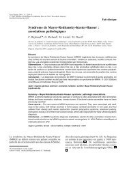
autoamputation should be considered when evaluating<br />
a prepubertal girl with a mobile pelvic mass.<br />
doi:10.1016/j.jpag.2008.01.053<br />
Isolated Transient Neonatal<br />
Clitoromegaly with<br />
Hyperandrogenism of<br />
Unknown Etiology<br />
Tania Dumont, MD, Nathalie Fleming, MD,<br />
FRCSC, and Amanda Black, MD, FRCSC<br />
Children’s Hospital of Eastern Ontario (University of Ottawa),<br />
Ottawa, Ontario, <strong>Canada</strong><br />
Background: Neonatal clitoromegaly is usually attributed<br />
to androgen stimulation secondary to congenital adrenal<br />
hyperplasia or in utero exposure. We present a case<br />
of transient, isolated neonatal clitoromegaly associated<br />
with increased androgen levels that spontaneously resolved<br />
when androgen levels normalized. No cause of<br />
the hyperandrogenism has been found.<br />
Case:M.S.wasdeliveredat25þ 5 weeks gestational<br />
age by caesarean section <strong>for</strong> antepartum hemorrhage<br />
secondary to placenta previa percreta. Birthweight was<br />
775 g.<br />
Clitoromegaly was first documented at three months<br />
of age. The clitoris measured 18 mm in length and 13<br />
mm in width. There was no labial hyperpigmentation<br />
or rugation and a separate single urethra, a patent vaginal<br />
opening, and a patent anus were seen. No pubic<br />
hair, axillary hair, acne, or breast tissue was seen.<br />
Initial bloodwork documented extremely high levels<br />
of free testosterone (117 pmol/L), DHEAS (O27 umol/<br />
L), and elevated 17-OH progesterone (19.3 nmol/L).<br />
Electrolytes were normal and congenital adrenal<br />
hyperplasia (CAH) was ruled out with a subsequent<br />
ACTH stimulation test (17-OHP 15.5 nmol/L, cortisol<br />
832 nmol/L). A normal 11-deoxycorticosterone level<br />
(12 ng/dl) ruled out an 11-beta-hydroxylase deficiency.<br />
A normal 46 XX karyotype with no mosaicism ruled<br />
out testicular feminization. Mullerian inhibiting substance<br />
(MIS) level was less than 0.1 ng/mL, indicating<br />
no ovotesticular tissue. An abdominal-pelvic ultrasound<br />
documented two normal ovaries, a uterus, and<br />
no testes. No adrenal gland abnormalities were seen.<br />
Beta-HCG was less than 1 IU/L. An initially elevated<br />
alpha-feta protein (2979 ug/L) and liver function tests<br />
were thought to be a result of total parental nutrition.<br />
These levels declined over time.<br />
In utero exposure to androgens was ruled out on maternal<br />
history. There was no maternal history of drug use,<br />
hyperandrogenism, or virilization during pregnancy.<br />
Multiple radiologic investigations during the pregnancy,<br />
per<strong>for</strong>med because of the placenta percreta, did not<br />
detect ovarian cysts or androgen secreting tumors.<br />
Two weeks after the initial bloodwork, androgen<br />
levels had decreased (free testosterone 30 pmol/L,<br />
DHEAS 20 umol/L). One month later, androgen<br />
levels had completely normalized without therapy<br />
(free testosterone 8 pmol/L, DHEAS 5.7 umol/L).<br />
At five months of age, the clitoris measured 10 mm<br />
in length and 5 mm in width.<br />
Comments: Excessive growth of the clitoris in a female<br />
infant suggests androgen exposure. In most cases,<br />
a source of androgen production is determined. This<br />
case demonstrates the occurrence of hyperandrogenism<br />
and clitoromegaly in an ex-premature female infant<br />
that resolved spontaneously without therapy. Only<br />
one other case of hyperandrogenism and clitoromegaly<br />
that resolved spontaneously in a preterm female infant<br />
has been reported. The clitoromegaly in that case was<br />
driven by high testosterone levels that were postulated<br />
to be a result of repeated blood transfusions from an<br />
adult male. The etiology of the hyperandrogenism<br />
and resultant clitoromegaly in our case is not known.<br />
doi:10.1016/j.jpag.2008.01.054<br />
<strong>MRKH</strong> with <strong>Vagina</strong>l<br />
Remnants: <strong>Options</strong> <strong>for</strong><br />
<strong>Creating</strong> a <strong>Functional</strong> <strong>Vagina</strong><br />
Nia L. Robinson, MD 1,3 ,<br />
and Marc R. Laufer, MD 1,2,3<br />
1 Department of Obstetrics and Gynecology, Brigham and<br />
Women’s Hospital;<br />
2 Division of Gynecology, Children’s<br />
Hospital Boston; and 3 Harvard Medical School, Boston,<br />
Massachusetts
NASPAG 22 nd Annual Clinical Meeting<br />
93<br />
Background: Mayer-Rokitansky-Kuster-Hauser<br />
(<strong>MRKH</strong>) syndrome, also known as mullerian aplasia<br />
or vaginal agenesis, typically manifests as primary<br />
amenorrhea and may present with pelvic pain.<br />
Although most often diagnosed in adolescence, the<br />
initial age of presentation varies and may be further<br />
complicated by misdiagnosis. This case reports an<br />
adult female with <strong>MRKH</strong> and native vaginal remnants<br />
and proposes a novel surgical approach which can be<br />
applied to an adolescent population.<br />
Case: A 43 year old non-sexually active woman was<br />
referred <strong>for</strong> primary amenorrhea. Thelarche and pubarche<br />
occurred normally; she complained of pelvic<br />
pain and a few episodes of bleeding of unclear etiology.<br />
Chromosomal analysis was 46 XX. MRI revealed normal<br />
ovaries, absent uterus, small cervix, and a small<br />
hematocolpos. A laparoscopy was notable <strong>for</strong> the presence<br />
of bilateral ovarian tissue and absence of a uterus<br />
or cervix. A small dimple 2e3 cm inferior to the urethra<br />
was visualized, however unable to be probed. The<br />
working diagnosis was vaginal agenesis.<br />
Treatment options <strong>for</strong> the creation of a functional vagina<br />
included utilization of dilators or a skin graft. The<br />
patient opted <strong>for</strong> a McIndoe vaginoplasty with a Repli<strong>for</strong>m<br />
graft. A transverse incision was made at the appropriate<br />
location of the introitus. During the dissection to<br />
create a vaginal space, two narrow lateral vaginal tracts<br />
were visualized. Each tract was lined by vaginal mucosa<br />
and contained mucus. Given concern about scar<br />
tissue <strong>for</strong>mation within the created vaginal space, the<br />
Repli<strong>for</strong>m graft was utilized instead of proceeding with<br />
placement of a mold without a graft. Routine post-<br />
McIndoe care was per<strong>for</strong>med and the patient was discharged<br />
home with a Mylex dilator in place. Two weeks<br />
post-operatively the patient complained of vaginal<br />
burning and tissue protruding from the vagina. Examination<br />
revealed sloughing of part of the Repli<strong>for</strong>m graft<br />
from the lateral areas of native vagina, with attachment<br />
of the graft anteriorly and posteriorly in the midline.<br />
The length of the vaginal canal was maintained and<br />
healthy appearing vaginal tissue was visualized distally.<br />
Excess graft tissue was excised. Long term treatment<br />
plan was continuous use of flexible vaginal<br />
dilators <strong>for</strong> at least 3 months.<br />
Comments: This case describes a novel ‘‘hybrid’’<br />
method to construct a functional vagina in a woman<br />
with <strong>MRKH</strong> and blind vaginal remnants. We utilized<br />
a graft to epithelialize the newly created vagina while<br />
the native vaginal tissue was able to proliferate and incorporate<br />
into the neovagina. This surgical approach<br />
is a viable option <strong>for</strong> adolescents with <strong>MRKH</strong> and<br />
the presence of native vaginal remnants, and should<br />
be considered in patients with <strong>MRKH</strong>, pelvic pain,<br />
and episodes of bleeding.<br />
doi:10.1016/j.jpag.2008.01.055<br />
Cervical Dysplasia and<br />
Associated Risk Factors<br />
in a Juvenile Detainee<br />
Population<br />
Vicki Scholten, MD, Sarah Gander, MD,<br />
Ivana Osswald, MD, and<br />
Richard VanWylick, MD, FRCPC<br />
Queen’s University, Kingston, Ontario, <strong>Canada</strong><br />
Background: <strong>Canada</strong> has recently licensed a new<br />
human papilloma virus (HPV) vaccine <strong>for</strong> use in adolescent<br />
females, aiming to decrease the incidence of<br />
oncogenic serotypes of HPV and associated cervical<br />
cancer. There are many known risk factors <strong>for</strong> cervical<br />
cancer, including infection with certain strains of<br />
HPV, a history of sexually transmitted infections,<br />
early age at first intercourse, multiple partners, and<br />
low socioeconomic status. The current screening<br />
method using the Papanicolaou (Pap) test is limited<br />
in detecting HPV infection and cervical cancer, with<br />
an overall sensitivity of only 70% when used routinely.<br />
We per<strong>for</strong>med a chart review to identify the<br />
juvenile detainee population as a high risk group <strong>for</strong><br />
HPV infection and subsequent risk <strong>for</strong> cervical cancer,<br />
and there<strong>for</strong>e an important target <strong>for</strong> primary<br />
HPV prevention.<br />
Methods: A retrospective chart review was conducted<br />
at the Sundance Detention Center in Kingston,<br />
Ontario, of all female detainees admitted between<br />
2003 and 2006. Data collection included the number<br />
of girls who had Pap tests, their results, and the incidence<br />
of sexually transmitted infections (STIs). In<strong>for</strong>mation<br />
about other risk factors, such as prostitution,<br />
number of sexual partners, and the use of contraception<br />
or protection, was also evaluated.<br />
Results: In total, 119 charts were reviewed. The<br />
patient ages ranged from 11 to 19 (average age 16).<br />
Of the 50 girls who had Pap smear results on record<br />
(42%), <strong>for</strong> a total of 57 Pap smears (as several girls<br />
had more than one), 46 (80.7%) were reported as normal,<br />
5 (8.8%) were reported as ASCUS (atypical<br />
squamous cells of unknown significance), and 6<br />
(10.5%) were reported as LSIL (low-grade squamous<br />
intraepithelial lesion). 1 girl was tested <strong>for</strong> HPV and<br />
was positive. The prevalence of STIs varied: 4% of<br />
those tested were positive <strong>for</strong> Gonorrhea, 10% <strong>for</strong><br />
Chlamydia, 32% <strong>for</strong> Bacterial vaginosis, 5% <strong>for</strong> Trichomonas,<br />
and there were no positive tests <strong>for</strong> Syphilis.<br />
77 of the girls were tested <strong>for</strong> HIV, Hepatitis B<br />
and Hepatitis C with no positive results, however 4<br />
girls had clinical evidence of Genital Herpes or Pelvic<br />
Inflammatory Disease. 75 (63%) of the girls reported