Placenta Previa, Placenta Accreta, and Vasa Previa
Placenta Previa, Placenta Accreta, and Vasa Previa
Placenta Previa, Placenta Accreta, and Vasa Previa

Create successful ePaper yourself
Turn your PDF publications into a flip-book with our unique Google optimized e-Paper software.
Clinical Expert Series<br />
Continuing medical education is available online at www.greenjournal.org<br />
<strong>Placenta</strong> <strong>Previa</strong>, <strong>Placenta</strong> <strong>Accreta</strong>, <strong>and</strong> <strong>Vasa</strong><br />
<strong>Previa</strong><br />
Yinka Oyelese, MD, <strong>and</strong> John C. Smulian, MD, MPH<br />
<strong>Placenta</strong> previa, placenta accreta, <strong>and</strong> vasa previa are important causes of bleeding in the second<br />
half of pregnancy <strong>and</strong> in labor. Risk factors for placenta previa include prior cesarean delivery,<br />
pregnancy termination, intrauterine surgery, smoking, multifetal gestation, increasing parity, <strong>and</strong><br />
maternal age. The diagnostic modality of choice for placenta previa is transvaginal ultrasonography,<br />
<strong>and</strong> women with a complete placenta previa should be delivered by cesarean. Small<br />
studies suggest that, when the placenta to cervical os distance is greater than 2 cm, women may<br />
safely have a vaginal delivery. Regional anesthesia for cesarean delivery in women with placenta<br />
previa is safe. Delivery should take place at an institution with adequate blood banking facilities.<br />
The incidence of placenta accreta is rising, primarily because of the rise in cesarean delivery<br />
rates. This condition can be associated with massive blood loss at delivery. Prenatal diagnosis by<br />
imaging, followed by planning of peripartum management by a multidisciplinary team, may help<br />
reduce morbidity <strong>and</strong> mortality. Women known to have placenta accreta should be delivered by<br />
cesarean, <strong>and</strong> no attempt should be made to separate the placenta at the time of delivery. The<br />
majority of women with significant degrees of placenta accreta will require a hysterectomy.<br />
Although successful conservative management has been described, there are currently insufficient<br />
data to recommend this approach to management routinely. <strong>Vasa</strong> previa carries a risk of<br />
fetal exsanguination <strong>and</strong> death when the membranes rupture. The condition can be diagnosed<br />
prenatally by ultrasound examination. Good outcomes depend on prenatal diagnosis <strong>and</strong><br />
cesarean delivery before the membranes rupture.<br />
(Obstet Gynecol 2006;107:927–41)<br />
Clinically important causes of bleeding in the<br />
second half of pregnancy <strong>and</strong> in labor include<br />
placenta previa, placenta accreta, <strong>and</strong> vasa previa.<br />
These conditions are associated with significant maternal<br />
<strong>and</strong> perinatal mortality <strong>and</strong> morbidity. This<br />
review presents a contemporary evidence-based approach<br />
to the management of these conditions.<br />
From the Division of Maternal Fetal Medicine, Department of Obstetrics,<br />
Gynecology, <strong>and</strong> Reproductive Sciences, UMDNJ-Robert Wood Johnson Medical<br />
School, Robert Wood Johnson University Hospital, New Brunswick, New Jersey.<br />
Corresponding author: Yinka Oyelese, MD, Division of Maternal Fetal Medicine,<br />
Department of Obstetrics, Gynecology, <strong>and</strong> Reproductive Sciences, UMDNJ-<br />
Robert Wood Johnson Medical School, 125 Paterson Street, New Brunswick, NJ<br />
08901-1977; e-mail: yinkamd@aol.com.<br />
© 2006 by The American College of Obstetricians <strong>and</strong> Gynecologists. Published<br />
by Lippincott Williams & Wilkins.<br />
ISSN: 0029-7844/06<br />
STUDY SELECTION<br />
We performed a thorough MEDLINE search using<br />
the keywords “placenta previa,” “placenta accreta,”<br />
“placenta percreta,” “placenta increta,” <strong>and</strong> “vasa<br />
previa.” Further articles were identified by crossreferencing.<br />
We were particularly interested in articles<br />
that dealt with the incidence, clinical implications,<br />
diagnosis, <strong>and</strong> management of these conditions.<br />
Only 5 r<strong>and</strong>omized controlled studies dealt with the<br />
management of placenta previa. None have specifically<br />
addressed the diagnosis or management of placenta<br />
accreta or vasa previa. The majority of publications<br />
on placenta previa, placenta accreta, <strong>and</strong> vasa<br />
previa are cohort or case-control studies, or case<br />
series/reports. Frequently no controls were available;<br />
a large proportion of studies were descriptive. Thus,<br />
the levels of evidence for most studies are II-2, II-3,<br />
VOL. 107, NO. 4, APRIL 2006 OBSTETRICS & GYNECOLOGY 927
<strong>and</strong> III. In cases where the literature does not provide<br />
convincing evidence for management of these conditions,<br />
we have described our experience <strong>and</strong> techniques.<br />
PLACENTA PREVIA<br />
Definition<br />
The term placenta previa refers to a placenta that<br />
overlies or is proximate to the internal os of the<br />
cervix. The placenta normally implants in the upper<br />
uterine segment. In placenta previa, the placenta<br />
either totally or partially lies within the lower uterine<br />
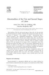
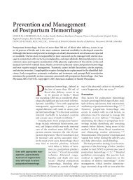
segment. Traditionally, placenta previa has been categorized<br />
into 4 types (Fig. 1):<br />
1. Complete placenta previa, where the placenta<br />
completely covers the internal os.<br />
2. Partial placenta previa, where the placenta<br />
partially covers the internal os. Thus, this scenario<br />
occurs only when the internal os is dilated to some<br />
degree.<br />
3. Marginal placenta previa, which just reaches the<br />
internal os, but does not cover it.<br />
4. Low-lying placenta, which extends into the lower<br />
uterine segment but does not reach the internal os.<br />
Clinical Importance<br />
Morbidities associated with placenta previa include<br />
antepartum bleeding (relative risk [RR] 9.81, 95%<br />
confidence interval [CI] 8.92–10.79), need for hysterectomy<br />
(RR 33.26, 95% CI 18.19–60.89), morbid<br />
adherence of the placenta, intrapartum hemorrhage<br />
(RR 2.48, 95% CI 1.55–3.98), postpartum hemorrhage<br />
(RR 1.86, 95% CI 1.46–2.36), blood transfusion<br />
(RR 10.05, 95% CI 7.45–13.55), septicemia (RR 5.5,<br />
95% CI 1.31–23.54), <strong>and</strong> thrombophlebitis (RR 4.85,<br />
95% CI 1.50–15.69). 1 In the United States, maternal<br />
mortality occurs in 0.03% of cases of placenta previa. 2<br />
Women with placenta previa may suffer considerable<br />
emotional distress because of recurrent bleeding<br />
along with hospitalizations that frequently occur in<br />
the second half of pregnancy. <strong>Placenta</strong> previa is also<br />
associated with an increase in preterm birth <strong>and</strong><br />
perinatal mortality <strong>and</strong> morbidity. 3 Finally, there is a<br />
higher rate of congenital malformations among<br />
women with placenta previa, although the precise<br />
mechanisms for these are unclear. 3<br />
Incidence <strong>and</strong> Risk Factors<br />
<strong>Placenta</strong> previa complicates approximately 0.3–0.5%<br />
of pregnancies. 2 A United States population-based<br />
study for the years 1979–1987 found the overall<br />
annual incidence of placenta previa to be 4.8 per<br />
1,000 deliveries (0.48%). 2 Several studies have found<br />
that risk factors for placenta previa include a history<br />
of prior cesarean delivery, 4 termination of pregnancy<br />
or uterine surgery, 4,5 smoking, 6 increasing age, 7 multiparity,<br />
7 cocaine, 8 <strong>and</strong> multiple pregnancy. 9 The likelihood<br />
of placenta previa increases in a dose-response<br />
fashion with a greater number of prior cesarean<br />
deliveries <strong>and</strong> with greater parity, with relative risks of<br />
previa rising from 4.5 (95% CI 3.6–5.5) in women<br />
with one prior cesarean delivery to 44.9 (95% CI<br />
13.5–149.5) in women with 4 prior cesarean deliveries,<br />
respectively. 7<br />
Pathophysiology<br />
It is unclear why some placentas implant in the lower<br />
uterine segment rather than in the fundus. 10 It does<br />
appear that uterine scarring may predispose to placental<br />
implantation in the lower segment. With the<br />
progression of pregnancy, more than 90% of these<br />
low-lying placentas identified early in pregnancy will<br />
appear to move away from the cervix <strong>and</strong> out of the<br />
lower uterine segment. Although the term “placental<br />
migration” has been used, most authorities do not<br />
believe the placenta moves. 10 Rather, it is felt that the<br />
placenta grows preferentially toward a better vascularized<br />
fundus (trophotropism), whereas the placenta<br />
overlying the less well vascularized cervix may undergo<br />
atrophy. 10 In some cases, this atrophy leaves<br />
vessels running through the membranes, unsupported<br />
by placental tissue or cord (vasa previa). 10 In cases<br />
where the atrophy is incomplete, a succenturiate lobe<br />
may develop. The apparent movement of the placenta<br />
may also be due to the development of the<br />
lower uterine segment. Contractions <strong>and</strong> cervical<br />
effacement <strong>and</strong> dilation that occur in the third trimester<br />
cause separation of the placenta, which leads to<br />
small amounts of bleeding. This bleeding may stimulate<br />
further uterine contractions, which, in turn, stimulates<br />
further placental separation <strong>and</strong> bleeding.<br />
Rarely are these initial bleeds a major problem,<br />
although they may be a reason for hospitalization. In<br />
labor, as the cervix dilates <strong>and</strong> effaces, there is usually<br />
placental separation <strong>and</strong> unavoidable bleeding.<br />
Diagnostic Approach<br />
The classic clinical presentation of placenta previa is<br />
painless bleeding in the late second trimester or early<br />
third trimester. However, some patients with placenta<br />
previa will experience painful bleeding, possibly the<br />
consequence of uterine contractions or placental separation,<br />
whereas others will experience no bleeding at<br />
all before labor. <strong>Placenta</strong> previa may also lead to an<br />
unstable lie or malpresentation in late pregnancy.<br />
The majority of cases of placenta previa are<br />
928 Oyelese <strong>and</strong> Smulian <strong>Placenta</strong> <strong>Previa</strong>, <strong>Accreta</strong>, <strong>and</strong> <strong>Vasa</strong> <strong>Previa</strong> OBSTETRICS & GYNECOLOGY
diagnosed during routine sonography in asymptomatic<br />
women, usually during the second trimester.<br />
Although transabdominal sonography is frequently<br />
used for placental location, this technique lacks some<br />
precision in diagnosing placenta previa. 11,12 Numerous<br />
studies have demonstrated the accuracy of transvaginal<br />
sonography for the diagnosis of placenta<br />
previa, uniformly finding that transvaginal sonography<br />
is superior to transabdominal sonography for this<br />
indication (Fig. 2). 11,12 False-positive <strong>and</strong> -negative<br />
rates for the diagnosis of placenta previa using transabdominal<br />
sonography range from 2% to 25%. 11 A<br />
study by Smith <strong>and</strong> colleagues 11 of 131 women believed<br />
to have a placenta previa by transabdominal<br />
sonography found that anatomic l<strong>and</strong>marks crucial<br />
for accurate diagnosis were poorly recognized in 50%<br />
of cases. In 26% of the cases of suspected placenta<br />
previa, the initial diagnosis was changed after transvaginal<br />
sonography because it was incorrect.<br />
The superiority of transvaginal sonography over<br />
transabdominal sonography can be attributed to several<br />
factors:<br />
1. The transabdominal approach requires bladder<br />
filling, which results in approximation of the anterior<br />
<strong>and</strong> posterior walls of the lower uterine segment, with<br />
the result that a normally situated placenta may<br />
falsely appear to be a previa.<br />
2. Vaginal probes are closer to the region of<br />
interest, <strong>and</strong> typically of higher frequency, <strong>and</strong> therefore<br />
obtain higher resolution images than transabdominal<br />
probes.<br />
3. The internal cervical os <strong>and</strong> the lower placental<br />
edge frequently cannot be imaged adequately by the<br />
transabdominal approach. The position of the internal<br />
os is assumed rather than actually seen.<br />
4. The fetal head may obscure views of the lower<br />
placental edge when using the transabdominal approach,<br />
<strong>and</strong> a posterior placenta previa may not be<br />
adequately imaged.<br />
The improved accuracy of transvaginal sonography<br />
over transabdominal sonography means that<br />
fewer false-positive diagnoses are made; thus, the rate<br />
of placenta previa is significantly lower when using<br />
transvaginal sonography than when using transabdominal<br />
sonography. 11,13 Lauria <strong>and</strong> colleagues, 13 performing<br />
routine transvaginal sonography, found an<br />
incidence of placenta previa of only 1.1% at 15–20<br />
weeks, considerably lower than the second trimester<br />
placenta previa incidence of 15–20% reported by<br />
previous investigators using transabdominal sonography.<br />
14 Numerous studies have demonstrated the<br />
safety of transvaginal sonography for the diagnosis of<br />
placenta previa. 12,15 Importantly, this imaging tech-<br />
nique does not lead to an increase in bleeding. 15 This<br />
is for 2 main reasons: 1) the vaginal probe is introduced<br />
at an angle that places it against the anterior<br />
fornix <strong>and</strong> anterior lip of the cervix, unlike a digital<br />
examination, where articulation of the h<strong>and</strong> allows<br />
introduction of the examining finger through the<br />
cervix (Fig. 3); 15 2) the optimal distance for visualization<br />
of the cervix is 2–3 cm away from the cervix, so<br />
the probe is generally not advanced sufficiently to<br />
make contact with the placenta. 15 Nonetheless, the<br />
examination should be performed by personnel experienced<br />
in transvaginal sonography, <strong>and</strong> the transvaginal<br />
probe should always be inserted carefully,<br />
with the examiner looking at the monitor to avoid<br />
putting the probe in the cervix.<br />
Translabial sonography has been suggested as an<br />
alternative to transvaginal sonography <strong>and</strong> has been<br />
shown to be superior to transabdominal sonography<br />
for placental location. 16 However, because transvaginal<br />
sonography is accurate, safe, <strong>and</strong> well tolerated, it<br />
should be the imaging modality of choice.<br />
Several studies have demonstrated that the majority<br />
of placentas that are in the lower uterine<br />
segment in the second trimester will no longer be in<br />
the region of the cervix by the time of delivery (Table<br />
1). 13,17–21 Persistence to term can be predicted based<br />
on whether or not the placenta overlaps the internal<br />
os in the second trimester, <strong>and</strong> to what extent. 13,17–21<br />
The later in pregnancy that placenta previa is diagnosed,<br />
the higher the likelihood of persistence to<br />
delivery. 22 Women who at 20 weeks have a low-lying<br />
placenta that does not overlie the internal os will not<br />
have a placenta previa at term <strong>and</strong> need no further<br />
sonographic examinations for placental location.<br />
However, the presence of a low-lying placenta in the<br />
second trimester is a risk factor for developing a vasa<br />
previa, <strong>and</strong> therefore, in these cases, a sonogram<br />
should be performed later in pregnancy to exclude<br />
that condition.<br />
Management<br />
In the past, suspected placenta previa was managed<br />
by vaginal examination <strong>and</strong> immediate cesarean delivery<br />
if placenta previa was confirmed. It was believed<br />
that the first bleed (usually occurring in the<br />
early third trimester) would lead to maternal death.<br />
However, MacAfee 23 showed that, in the absence of<br />
interference, this almost never happened, <strong>and</strong> that the<br />
high perinatal mortality from placenta previa was<br />
primarily due to prematurity, which could be reduced<br />
considerably by conservative expectant management<br />
<strong>and</strong> delivery as close to term as possible.<br />
VOL. 107, NO. 4, APRIL 2006 Oyelese <strong>and</strong> Smulian <strong>Placenta</strong> <strong>Previa</strong>, <strong>Accreta</strong>, <strong>and</strong> <strong>Vasa</strong> <strong>Previa</strong> 929
Women who present with bleeding in the second<br />
half of pregnancy should have a sonographic examination<br />
(preferably by the transvaginal approach) for<br />
placental location prior to any attempt to perform a<br />
digital examination. Digital vaginal examination with<br />
a placenta previa may provoke catastrophic hemorrhage<br />
<strong>and</strong> should not be performed.<br />
It is reasonable to hospitalize women with placenta<br />
previa while they are having an acute bleeding<br />
episode or uterine contractions. One to two wide-bore<br />
intravenous cannulas should be inserted <strong>and</strong> blood<br />
taken for a full blood count <strong>and</strong> type <strong>and</strong> screen. In<br />
the absence of massive bleeding or other complications,<br />
coagulation studies are not helpful. The blood<br />
bank must be capable of making available at least 4<br />
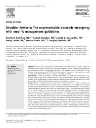
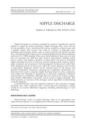
Fig. 2. Transvaginal sonogram of a complete placenta<br />
previa (PP). Note that both the placenta <strong>and</strong> the internal<br />
cervical os (arrow) are clearly depicted. A, anterior lip of<br />
cervix; P, posterior lip of cervix. The placenta just overlaps<br />
the internal os. One can see how this could become a<br />
partial placenta previa covering just the anterior lip of the<br />
cervix if cervical dilation were to occur.<br />
Oyelese. <strong>Placenta</strong> <strong>Previa</strong>, <strong>Accreta</strong>, <strong>and</strong> <strong>Vasa</strong> <strong>Previa</strong>. Obstet<br />
Gynecol 2006.<br />
Fig. 1. Types of placenta previa.<br />
Illustration: John Yanson.<br />
Oyelese. <strong>Placenta</strong> <strong>Previa</strong>, <strong>Accreta</strong>,<br />
<strong>and</strong> <strong>Vasa</strong> <strong>Previa</strong>. Obstet Gynecol<br />
2006.<br />
units of compatible packed red blood cells <strong>and</strong> coagulation<br />
factors at short notice. Rh immune globulin<br />
should be administered to Rh-negative women. A<br />
Kleihauer-Bettke test for quantification of fetal-maternal<br />
transfusion should also be performed in Rhnegative<br />
women because the mother may require<br />
increased doses of Rh immune globulin.<br />
Small studies have suggested a benefit of tocolytic<br />
therapy for women with placenta previa who are<br />
having contractions. 24,25 Contractions may lead to<br />
Fig. 3. Diagram demonstrating the technique for transvaginal<br />
sonography of placenta previa. T, transvaginal transducer;<br />
A, anterior lip of cervix; P, posterior lip of cervix.<br />
Complete placenta previa is shown completely covering<br />
the internal os (arrow). The transvaginal transducer lies<br />
within the vagina, about 2 cm from the anterior lip of the<br />
cervix. The angle between the transducer <strong>and</strong> the cervical<br />
canal is 35 degrees, demonstrating why the probe does not<br />
enter the cervix. Illustration: John Yanson.<br />
Oyelese. <strong>Placenta</strong> <strong>Previa</strong>, <strong>Accreta</strong>, <strong>and</strong> <strong>Vasa</strong> <strong>Previa</strong>. Obstet<br />
Gynecol 2006.<br />
930 Oyelese <strong>and</strong> Smulian <strong>Placenta</strong> <strong>Previa</strong>, <strong>Accreta</strong>, <strong>and</strong> <strong>Vasa</strong> <strong>Previa</strong> OBSTETRICS & GYNECOLOGY
Table 1. Studies of Second Trimester Transvaginal Sonography in the Prediction of <strong>Placenta</strong> <strong>Previa</strong> at<br />
Delivery<br />
Author<br />
Gestational Age at<br />
Sonogram (wk)<br />
Number of<br />
Women<br />
cervical effacement <strong>and</strong> changes in the lower uterine<br />
segment, provoking bleeding which, in turn, stimulates<br />
contractions, creating a vicious cycle. Sharma<br />
<strong>and</strong> colleagues 24 carried out a small r<strong>and</strong>omized study<br />
using the �-adrenergic ritodrine <strong>and</strong> found a significant<br />
prolongation in pregnancy <strong>and</strong> higher birth<br />
weights in women treated with ritodrine when compared<br />
with women treated with placebo. Similarly,<br />
Besinger <strong>and</strong> colleagues, 25 in a retrospective study,<br />
found that use of intravenous magnesium sulfate<br />
<strong>and</strong>/or oral or subcutaneous terbutaline in women<br />
with symptomatic placenta previa was associated with<br />
greater prolongation of pregnancy <strong>and</strong> higher birth<br />
weight than in women who were not treated with<br />
tocolytics. Thus, cautious use of tocolytics in women<br />
with placenta previa who are having contractions,<br />
when both mother <strong>and</strong> fetus are stable, appears<br />
reasonable.<br />
Steroids should be administered in women between<br />
24 <strong>and</strong> 34 weeks of gestation, generally at the<br />
time of admission for bleeding, to promote fetal lung<br />
maturation. The patient <strong>and</strong> her family should have a<br />
neonatology consultation so that the management of<br />
the infant after birth may be discussed. In women<br />
who have a history of cesarean delivery or uterine<br />
surgery, detailed sonography should be performed to<br />
exclude placenta accreta. Because prematurity is the<br />
main cause of perinatal mortality associated with<br />
placenta previa, it is desirable to prolong gestation as<br />
long as safely possible. Therefore, before 32 weeks of<br />
gestation, moderate-to-severe bleeding when there is<br />
no maternal or fetal compromise may be managed<br />
aggressively with blood transfusions, rather than resorting<br />
to delivery. 26 When the patient has had no<br />
further bleeding for 48 hours, she may be considered<br />
for discharge as long as there are appropriate home<br />
conditions to allow outpatient management. Specifically,<br />
the patient should have access to a telephone,<br />
have a responsible adult <strong>and</strong> transportation available<br />
at all times, <strong>and</strong> must live within reasonable distance<br />
of a hospital. She should return to the hospital imme-<br />
Incidence of <strong>Placenta</strong> <strong>Previa</strong><br />
at First- or Second-Trimester<br />
Sonography [n (%)]<br />
Incidence at<br />
Delivery [n (%)]<br />
Becker 17<br />
20–23 8,650 99 (1.1) 28 (0.32)<br />
Taipale21 18–23 3,969 57 (1.5) 5 (0.14)<br />
Hill19 9–13 1,252 77 (6.2) 4 (0.31)<br />
Mustafa20 20–24 203 8 (3.9) 4 (1.9)<br />
Lauria13 15–20 2,910 36 (1.2) 5 (0.17)<br />
Rosati18 10–16 2,158 105 (4.9) 8 (0.37)<br />
diately if she experiences bleeding or contractions.<br />
Although there are no data to support the efficacy of<br />
avoidance of intercourse <strong>and</strong> excessive activity, common<br />
sense suggests that these should be avoided. Similarly,<br />
bedrest is often advised, but there is no evidence<br />
that demonstrates that this practice is beneficial.<br />
Outpatient Versus Inpatient Management<br />
Whether women with placenta previa should be<br />
managed as inpatients or outpatients has been a<br />
matter of controversy. A few retrospective studies<br />
have addressed this issue <strong>and</strong> have found no difference<br />
in outcomes, whether patients were managed in<br />
hospital or at home, <strong>and</strong> found that outpatient management<br />
may be associated with lower costs. 27,28<br />
These studies concluded that outpatient management<br />
of selected women with placenta previa was safe.<br />
However, in another retrospective study, D’Angelo<br />
<strong>and</strong> Irwin 29 found an increase in perinatal mortality,<br />
lower gestational age at delivery, increased neonatal<br />
hospitalization duration, <strong>and</strong> neonatal morbidity<br />
among women who were managed as outpatients<br />
when compared with those managed expectantly as<br />
inpatients. In one of the few prospective r<strong>and</strong>omized<br />
studies dealing with placenta previa, Wing et al 30<br />
r<strong>and</strong>omized 53 women with placenta previa at gestational<br />
ages between 24 <strong>and</strong> 36 weeks, who had been<br />
initially stabilized in hospital, to inpatient or outpatient<br />
management <strong>and</strong> found no significant difference<br />
in outcomes. Thus, women who are stable <strong>and</strong> asymptomatic,<br />
<strong>and</strong> who are reliable <strong>and</strong> have quick access to<br />
hospital, may be considered for outpatient management.<br />
Cerclage<br />
Arias 31 r<strong>and</strong>omized 25 women who were admitted to<br />
hospital with symptomatic placenta previa at 24–30<br />
weeks gestation to cerclage or no cerclage <strong>and</strong> found<br />
a higher mean birth weight <strong>and</strong> gestational age at<br />
delivery <strong>and</strong> fewer neonatal complications in the<br />
cerclage group. Women with cerclage had lower<br />
hospitalization costs <strong>and</strong> fewer bleeding episodes.<br />
VOL. 107, NO. 4, APRIL 2006 Oyelese <strong>and</strong> Smulian <strong>Placenta</strong> <strong>Previa</strong>, <strong>Accreta</strong>, <strong>and</strong> <strong>Vasa</strong> <strong>Previa</strong> 931
However, in a later study, Cobo <strong>and</strong> colleagues 32<br />
r<strong>and</strong>omized 39 women with placenta previa at 24–30<br />
weeks to cerclage or no cerclage <strong>and</strong> found no<br />
statistically significant differences in gestational age at<br />
delivery, prolongation of pregnancy, or in amount of<br />
blood lost between the 2 groups. In view of the lack of<br />
convincing data to support cerclage in these women,<br />
cerclage should not be performed for treatment of<br />
placenta previa.<br />
Mode of Delivery<br />
There is consensus that a placenta previa that totally<br />
or partially overlies the internal cervical os requires<br />
delivery by cesarean. However, the mode of delivery<br />
when the placenta lies in proximity to the internal os<br />
is more controversial. Three small retrospective studies<br />
using transvaginal or translabial sonography have<br />
evaluated the role of ultrasonography in determining<br />
the optimal mode of delivery for women whose<br />
placentas were in proximity to the internal cervical<br />
os. 33–35 All 3 studies found that women in whom the<br />
distance between the lower placental edge <strong>and</strong> the<br />
internal cervical os was greater than 2 cm could safely<br />
have a vaginal delivery. Conversely, among women<br />
with a placenta-internal os distance less than 2 cm, the<br />
overwhelming majority required cesarean delivery,<br />
usually for bleeding. However, in none of these<br />
studies were the clinicians blinded to the results of the<br />
scan, <strong>and</strong> this may have influenced obstetric management.<br />
Furthermore, these studies had relatively small<br />
numbers. Nonetheless, the studies suggest that<br />
women with placenta previa should have a transvaginal<br />
sonogram in the late third trimester, <strong>and</strong> that<br />
those with a placental edge to internal os distance of<br />
less than 2 cm should be delivered by cesarean. It has<br />
been our experience that women with a placentainternal<br />
os distance of less than 2 cm who undergo a<br />
trial of labor almost invariably experience significant<br />
bleeding during labor, necessitating cesarean delivery.<br />
Consequently, it is now our practice to deliver<br />
these women by elective cesarean. Women whose<br />
placentas are 2 cm or more from the os undergo a<br />
normal labor. It is important though to realize that, in<br />
women with a placenta that extends into the noncontractile<br />
lower uterine segment who have a vaginal<br />
delivery, there is potential for postpartum hemorrhage.<br />
When there is an anterior placenta previa, there is<br />
a considerable likelihood of incising through the<br />
placenta during delivery. This could lead to significant<br />
maternal <strong>and</strong> fetal blood loss <strong>and</strong> also to difficulty<br />
with delivery, but this rarely constitutes a significant<br />
problem. Alternative strategies have been<br />
proposed <strong>and</strong> used to avoid incision into the placenta.<br />
These include use of a fundal vertical uterine incision,<br />
especially in women who have no desire for further<br />
childbearing. 36 This may especially be useful when<br />
there is a complete placenta previa with a fetal<br />
transverse lie with the fetal back down. Sonography<br />
before surgery for placental location enables the<br />
surgeon to plan the most appropriate incision. 36 Generally,<br />
we perform a lower segment transverse uterine<br />
incision, incising the placenta when it is unavoidable.<br />
The infant is delivered as rapidly as possible, <strong>and</strong> the<br />
cord is clamped immediately to avoid hemorrhage<br />
from fetal vessels.<br />
Timing of Delivery<br />
As gestational age advances, there is an increased risk<br />
of significant bleeding, necessitating delivery. It is<br />
preferable to perform a cesarean delivery for placenta<br />
previa under controlled scheduled conditions rather<br />
than as an emergency. Therefore, in a stable patient,<br />
it is reasonable to perform a cesarean delivery at<br />
36–37 weeks of gestation, after documentation of fetal<br />
lung maturity by amniocentesis. If the amniocentesis<br />
does not demonstrate lung maturity, we deliver the<br />
women by elective cesarean at 38 weeks, without<br />
repeating the amniocentesis, if they remain stable, or<br />
earlier if bleeding occurs or the patient goes into<br />
labor.<br />
Anesthesia for Delivery<br />
In the past, it was generally recommended that cesarean<br />
deliveries for placenta previa be performed under<br />
general anesthetic. 37 It was believed that this allowed<br />
more controlled surgery. At least 2 studies, including<br />
a prospective r<strong>and</strong>omized trial, have found that cesarean<br />
deliveries for placenta previa performed under<br />
general anesthetic were associated with significantly<br />
greater estimated blood loss <strong>and</strong> greater requirements<br />
for blood transfusion than those performed under<br />
regional anesthesia, 38,39 possibly due to increased uterine<br />
relaxation associated with general anesthetic. Otherwise,<br />
there was no difference in the incidence of<br />
intraoperative or anesthesia complications between<br />
regional <strong>and</strong> general anesthesia. A survey of anesthesiologists<br />
in the United Kingdom found a wide variety<br />
of opinions regarding whether general or regional<br />
anesthesia should be used for cesarean for placenta<br />
previa. However, anesthesiologists who did more<br />
obstetric anesthesia were more likely to employ regional<br />
anesthesia. 40 Another U.K. survey found that,<br />
60% of the time, anesthesiologists used regional anesthesia<br />
for cesarean for placenta previa. 37 At our<br />
institution, we generally perform cesarean deliveries<br />
for placenta previa under regional anesthesia.<br />
932 Oyelese <strong>and</strong> Smulian <strong>Placenta</strong> <strong>Previa</strong>, <strong>Accreta</strong>, <strong>and</strong> <strong>Vasa</strong> <strong>Previa</strong> OBSTETRICS & GYNECOLOGY
PLACENTA ACCRETA<br />
Definition<br />
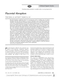
<strong>Placenta</strong> accreta refers to a placenta that is abnormally<br />
adherent to the uterus (Fig. 4). When the<br />
placenta invades the myometrium, the term placenta<br />
increta is used, whereas placenta percreta refers to a<br />
placenta that has invaded through the myometrium<br />
<strong>and</strong> serosa, sometimes into adjacent organs, such as<br />
the bladder. The term placenta accreta is often used<br />
interchangeably as a general term to describe all of<br />
these conditions.<br />
Clinical Significance<br />
<strong>Placenta</strong> accreta may lead to massive obstetric hemorrhage,<br />
resulting in such complications as disseminated<br />
intravascular coagulopathy, need for hysterectomy,<br />
surgical injury to the ureters, bladder, <strong>and</strong> other<br />
viscera, adult respiratory distress syndrome, renal<br />
failure, <strong>and</strong> even death. 41,42 The average blood loss at<br />
delivery in women with placenta accreta is 3,000–<br />
5,000 mL. 41 Indeed, in several centers, placenta accreta<br />
has become the leading reason for cesarean<br />
hysterectomy. 43 Rarely, placenta accreta may lead to<br />
spontaneous uterine rupture in the second or third<br />
trimester, resulting in intraperitoneal hemorrhage, a<br />
life-threatening emergency. 44 Minor degrees of placenta<br />
accreta may occur, which may lead to slightly<br />
heavier postpartum bleeding, but may not require the<br />
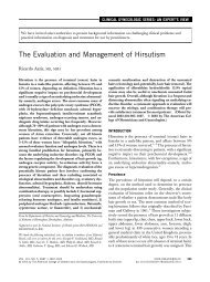
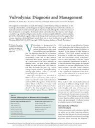
Fig. 4. Hysterectomy specimen demonstrating placenta<br />
accreta. This placenta accreta was diagnosed prenatally.<br />
The placenta (p) has invaded the myometrium (arrow) <strong>and</strong><br />
after hysterectomy could not be separated from the uterus.<br />
There were no planes of demarcation between placenta<br />
<strong>and</strong> myometrium. cx, cervix; f, uterine fundus; c, umbilical<br />
cord.<br />
Oyelese. <strong>Placenta</strong> <strong>Previa</strong>, <strong>Accreta</strong>, <strong>and</strong> <strong>Vasa</strong> <strong>Previa</strong>. Obstet<br />
Gynecol 2006.<br />
aggressive management that is often employed with<br />
more extensive placenta accreta.<br />
Incidence <strong>and</strong> Risk Factors<br />
Miller <strong>and</strong> colleagues, 45 reviewing 155,670 deliveries<br />
at their hospital between 1985 <strong>and</strong> 1994, found that<br />
62 (one in 2,510) were complicated by placenta<br />
accreta. The incidence of placenta accreta is increasing,<br />
primarily as a consequence of rising cesarean<br />
delivery rates. A recent study by Wu <strong>and</strong> colleagues 46<br />
looking at placenta accreta over a 20-year period<br />
(1982–2002) found an incidence of 1 in 533 pregnancies<br />
at their institution. <strong>Placenta</strong> accreta occurs most<br />
frequently in women with one or more prior cesarean<br />
deliveries who have a placenta previa in the current<br />
pregnancy. Clarke <strong>and</strong> colleagues 47 found that, in the<br />
presence of a placenta previa, the risk of having<br />
placenta accreta increased from 24% in women with<br />
one prior cesarean delivery to 67% in women with 3<br />
or more prior cesareans.<br />
It has been proposed that the abnormality of the<br />
placental-uterine interface in women with placenta<br />
accreta will lead to leakage of fetal alpha-fetoprotein<br />
into the maternal circulation, resulting in elevated<br />
levels of maternal serum alpha-fetoprotein<br />
(MSAFP). 48 Kupferminc <strong>and</strong> colleagues, 49 reviewing<br />
44 cases of women who had cesarean hysterectomies,<br />
found that 9 of the 20 (45%) with placenta accreta had<br />
elevated MSAFP levels (between 2.7 <strong>and</strong> 40.3 multiples<br />
of the median [MoMs]), whereas the controls all<br />
had MSAFP levels within normal limits (� 2.0<br />
MoMs). Similarly, Zelop <strong>and</strong> colleagues 48 found elevated<br />
second-trimester MSAFP levels (between 2.3<br />
<strong>and</strong> 5.5 MoMs) in 45% of 11 women with placenta<br />
accreta, whereas none of the controls who had placenta<br />
previa without accreta had MSAFP elevations.<br />
Although these studies are small, they suggest that<br />
women with elevated MSAFP levels with no other<br />
obvious cause should be considered at increased risk<br />
of placenta accreta.<br />
Pathophysiology<br />
<strong>Placenta</strong> accreta is thought to be due to an absence or<br />
deficiency of Nitabuch’s layer or the spongiosus layer<br />
of the decidua. 10 Benirschke <strong>and</strong> Kaufmann 10 suggest<br />
that this is the consequence of failure of reconstitution<br />
of the endometrium/decidua basalis after repair of a<br />
cesarean incision. Histology usually shows that the<br />
trophoblast has invaded the myometrium without<br />
intervening decidua. 10 This becomes a problem at<br />
delivery when the placenta does not separate <strong>and</strong><br />
massive bleeding ensues (Fig. 5).<br />
VOL. 107, NO. 4, APRIL 2006 Oyelese <strong>and</strong> Smulian <strong>Placenta</strong> <strong>Previa</strong>, <strong>Accreta</strong>, <strong>and</strong> <strong>Vasa</strong> <strong>Previa</strong> 933
Fig. 5. Grayscale sonogram of placenta percreta. Note the<br />
prominent placental lacunae (arrows) giving the lower<br />
uterine segment a “moth-eaten” appearance. The diagnosis<br />
was confirmed at delivery. p, placenta; h, fetal head; b,<br />
bladder.<br />
Oyelese. <strong>Placenta</strong> <strong>Previa</strong>, <strong>Accreta</strong>, <strong>and</strong> <strong>Vasa</strong> <strong>Previa</strong>. Obstet<br />
Gynecol 2006.<br />
Diagnostic Approach<br />
It is important to make the diagnosis of placenta<br />
accreta prenatally because this allows effective management<br />
planning to minimize morbidity. This diagnosis<br />
is usually made by ultrasonography or magnetic<br />
resonance imaging (MRI). <strong>Placenta</strong> accreta should be<br />
suspected in women who have both a placenta previa<br />
<strong>and</strong> a history of cesarean delivery or other uterine<br />
surgery. 41,50 Vigilance is particularly indicated when<br />
the placenta is anterior <strong>and</strong> overlies the cesarean scar.<br />
Ultrasonography<br />
Several studies have documented the efficacy of<br />
sonography in the diagnosis of placenta accreta. 50–52<br />
Comstock, 50 in a recent review, described the sonographic<br />
features suggestive of placenta accreta. These<br />
include irregularly shaped placental lacunae (vascular<br />
spaces) within the placenta, thinning of the myometrium<br />
overlying the placenta, loss of the retroplacental<br />
“clear space,” protrusion of the placenta into the<br />
bladder, increased vascularity of the uterine serosabladder<br />
interface, <strong>and</strong>, on Doppler ultrasonography,<br />
turbulent blood flow through the lacunae (Figs. 5, 6). 51<br />
In a previous study, Comstock <strong>and</strong> colleagues 51 had<br />
found, at 15–20 weeks of gestation, that the presence<br />
of lacunae in the placenta was the most predictive<br />
sonographic sign of placenta accreta, with a sensitivity<br />
of 79% <strong>and</strong> a positive predictive value of 92%. These<br />
lacunae may give the placenta a “moth-eaten” or<br />
“Swiss cheese” appearance (Fig. 5). The risk of placenta<br />
accreta increases with an increased number of<br />
lacunae. 52 Obliteration of the retroplacental “clear<br />
space,” which is the finding most commonly thought<br />
to be associated with placenta accreta, had only a 57%<br />
sensitivity <strong>and</strong> a false-positive rate of 48.4%. 51 After 20<br />
weeks of gestation, the sensitivity of these findings<br />
increased, with values of 93% <strong>and</strong> 80% for lacunae<br />
<strong>and</strong> obliteration of the retroplacental clear space,<br />
respectively. 51 The investigators found that a sonographic<br />
appearance of apparent bulging into the<br />
bladder may occur in cases of placenta accreta without<br />
increta or percreta. 50 Thus, this finding may not<br />
reliably differentiate between cases in which the<br />
placenta has invaded the bladder <strong>and</strong> cases in which<br />
it has not. 50<br />
Power <strong>and</strong> color Doppler are often used for the<br />
diagnosis of placenta accreta, demonstrating turbulent<br />
flow through placental lacunae (Fig. 6). 53 However, in<br />
the majority of cases, this imaging modality does not<br />
significantly improve the diagnosis over that achieved<br />
by grayscale sonography alone. Thus, in the majority<br />
of clinical situations, Doppler should not be the<br />
primary technique used to diagnose placenta accreta.<br />
A retrospective review of images of first-trimester<br />
sonograms of cases of placenta accreta found that, in<br />
all the cases, the gestational sac was in the lower<br />
uterine segment <strong>and</strong> that the gestational sac was<br />
abnormally close to the uterine scar, suggesting attachment<br />
to the scar. 54 This finding in the first trimester,<br />
therefore, in women with a prior cesarean delivery,<br />
should lead to a suspicion of placenta accreta.<br />
Magnetic Resonance Imaging<br />
Several articles have described the use of MRI in the<br />
diagnosis of placenta accreta. 55–57 Most were retrospective,<br />
limited to a few cases, <strong>and</strong> lacked pathologic<br />
correlation. 56 Although most studies have suggested<br />
reasonable diagnostic accuracy of MRI for placenta<br />
accreta, it appears that MRI is no more sensitive than<br />
ultrasonography for diagnosing placenta accreta. 50,57<br />
Ultrasonography is readily available in most centers,<br />
whereas MRI is costly <strong>and</strong> relatively inaccessible.<br />
Therefore, at the present time, sonography is the<br />
primary imaging modality for diagnosing accreta.<br />
However, when there is a posterior placenta accreta,<br />
ultrasonography may be less than adequate, <strong>and</strong> MRI<br />
934 Oyelese <strong>and</strong> Smulian <strong>Placenta</strong> <strong>Previa</strong>, <strong>Accreta</strong>, <strong>and</strong> <strong>Vasa</strong> <strong>Previa</strong> OBSTETRICS & GYNECOLOGY
may be superior to ultrasonography for this specific<br />
indication. 50,57<br />
Therapeutic Approach<br />
It is generally accepted that placenta accreta is ideally<br />
treated by total abdominal hysterectomy. In addition,<br />
there is almost universal consensus that the placenta<br />
should be left in place; attempts to detach the placenta<br />
frequently result in massive hemorrhage. However,<br />
the physician should be aware that focal placenta<br />
accreta may exist that may not require such aggressive<br />
therapy. It is better to perform surgery for<br />
placenta accreta under elective, controlled conditions<br />
rather than as an emergency without adequate preparation.<br />
Therefore, scheduled delivery at 36–37<br />
weeks of gestation, after documentation of fetal lung<br />
maturity by amniocentesis, seems reasonable. If amniocentesis<br />
fails to document fetal lung maturity, the<br />
patient, if stable, should be delivered by cesarean by<br />
38 weeks, or earlier, if she bleeds or goes into labor. A<br />
study comparing emergency with elective peripartum<br />
hysterectomy found that women in the emergency<br />
hysterectomy group had greater intraoperative blood<br />
loss, were more likely to have intraoperative hypotension,<br />
<strong>and</strong> were more likely to receive blood transfusions<br />
than women who had elective obstetric hysterectomies.<br />
58<br />
Prevention of complications ideally requires a<br />
multidisciplinary team approach. The patient<br />
should be counseled preoperatively about the need<br />
for hysterectomy <strong>and</strong> the likely requirement for<br />
transfusion of blood <strong>and</strong> blood products. 59 Although<br />
scheduled delivery should be the goal,<br />
contingency plans should be made for possible<br />
emergent delivery if necessary. It is important that<br />
delivery be performed by an experienced obstetric<br />
surgeon, with other surgical specialties such as<br />
urology <strong>and</strong> gynecological oncology readily available<br />
if required. It is not unusual for the lower<br />
uterine segment to be markedly enlarged <strong>and</strong> vascular,<br />
with distortion of normal anatomy <strong>and</strong> tissue<br />
planes. Preoperative cystoscopy with placement of<br />
ureteric stents may help prevent urinary tract injury.<br />
At our center, we usually insert a 3-way Foley<br />
catheter in the bladder via the urethra, allowing<br />
simultaneous irrigation <strong>and</strong> drainage of the bladder<br />
during the surgery. In instances where tissue plane<br />
identification is difficult because of adhesions or the<br />
invasive placenta, we have the option of distending<br />
the bladder to aid in its identification <strong>and</strong> then<br />
emptying it to avoid injury while we proceed with<br />
surgery. Use of a vertical skin incision facilitates<br />
adequate exposure. Generally, a vertical incision in<br />
the uterus allows delivery of the infant while avoiding<br />
the placenta. There should be no attempt to<br />
detach the placenta from the uterine wall. The<br />
edges of the uterine incision should be oversewn for<br />
hemostasis, after which a total abdominal hysterectomy<br />
should be performed. Although some have<br />
advocated supracervical hysterectomy, in the majority<br />
of cases the lower uterine segment is involved<br />
in the morbid adhesion <strong>and</strong> therefore needs to be<br />
removed.<br />
It is important to minimize blood loss <strong>and</strong> ensure<br />
that the blood lost is replaced promptly <strong>and</strong> adequately.<br />
59 Because of the large volumes of blood that are<br />
typically lost, as well as the replacement with packed<br />
red blood cells, these patients are at risk of disseminated<br />
intravascular coagulopathy. Thus, coagulation<br />
factors should be replaced liberally, adequately, <strong>and</strong><br />
quickly. Donor-directed blood transfusions <strong>and</strong> use of<br />
a blood cell saver may reduce the need for transfusion<br />
with blood from another donor. 59 Some centers use<br />
acute normovolemic hemodilution to reduce the need<br />
for blood. 41 The role of experienced anesthesiology<br />
personnel who are skilled in obstetric anesthesia<br />
cannot be overemphasized, <strong>and</strong> they should be involved<br />
in preoperative assessment of the patient. 59<br />
Regional anesthesia has been shown to be safe in the<br />
management of placenta accreta.<br />
Balloon Catheter Occlusion <strong>and</strong> Embolization<br />
Balloon catheter occlusion or embolization of the<br />
pelvic vessels decreases blood flow to the uterus <strong>and</strong><br />
potentially leads to reduced blood loss <strong>and</strong> makes it<br />
possible to perform surgery under easier, more controlled<br />
circumstances, with less profuse hemorrhage.<br />
60–62 Two different approaches have been described.<br />
In one approach, several investigators<br />
preoperatively place occlusive balloon catheters in<br />
the internal iliac arteries. These catheters are inflated<br />
after delivery of the fetus, allowing surgery under<br />
controlled circumstances, <strong>and</strong> are deflated after the<br />
surgery. In the other major approach, catheters with<br />
or without balloons are placed preoperatively in the<br />
internal iliac arteries, <strong>and</strong> embolization of the vessels<br />
is performed after delivery of the fetus <strong>and</strong> before<br />
hysterectomy. These studies are for the most part<br />
retrospective <strong>and</strong> limited by small numbers. Levine<br />
<strong>and</strong> colleagues 62 did not find that pelvic vessel embolization<br />
improved surgical outcomes when compared<br />
with women who did not have embolization. Kidney<br />
et al 61 reported 5 cases of placenta accreta where<br />
prophylactic hypogastric artery balloon catheter embolization<br />
was performed after the cesarean delivery<br />
<strong>and</strong> before hysterectomy. These authors suggested<br />
VOL. 107, NO. 4, APRIL 2006 Oyelese <strong>and</strong> Smulian <strong>Placenta</strong> <strong>Previa</strong>, <strong>Accreta</strong>, <strong>and</strong> <strong>Vasa</strong> <strong>Previa</strong> 935
that embolization was both effective <strong>and</strong> safe, but<br />
there was no comparison group. A study by Alvarez<br />
<strong>and</strong> colleagues 60 found that elective embolization<br />
resulted in improved outcomes when compared with<br />
embolizations done emergently. At our center, the patient<br />
has occlusive balloon catheters placed in the anterior<br />
branch of the internal iliac arteries before surgery.<br />
After delivery of the infant, the balloons are inflated <strong>and</strong><br />
embolization is performed before hysterectomy.<br />
Management Without Hysterectomy<br />
Hysterectomy removes any prospect of future fertility<br />
<strong>and</strong> is associated with considerable morbidity<br />
<strong>and</strong> potential mortality, including that of surgical<br />
injury, given the distorted tissue planes <strong>and</strong> the<br />
need to operate in what is sometimes a blood-filled<br />
field. To minimize these complications <strong>and</strong> preserve<br />
fertility, recently there has been some interest<br />
in attempting to conserve the uterus <strong>and</strong> avoid<br />
hysterectomy. 63–66 Generally, in these cases, the<br />
placenta is left in situ, with no attempt at removal.<br />
Adjunctive procedures include embolization of the<br />
internal iliac vessels, treatment with methotrexate,<br />
resection of the affected segment of the uterus, use<br />
of uterine compression sutures, <strong>and</strong> oversewing of<br />
the placental bed. 63–66 A problem with several of<br />
these reports is that varying criteria are used for the<br />
diagnosis of placenta accreta, <strong>and</strong> in most cases,<br />
there was no pathologic confirmation of the diagnosis.<br />
56,65 Thus, it is possible that some cases did not<br />
have a placenta accreta. A further problem is that,<br />
in several cases, the patients developed severe<br />
hemorrhage necessitating either emergency surgical<br />
intervention or embolization. 64,67 It is preferable<br />
to deal with massive hemorrhage in a controlled<br />
setting with all resources available, rather than to<br />
have to deal with it as an emergency at an unpredictable<br />
time. Conservative management also carries<br />
the risk of intrauterine infection, which could<br />
potentially be life threatening.<br />
Nevertheless, conservative management may<br />
have a limited role in carefully selected patients who<br />
desire future fertility. It has been suggested that<br />
delayed surgery leads to a less vascular surgical field<br />
<strong>and</strong> may have potential benefits when there is bladder<br />
involvement. 42 Women offered conservative management<br />
should be counseled extensively that the outcomes<br />
are unpredictable <strong>and</strong> that there is a significant<br />
risk of serious complications including death. It is<br />
possible that, in the future, conservative management<br />
will assume a more important role in the management<br />
of placenta accreta. However, at the present time, this<br />
option cannot be recommended as a mainstay of<br />
therapy. Further studies are required to identify<br />
women who may be ideal c<strong>and</strong>idates for conservative<br />
management <strong>and</strong> to define the risks associated with<br />
this approach.<br />
Methotrexate Therapy<br />
Methotrexate, a folate antagonist, has been proposed<br />
as a conservative treatment for placenta accreta. 63<br />
Methotrexate acts primarily against rapidly dividing<br />
cells <strong>and</strong> therefore is effective against proliferating<br />
trophoblast. However, more recently, others have<br />
argued that, after delivery of the fetus, the placenta is<br />
no longer dividing <strong>and</strong> therefore methotrexate is of<br />
no value. Mussalli <strong>and</strong> colleagues 68 reported 3 cases of<br />
suspected placenta accreta managed conservatively<br />
with methotrexate therapy. In 2 of the 3 cases, uterine<br />
conservation was possible. However, the use of methotrexate<br />
did not prevent delayed hemorrhage. At<br />
least 2 reports have documented failed conservative<br />
treatment of placenta accreta with methotrexate. 64,67<br />
No large studies have compared methotrexate with<br />
no methotrexate in the treatment of placenta accreta.<br />
Therefore, at the present time, there are no convincing<br />
data for or against the use of methotrexate for<br />
accreta.<br />
Bladder Involvement<br />
The bladder is the most frequently involved extrauterine<br />
organ when there is a placenta percreta.<br />
Bladder involvement is associated with significant<br />
morbidity. 69–72 Washecka <strong>and</strong> Behling 73 carried out a<br />
meta-analysis of 54 reported cases of placenta percreta<br />
with bladder involvement. They found that<br />
predelivery hematuria was only present in 17 cases<br />
(31%). Although cystoscopy was performed in 12 of<br />
these patients, in no case did it help in making the<br />
diagnosis. In 33% of the cases, the diagnosis was made<br />
prenatally by ultrasonography or MRI. The maternal<br />
morbidity was high, with 39 urologic complications.<br />
These included laceration of the bladder (26%), urinary<br />
fistula (13%), gross hematuria (9%), ureteral<br />
transaction (6%), <strong>and</strong> small capacity bladder (4%).<br />
Partial cystectomy was necessary in 24 cases (44%).<br />
There were 3 maternal deaths (5.6%) <strong>and</strong> 14 fetal<br />
deaths (25.9%).<br />
Management of the patient with bladder involvement<br />
requires careful perioperative planning <strong>and</strong> should<br />
involve a urogynecologist, a urologist, <strong>and</strong>/or a gynecological<br />
oncologist. Preoperative cystoscopy <strong>and</strong> placement<br />
of ureteric stents may aid in identification of the<br />
ureters, leading to a reduced risk of damage or injury to<br />
these structures. Involvement of the bladder may require<br />
resection of the bladder <strong>and</strong>, occasionally, of the<br />
936 Oyelese <strong>and</strong> Smulian <strong>Placenta</strong> <strong>Previa</strong>, <strong>Accreta</strong>, <strong>and</strong> <strong>Vasa</strong> <strong>Previa</strong> OBSTETRICS & GYNECOLOGY
Fig. 6. Color Doppler of placenta percreta (same patient as<br />
in Fig. 5). Note the vascularity of the bladder wall (b). At<br />
surgery, the bladder wall was involved. p, placenta; f, fetus.<br />
Oyelese. <strong>Placenta</strong> <strong>Previa</strong>, <strong>Accreta</strong>, <strong>and</strong> <strong>Vasa</strong> <strong>Previa</strong>. Obstet<br />
Gynecol 2006.<br />
Fig. 7. <strong>Placenta</strong> after delivery showing vasa previa. Vessels<br />
are seen running unprotected through the membranes. p,<br />
placenta<br />
Oyelese. <strong>Placenta</strong> <strong>Previa</strong>, <strong>Accreta</strong>, <strong>and</strong> <strong>Vasa</strong> <strong>Previa</strong>. Obstet<br />
Gynecol 2006.<br />
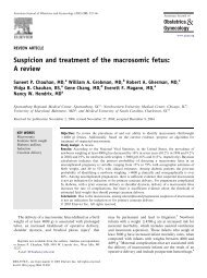
Fig. 8. <strong>Vasa</strong> previa. Transvaginal ultrasonography with<br />
color Doppler showing the fetal vessels running over<br />
internal os of the cervix (arrow). h, fetal head.<br />
Oyelese. <strong>Placenta</strong> <strong>Previa</strong>, <strong>Accreta</strong>, <strong>and</strong> <strong>Vasa</strong> <strong>Previa</strong>. Obstet<br />
Gynecol 2006.<br />
ureters. Intentional cystotomy may be helpful in identifying<br />
the extent of involvement <strong>and</strong> location of the<br />
ureters (Bakri YN, Sundin T. Cystotomy for placenta<br />
previa percreta with bladder invasion [letter]. Urology<br />
1992;40:580).<br />
VASA PREVIA<br />
Definition<br />
<strong>Vasa</strong> previa refers to fetal vessels running through the<br />
membranes over the cervix <strong>and</strong> under the fetal presenting<br />
part, unprotected by placenta or umbilical<br />
cord. 74 The condition usually results either from a<br />
velamentous insertion of the cord into the membranes<br />
rather than the placenta (Fig. 7) or from vessels<br />
running between lobes of a placenta with one or more<br />
accessory lobes. 74,75<br />
Clinical Importance<br />
<strong>Vasa</strong> previa is a condition which, undiagnosed, is<br />
associated with a perinatal mortality of approximately<br />
60%. 76 The condition is important because, when the<br />
membranes rupture, spontaneously or artificially, the<br />
fetal vessels running through the membranes have a<br />
high risk of concomitant rupture, frequently resulting<br />
in fetal exsanguination <strong>and</strong> death. 74,77 Because the<br />
fetal blood volume is only about 80–100 mL/kg, loss<br />
of even small amounts of blood could prove disastrous<br />
to the fetus. Pressure on the unprotected vessels<br />
by the presenting part could lead to fetal asphyxia <strong>and</strong><br />
death.<br />
VOL. 107, NO. 4, APRIL 2006 Oyelese <strong>and</strong> Smulian <strong>Placenta</strong> <strong>Previa</strong>, <strong>Accreta</strong>, <strong>and</strong> <strong>Vasa</strong> <strong>Previa</strong> 937
Incidence <strong>and</strong> Risk Factors<br />
The estimated incidence of vasa previa is approximately<br />
1 in 2,500 deliveries. 74 Risk factors for the<br />
condition include a second-trimester low-lying placenta<br />
(even if the “low-lying” placenta or placenta<br />
previa resolves in the third trimester), 78 pregnancies in<br />
which the placenta has accessory lobes, multiple<br />
pregnancies, <strong>and</strong> pregnancies resulting from in vitro<br />
fertilization. 79<br />
Pathophysiology<br />
The pathophysiology of vasa previa was discussed<br />
earlier under the pathophysiology of placenta previa.<br />
Diagnostic Approach<br />
<strong>Vasa</strong> previa is most commonly diagnosed when rupture<br />
of the membranes is accompanied by vaginal<br />
bleeding <strong>and</strong> fetal distress or death. The diagnosis is<br />
often confirmed only when the placenta is inspected<br />
after delivery. Consequently, until recently, most obstetricians<br />
have been resigned to the belief that the<br />
death of a fetus from a ruptured vasa previa is<br />
unavoidable. Very rarely (<strong>and</strong> fortuitously), vasa previa<br />
may be diagnosed during a digital cervical examination<br />
when the examiner’s fingers palpate fetal<br />
vessels running through the membranes. Use of an<br />
amnioscope in this situation may allow direct visualization<br />
of the vessels. When bleeding occurs in pregnancy<br />
or during labor, a test to determine the presence<br />
of fetal blood cells in the vaginal blood, such as<br />
the Apt test or Kleihauer-Bettke test, may aid in the<br />
diagnosis of vasa previa. 74 However, when acute<br />
bleeding occurs from a ruptured vasa previa, emergent<br />
delivery is frequently indicated, <strong>and</strong> there may<br />
be no time to test for fetal blood cells. Whenever<br />
bleeding accompanies rupture of the membranes in<br />
labor, especially if there are associated fetal heart rate<br />
decelerations, fetal bradycardia, or a sinusoidal fetal<br />
heart rate pattern, the obstetrician should have a high<br />
index of suspicion for a ruptured vasa previa. 80,81 In<br />
these situations, most frequently, immediate delivery<br />
by cesarean is indicated. Even when the neonate has<br />
lost considerable blood, immediate transfusion may<br />
be lifesaving. 82<br />
Numerous reports <strong>and</strong> studies have demonstrated<br />
that vasa previa can be diagnosed prenatally with<br />
ultrasonography. 75,83 The grayscale sonographic appearance<br />
of vasa previa is of linear echolucent structures<br />
overlying the cervix. 83 When color or power<br />
Doppler is used, flow can be demonstrated through<br />
these vessels, <strong>and</strong> pulsed Doppler will demonstrate a<br />
fetal umbilical arterial or venous waveform (Fig. 8). It<br />
is important to differentiate a vasa previa from a funic<br />
presentation. In the latter, the vessels will move when<br />
the patient changes position, especially when the<br />
patient is placed in the Trendelenburg position. Conversely,<br />
the vessels do not move when there is a vasa<br />
previa. The majority of prenatally diagnosed cases of<br />
vasa previa are detected incidentally in women who<br />
have transvaginal sonography for evaluation of lowlying<br />
placentas. However, studies have demonstrated<br />
that the majority of cases of vasa previa in asymptomatic<br />
women can be diagnosed prenatally through a<br />
policy of routinely evaluating the placental cord insertion<br />
when an ultrasound examination is performed<br />
<strong>and</strong> considering vaginal sonography with color Doppler<br />
if the placental cord insertion cannot be identified<br />
or if there is a low-lying placenta or a suspected<br />
succenturiate placental lobe. 75,83,84<br />
At least 4 studies have prospectively evaluated<br />
the use of ultrasonography in routine screening for<br />
vasa previa in large populations. 75,83–85 These studies<br />
found that sonographic identification of placental<br />
cord insertion was accurate <strong>and</strong> sensitive <strong>and</strong> added<br />
little or no extra time to the duration of the obstetric<br />
sonographic examination. In all the prenatally diagnosed<br />
cases, the neonatal survival of infants without<br />
congenital malformations was 100%.<br />
Therapeutic Approach<br />
Good outcomes with vasa previa depend on prenatal<br />
diagnosis <strong>and</strong> delivery by cesarean before rupture of<br />
the membranes. We previously carried out a multicenter<br />
retrospective study of 155 cases of vasa previa,<br />
evaluating the impact of prenatal diagnosis on outcomes<br />
of pregnancies complicated by vasa previa. 76<br />
In 61 of these cases, the diagnosis was made prenatally.<br />
We determined that, in the absence of prenatal<br />
diagnosis, the perinatal mortality was 56%, whereas<br />
97% of fetuses survived when the diagnosis was made<br />
prenatally. 76 Among survivors, when the diagnosis<br />
was not made prenatally, the median 1- <strong>and</strong> 5-minute<br />
Apgar scores were only 1 <strong>and</strong> 4, respectively, compared<br />
with 8 <strong>and</strong> 9, respectively, when the condition<br />
was diagnosed prenatally. 76 Two thirds of women had<br />
a low-lying placenta in the second trimester, whereas,<br />
by the time of delivery, only one third of these (20%)<br />
had a low-lying placenta. In one third of cases, the<br />
placenta was bi-lobed. The main predictors of survival<br />
were prenatal diagnosis <strong>and</strong> gestational age at<br />
delivery.<br />
Consideration should be given to hospitalization<br />
at about 30–32 weeks <strong>and</strong> administration of corticosteroids<br />
to promote fetal lung maturation. Hospitalization<br />
allows proximity to the operating room for<br />
938 Oyelese <strong>and</strong> Smulian <strong>Placenta</strong> <strong>Previa</strong>, <strong>Accreta</strong>, <strong>and</strong> <strong>Vasa</strong> <strong>Previa</strong> OBSTETRICS & GYNECOLOGY
emergent cesarean delivery if the membranes rupture.<br />
Approximately 10% of women will rupture their<br />
membranes before the onset of labor, so this risk is<br />
significant. However, in selected asymptomatic patients,<br />
there may be a role for outpatient management,<br />
especially if the patient has no signs of labor or<br />
uterine activity <strong>and</strong> has a long-closed cervix on<br />
transvaginal sonography. Delivery should occur at an<br />
institution where there are adequate facilities for<br />
neonatal resuscitation that might include emergent<br />
blood transfusions. It is preferable that, before surgery,<br />
the surgeon is aware of the position of the fetal<br />
vessels <strong>and</strong> plans the incision to avoid lacerating these<br />
vessels. We have previously described the use of<br />
3-dimensional ultrasonography with power Doppler<br />
angiography to map out the fetal vessels <strong>and</strong> thereby<br />
make the optimal uterine incision. 86,87 It is desirable to<br />
deliver the fetus en caul, with intact membranes,<br />
avoiding incising the membranes.<br />
A gestational age of between 35 <strong>and</strong> 36 weeks is<br />
the optimal age for cesarean delivery in women with<br />
vasa previa, with a reasonable tradeoff between prematurity<br />
with the risk of respiratory distress syndrome<br />
<strong>and</strong> that of rupture of the membranes with the risk of<br />
fetal exsanguination <strong>and</strong> death. 76 Although amniocentesis<br />
is generally recommended before elective cesarean<br />
delivery before 39 weeks in most conditions, in<br />
vasa previa, if the membranes rupture, the risks of<br />
fetal death or adverse outcome are so severe that we<br />
feel it is justifiable to deliver these women by 36<br />
weeks without amniocentesis documentation of lung<br />
maturity<br />
We can think of no other condition in which<br />
prenatal diagnosis <strong>and</strong> appropriate perinatal management<br />
makes such a dramatic impact on the difference<br />
between survival <strong>and</strong> death for an otherwise healthy<br />
infant. Thus, especially because it adds little in terms<br />
of time to the routine obstetric sonogram, it is our<br />
opinion that screening for vasa previa should be<br />
routine.<br />
CONCLUSION<br />
Achieving optimal outcomes with placenta previa,<br />
placenta accreta, <strong>and</strong> vasa previa depends on prenatal<br />
diagnosis <strong>and</strong> appropriate management at the time of<br />
delivery. Advances in ultrasonography have made it<br />
possible to diagnose all 3 conditions with reasonable<br />
accuracy, which allows appropriate management<br />
planning. Women with these conditions should be<br />
considered at high risk <strong>and</strong> should be delivered at<br />
institutions with skilled personnel, adequate blood<br />
transfusion facilities, <strong>and</strong> good neonatal resources.<br />
REFERENCES<br />
1. Crane JM, Van den Hof MC, Dodds L, Armson BA, Liston R.<br />
Maternal complications with placenta previa. Am J Perinatol<br />
2000;17:101–5.<br />
2. Iyasu S, Saftlas AK, Rowley DL, Koonin LM, Lawson HW,<br />
Atrash HK. The epidemiology of placenta previa in the United<br />
States, 1979 through 1987. Am J Obstet Gynecol 1993;168:<br />
1424–9.<br />
3. Crane JM, van den Hof MC, Dodds L, Armson BA, Liston R.<br />
Neonatal outcomes with placenta previa. Obstet Gynecol<br />
1999;93:541–4.<br />
4. Ananth CV, Smulian JC, Vintzileos AM. The association of<br />
placenta previa with history of cesarean delivery <strong>and</strong> abortion:<br />
a metaanalysis. Am J Obstet Gynecol 1997;177:1071–8.<br />
5. Barrett JM, Boehm FH, Killam AP. Induced abortion: a risk<br />
factor for placenta previa. Am J Obstet Gynecol 1981;141:<br />
769–72.<br />
6. Ananth CV, Savitz DA, Luther ER. Maternal cigarette smoking<br />
as a risk factor for placental abruption, placenta previa, <strong>and</strong><br />
uterine bleeding in pregnancy. Am J Epidemiol 1996;144:<br />
881–9.<br />
7. Ananth CV, Wilcox AJ, Savitz DA, Bowes WA Jr, Luther ER.<br />
Effect of maternal age <strong>and</strong> parity on the risk of uteroplacental<br />
bleeding disorders in pregnancy. Obstet Gynecol 1996;88:<br />
511–6.<br />
8. Macones GA, Sehdev HM, Parry S, Morgan MA, Berlin JA.<br />
The association between maternal cocaine use <strong>and</strong> placenta<br />
previa. Am J Obstet Gynecol 1997;177:1097–100.<br />
9. Ananth CV, Demissie K, Smulian JC, Vintzileos AM. <strong>Placenta</strong><br />
previa in singleton <strong>and</strong> twin births in the United States, 1989<br />
through 1998: a comparison of risk factor profiles <strong>and</strong> associated<br />
conditions. Am J Obstet Gynecol 2003;188:275–81.<br />
10. Benirschke K, Kaufmann P. Pathology of the human placenta.<br />
4th ed. New York (NY): Springer; 2000.<br />
11. Smith RS, Lauria MR, Comstock CH, Treadwell MC, Kirk JS,<br />
Lee W, et al. Transvaginal ultrasonography for all placentas<br />
that appear to be low-lying or over the internal cervical os.<br />
Ultrasound Obstet Gynecol 1997;9:22–4.<br />
12. Leerentveld RA, Gilberts EC, Arnold MJ, Wladimiroff JW.<br />
Accuracy <strong>and</strong> safety of transvaginal sonographic placental<br />
localization. Obstet Gynecol 1990;76:759–62.<br />
13. Lauria MR, Smith RS, Treadwell MC, Comstock CH, Kirk JS,<br />
Lee W, et al. The use of second-trimester transvaginal sonography<br />
to predict placenta previa. Ultrasound Obstet Gynecol<br />
1996;8:337–40.<br />
14. Varma TR. The implication of a low implantation of the<br />
placenta detected by ultrasonography in early pregnancy. Acta<br />
Obstet Gynecol Sc<strong>and</strong> 1981;60:265–8.<br />
15. Timor-Tritsch IE, Yunis RA. Confirming the safety of transvaginal<br />
sonography in patients suspected of placenta previa.<br />
Obstet Gynecol 1993;81:742–4.<br />
16. Hertzberg BS, Bowie JD, Carroll BA, Kliewer MA, Weber TM.<br />
Diagnosis of placenta previa during the third trimester: role of<br />
transperineal sonography. AJR Am J Roentgenol 1992;159:<br />
83–7.<br />
17. Becker RH, Vonk R, Mende BC, Ragosch V, Entezami M.<br />
The relevance of placental location at 20-23 gestational weeks<br />
for prediction of placenta previa at delivery: evaluation of 8650<br />
cases. Ultrasound Obstet Gynecol 2001;17:496–501.<br />
18. Rosati P, Guariglia L. Clinical significance of placenta previa<br />
detected at early routine transvaginal scan. J Ultrasound Med<br />
2000;19:581–5.<br />
VOL. 107, NO. 4, APRIL 2006 Oyelese <strong>and</strong> Smulian <strong>Placenta</strong> <strong>Previa</strong>, <strong>Accreta</strong>, <strong>and</strong> <strong>Vasa</strong> <strong>Previa</strong> 939
19. Hill LM, DiNofrio DM, Chenevey P. Transvaginal sonographic<br />
evaluation of first-trimester placenta previa. Ultrasound<br />
Obstet Gynecol 1995;5:301–3.<br />
20. Mustafa SA, Brizot ML, Carvalho MH, Watanabe L, Kahhale<br />
S, Zugaib M. Transvaginal ultrasonography in predicting placenta<br />
previa at delivery: a longitudinal study. Ultrasound<br />
Obstet Gynecol 2002;20:356–9.<br />
21. Taipale P, Hiilesmaa V, Ylostalo P. Transvaginal ultrasonography<br />
at 18-23 weeks in predicting placenta previa at delivery.<br />
Ultrasound Obstet Gynecol 1998;12:422–5.<br />
22. Dashe JS, McIntire DD, Ramus RM, Santos-Ramos R, Twickler<br />
DM. Persistence of placenta previa according to gestational<br />
age at ultrasound detection. Obstet Gynecol 2002;99:692–7.<br />
23. MacAfee C. <strong>Placenta</strong> previa: study of 174 cases. J Obstet<br />
Gynecol Br Emp 1945;52:313–24.<br />
24. Sharma A, Suri V, Gupta I. Tocolytic therapy in conservative<br />
management of symptomatic placenta previa. Int J Gynaecol<br />
Obstet 2004;84:109–13.<br />
25. Besinger RE, Moniak CW, Paskiewicz LS, Fisher SG, Tomich<br />
PG. The effect of tocolytic use in the management of symptomatic<br />
placenta previa. Am J Obstet Gynecol 1995;172:<br />
1770–5; discussion 1775–8.<br />
26. Cotton DB, Read JA, Paul RH, Quilligan EJ. The conservative<br />
aggressive management of placenta previa. Am J Obstet<br />
Gynecol 1980;137:687–95.<br />
27. Droste S, Keil K. Expectant management of placenta previa:<br />
cost-benefit analysis of outpatient treatment. Am J Obstet<br />
Gynecol 1994;170:1254–7.<br />
28. Mouer JR. <strong>Placenta</strong> previa: antepartum conservative management,<br />
inpatient versus outpatient. Am J Obstet Gynecol 1994;<br />
170:1683–5; discussion 1685–6.<br />
29. D’Angelo LJ, Irwin LF. Conservative management of placenta<br />
previa: a cost-benefit analysis. Am J Obstet Gynecol 1984;149:<br />
320–6.<br />
30. Wing DA, Paul RH, Millar LK. Management of the symptomatic<br />
placenta previa: a r<strong>and</strong>omized, controlled trial of inpatient<br />
versus outpatient expectant management. Am J Obstet<br />
Gynecol 1996;175:806–11.<br />
31. Arias F. Cervical cerclage for the temporary treatment of<br />
patients with placenta previa. Obstet Gynecol 1988;71:545–8.<br />
32. Cobo E, Conde-Agudelo A, Delgado J, Canaval H, Congote A.<br />
Cervical cerclage: an alternative for the management of placenta<br />
previa? Am J Obstet Gynecol 1998;179:122–5.<br />
33. Oppenheimer LW, Farine D, Ritchie JW, Lewinsky RM,<br />
Telford J, Fairbanks LA. What is a low-lying placenta? Am J<br />
Obstet Gynecol 1991;165:1036–8.<br />
34. Bhide A, Prefumo F, Moore J, Hollis B, Thilaganathan B.<br />
<strong>Placenta</strong>l edge to internal os distance in the late third trimester<br />
<strong>and</strong> mode of delivery in placenta praevia. BJOG 2003;110:<br />
860–4.<br />
35. Dawson WB, Dumas MD, Romano WM, Gagnon R, Gratton<br />
RJ, Mowbray RD. Translabial ultrasonography <strong>and</strong> placenta<br />
previa: does measurement of the os-placenta distance predict<br />
outcome? J Ultrasound Med 1996;15:441–6.<br />
36. Boehm FH, Fleischer AC, Barrett JM. Sonographic placental<br />
localization in the determination of the site of uterine incision<br />
for placenta previa. J Ultrasound Med 1982;1:311–4.<br />
37. Parekh N, Husaini SW, Russell IF. Caesarean section for<br />
placenta praevia: a retrospective study of anaesthetic management.<br />
Br J Anaesth 2000;84:725–30.<br />
38. Hong JY, Jee YS, Yoon HJ, Kim SM. Comparison of general<br />
<strong>and</strong> epidural anesthesia in elective cesarean section for placenta<br />
previa totalis: maternal hemodynamics, blood loss <strong>and</strong><br />
neonatal outcome. Int J Obstet Anesth 2003;12:12–6.<br />
39. Frederiksen MC, Glassenberg R, Stika CS. <strong>Placenta</strong> previa: a<br />
22-year analysis. Am J Obstet Gynecol 1999;180:1432–7.<br />
40. Bonner SM, Haynes SR, Ryall D. The anaesthetic management<br />
of Caesarean section for placenta praevia: a questionnaire<br />
survey. Anaesthesia 1995;50:992–4.<br />
41. Hudon L, Belfort MA, Broome DR. Diagnosis <strong>and</strong> management<br />
of placenta percreta: a review. Obstet Gynecol Surv<br />
1998;53:509–17.<br />
42. O’Brien JM, Barton JR, Donaldson ES. The management of<br />
placenta percreta: conservative <strong>and</strong> operative strategies. Am J<br />
Obstet Gynecol 1996;175:1632–8.<br />
43. Kastner ES, Figueroa R, Garry D, Maulik D. Emergency<br />
peripartum hysterectomy: experience at a community teaching<br />
hospital. Obstet Gynecol 2002;99:971–5.<br />
44. deRoux SJ, Prendergast NC, Adsay NV. Spontaneous uterine<br />
rupture with fatal hemoperitoneum due to placenta accreta<br />
percreta: a case report <strong>and</strong> review of the literature. Int J<br />
Gynecol Pathol 1999;18:82–6.<br />
45. Miller DA, Chollet JA, Goodwin TM. Clinical risk factors for<br />
placenta previa-placenta accreta. Am J Obstet Gynecol 1997;<br />
177:210–4.<br />
46. Wu S, Kocherginsky M, Hibbard JU. Abnormal placentation:<br />
twenty-year analysis. Am J Obstet Gynecol 2005;192:1458–61.<br />
47. Clark SL, Koonings PP, Phelan JP. <strong>Placenta</strong> previa/accreta <strong>and</strong><br />
prior cesarean section. Obstet Gynecol 1985;66:89–92.<br />
48. Zelop C, Nadel A, Frigoletto FD Jr, Pauker S, MacMillan M,<br />
Benacerraf BR. <strong>Placenta</strong> accreta/percreta/increta: a cause of<br />
elevated maternal serum alpha-fetoprotein. Obstet Gynecol<br />
1992;80:693–4.<br />
49. Kupferminc MJ, Tamura RK, Wigton TR, Glassenberg R,<br />
Socol ML. <strong>Placenta</strong> accreta is associated with elevated maternal<br />
serum alpha-fetoprotein. Obstet Gynecol 1993;82:266–9.<br />
50. Comstock CH. Antenatal diagnosis of placenta accreta: a<br />
review. Ultrasound Obstet Gynecol 2005;26:89–96.<br />
51. Comstock CH, Love JJ Jr, Bronsteen RA, Lee W, Vettraino<br />
IM, Huang RR, et al. Sonographic detection of placenta<br />
accreta in the second <strong>and</strong> third trimesters of pregnancy. Am J<br />
Obstet Gynecol 2004;190:1135–40.<br />
52. Finberg HJ, Williams JW. <strong>Placenta</strong> accreta: prospective sonographic<br />
diagnosis in patients with placenta previa <strong>and</strong> prior<br />
cesarean section. J Ultrasound Med 1992;11:333–43.<br />
53. Chou MM, Ho ES, Lee YH. Prenatal diagnosis of placenta<br />
previa accreta by transabdominal color Doppler ultrasound.<br />
Ultrasound Obstet Gynecol 2000;15:28–35.<br />
54. Comstock CH, Lee W, Vettraino IM, Bronsteen RA. The early<br />
sonographic appearance of placenta accreta. J Ultrasound Med<br />
2003;22:19–23; quiz 24–6.<br />
55. Thorp JM Jr, Wells SR, Wiest HH, Jeffries L, Lyles E.<br />
First-trimester diagnosis of placenta previa percreta by magnetic<br />
resonance imaging. Am J Obstet Gynecol 1998;178:<br />
616–8.<br />
56. Palacios Jaraquemada JM, Bruno CH. Magnetic resonance<br />
imaging in 300 cases of placenta accreta: surgical correlation of<br />
new findings. Acta Obstet Gynecol Sc<strong>and</strong> 2005;84:716–24.<br />
57. Levine D, Hulka CA, Ludmir J, Li W, Edelman RR. <strong>Placenta</strong><br />
accreta: evaluation with color Doppler US, power Doppler US,<br />
<strong>and</strong> MR imaging. Radiology 1997;205:773–6.<br />
58. Chestnut DH, Dewan DM, Redick LF, Caton D, Spielman FJ.<br />
Anesthetic management for obstetric hysterectomy: a multiinstitutional<br />
study. Anesthesiology 1989;70:607–10.<br />
59. <strong>Placenta</strong> accreta. ACOG Committee Opinion No. 266. American<br />
College of Obstetricians <strong>and</strong> Gynecologists. Obstet<br />
Gynecol 2002;99:169–70.<br />
940 Oyelese <strong>and</strong> Smulian <strong>Placenta</strong> <strong>Previa</strong>, <strong>Accreta</strong>, <strong>and</strong> <strong>Vasa</strong> <strong>Previa</strong> OBSTETRICS & GYNECOLOGY
60. Alvarez M, Lockwood CJ, Ghidini A, Dottino P, Mitty HA,<br />
Berkowitz RL. Prophylactic <strong>and</strong> emergent arterial catheterization<br />
for selective embolization in obstetric hemorrhage. Am J<br />
Perinatol 1992;9:441–4.<br />
61. Kidney DD, Nguyen AM, Ahdoot D, Bickmore D, Deutsch<br />
LS, Majors C. Prophylactic perioperative hypogastric artery<br />
balloon occlusion in abnormal placentation. AJR Am J Roentgenol<br />
2001;176:1521–4.<br />
62. Levine AB, Kuhlman K, Bonn J. <strong>Placenta</strong> accreta: comparison<br />
of cases managed with <strong>and</strong> without pelvic artery balloon<br />
catheters. J Matern Fetal Med 1999;8:173–6.<br />
63. Arulkumaran S, Ng CS, Ingemarsson I, Ratnam SS. Medical<br />
treatment of placenta accreta with methotrexate. Acta Obstet<br />
Gynecol Sc<strong>and</strong> 1986;65:285–6.<br />
64. Butt K, Gagnon A, Delisle MF. Failure of methotrexate <strong>and</strong><br />
internal iliac balloon catheterization to manage placenta percreta.<br />
Obstet Gynecol 2002;99:981–2.<br />
65. Kayem G, Davy C, Goffinet F, Thomas C, Clement D, Cabrol<br />
D. Conservative versus extirpative management in cases of<br />
placenta accreta. Obstet Gynecol 2004;104:531–6.<br />
66. Weinstein A, Ch<strong>and</strong>ra P, Schiavello H, Fleischer A. Conservative<br />
management of placenta previa percreta in a Jehovah’s<br />
Witness. Obstet Gynecol 2005;105 suppl:1247–50.<br />
67. Jaffe R, DuBeshter B, Sherer DM, Thompson EA, Woods JR<br />
Jr. Failure of methotrexate treatment for term placenta percreta.<br />
Am J Obstet Gynecol 1994;171:558–9.<br />
68. Mussalli GM, Shah J, Berck DJ, Elimian A, Tejani N, Manning<br />
FA. <strong>Placenta</strong> accreta <strong>and</strong> methotrexate therapy: three case<br />
reports. J Perinatol 2000;20:331–4.<br />
69. Silver LE, Hobel CJ, Lagasse L, Luttrull JW, Platt LD. <strong>Placenta</strong><br />
previa percreta with bladder involvement: new considerations<br />
<strong>and</strong> review of the literature. Ultrasound Obstet Gynecol 1997;<br />
9:131–8.<br />
70. Price FV, Resnik E, Heller KA, Christopherson WA. <strong>Placenta</strong><br />
previa percreta involving the urinary bladder: a report of two<br />
cases <strong>and</strong> review of the literature. Obstet Gynecol 1991;78<br />
suppl:508–11.<br />
71. Pelosi MA, 3rd, Pelosi MA. Modified cesarean hysterectomy for<br />
placenta previa percreta with bladder invasion: retrovesical lower<br />
uterine segment bypass. Obstet Gynecol 1999;93 suppl:830–3.<br />
72. Caliskan E, Tan O, Kurtaran V, Dilbaz B, Haberal A. <strong>Placenta</strong><br />
previa percreta with urinary bladder <strong>and</strong> ureter invasion. Arch<br />
Gynecol Obstet 2003;268:343–4.<br />
73. Washecka R, Behling A. Urologic complications of placenta<br />
percreta invading the urinary bladder: a case report <strong>and</strong> review<br />
of the literature. Hawaii Med J 2002;61:66–9.<br />
74. Oyelese KO, Turner M, Lees C, Campbell S. <strong>Vasa</strong> previa: an<br />
avoidable obstetric tragedy. Obstet Gynecol Surv 1999;54:<br />
138–45.<br />
75. Catanzarite V, Maida C, Thomas W, Mendoza A, Stanco L,<br />
Piacquadio KM. Prenatal sonographic diagnosis of vasa previa:<br />
ultrasound findings <strong>and</strong> obstetric outcome in ten cases. Ultrasound<br />
Obstet Gynecol 2001;18:109–15.<br />
76. Oyelese Y, Catanzarite V, Prefumo F, Lashley S, Schachter M,<br />
Tovbin Y, et al. <strong>Vasa</strong> previa: the impact of prenatal diagnosis<br />
on outcomes. Obstet Gynecol 2004;103:937–42.<br />
77. Oyelese KO, Schwarzler P, Coates S, Sanusi FA, Hamid R,<br />
Campbell S. A strategy for reducing the mortality rate from<br />
vasa previa using transvaginal sonography with color Doppler.<br />
Ultrasound Obstet Gynecol 1998;12:434–8.<br />
78. Francois K, Mayer S, Harris C, Perlow JH. Association of vasa<br />
previa at delivery with a history of second-trimester placenta<br />
previa. J Reprod Med 2003;48:771–4.<br />
79. Schachter M, Tovbin Y, Arieli S, Friedler S, Ron-El R,<br />
Sherman D. In vitro fertilization is a risk factor for vasa previa.<br />
Fertil Steril 2002;78:642–3.<br />
80. Antoine C, Young BK, Silverman F, Greco MA, Alvarez SP.<br />
Sinusoidal fetal heart rate pattern with vasa previa in twin<br />
pregnancy. J Reprod Med 1982;27:295–300.<br />
81. Gantt PA, Bird JS Jr, R<strong>and</strong>all GW. Sinusoidal fetal heart rate<br />
pattern with vasa previa. J Tenn Med Assoc 1990;83:393–4.<br />
82. Schellpfeffer MA. Improved neonatal outcome of vasa previa<br />
with aggressive intrapartum management: a report of two<br />
cases. J Reprod Med 1995;40:327–32.<br />
83. Lee W, Lee VL, Kirk JS, Sloan CT, Smith RS, Comstock CH.<br />
<strong>Vasa</strong> previa: prenatal diagnosis, natural evolution, <strong>and</strong> clinical<br />
outcome. Obstet Gynecol 2000;95:572–6.<br />
84. Sepulveda W, Rojas I, Robert JA, Schnapp C, Alcalde JL.<br />
Prenatal detection of velamentous insertion of the umbilical<br />
cord: a prospective color Doppler ultrasound study. Ultrasound<br />
Obstet Gynecol 2003;21:564–9.<br />
85. Nomiyama M, Toyota Y, Kawano H. Antenatal diagnosis of<br />
velamentous umbilical cord insertion <strong>and</strong> vasa previa with<br />
color Doppler imaging. Ultrasound Obstet Gynecol 1998;12:<br />
426–9.<br />
86. Canterino JC, Mondestin-Sorrentino M, Muench MV, Feld S,<br />
Baum JD, Fern<strong>and</strong>ez CO. <strong>Vasa</strong> previa: prenatal diagnosis <strong>and</strong><br />
evaluation with 3-dimensional sonography <strong>and</strong> power angiography.<br />
J Ultrasound Med 2005;24:721–5.<br />
87. Oyelese Y, Chavez MR, Yeo L, Giannina G, Kontopoulos EV,<br />
Smulian JC, et al. Three-dimensional sonographic diagnosis of<br />
vasa previa. Ultrasound Obstet Gynecol 2004;24:211–5.<br />
VOL. 107, NO. 4, APRIL 2006 Oyelese <strong>and</strong> Smulian <strong>Placenta</strong> <strong>Previa</strong>, <strong>Accreta</strong>, <strong>and</strong> <strong>Vasa</strong> <strong>Previa</strong> 941